. 2007 Sep;13(9):1453-68.
doi: 10.1261/rna.501707. Epub 2007 Jul 12.

FRET-detectable interactions between the ARE binding proteins, HuR and p37AUF1

Affiliations

FRET-detectable interactions between the ARE binding proteins, HuR and p37AUF1

Pamela S David et al. RNA. 2007 Sep.

Abstract

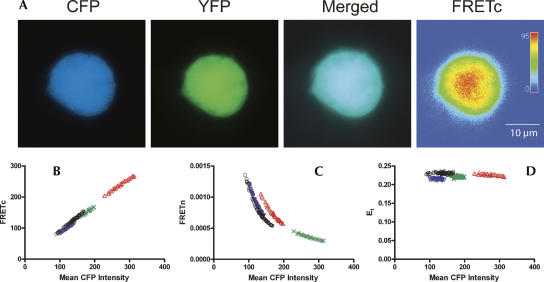
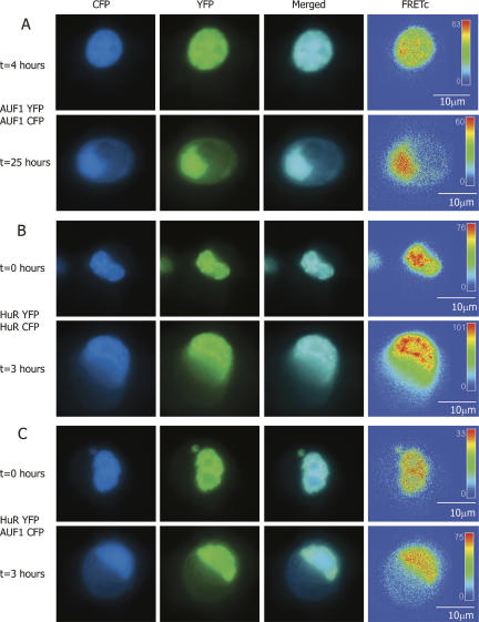
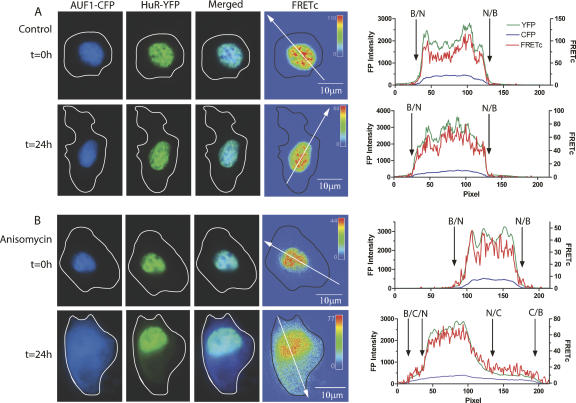
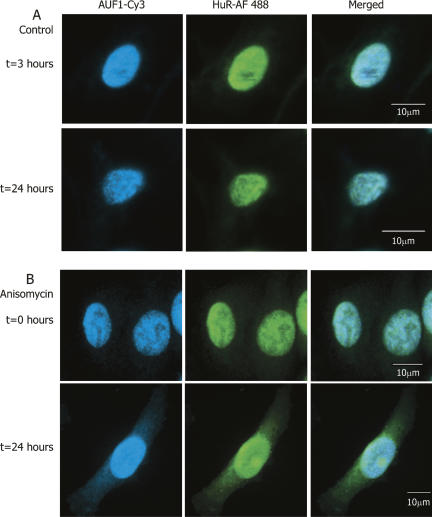
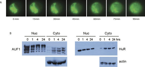
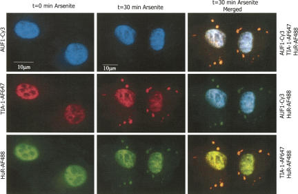
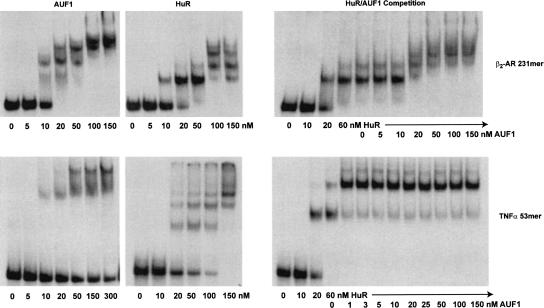
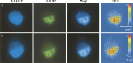
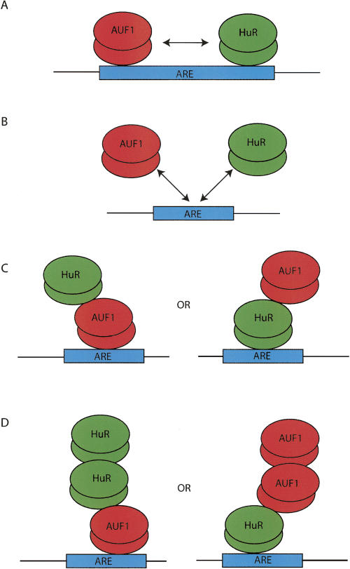
A number of highly regulated gene classes are regulated post-transcriptionally at the level of mRNA stability. A central feature in these mRNAs is the presence of A+U-rich elements (ARE) within their 3' UTRs. Two ARE binding proteins, HuR and AUF1, are associated with mRNA stabilization and destabilization, respectively. Previous studies have demonstrated homomultimerization of each protein and the capacity to bind simultaneous or competitively to a single ARE. To investigate this possibility further, cell biological and biophysical approaches were undertaken. Protein-protein interaction was monitored by fluorescence resonance energy transfer (FRET) and by immunocytochemistry in live and fixed cells using fluorescently labeled CFP/YFP fusion proteins of HuR and p37AUF1. Strong nuclear FRET between HuR/HuR and AUF1/AUF1 homodimers as well as HuR/AUF1 heterodimers was observed. Treatment with the MAP kinase activator, anisomycin, which commonly stabilizes ARE-containing mRNAs, caused rapid nuclear to cytoplasmic shuttling of HuR. AUF1 also underwent shuttling, but on a longer time scale. After shuttling, HuR/HuR, AUF1/AUF1, and HuR/AUF1, FRET was also observed in the cytoplasm. In further studies, arsenite rapidly induced the formation of stress granules containing HuR and TIA-1 but not AUF1. The current studies demonstrate that two mRNA binding proteins, HuR and AUF1, are colocalized and are capable of functional interaction in both the nucleus and cytoplasm. FRET-based detection of AUF1/HuR interaction may serve as a basis of opening up new dimensions in delineating the functional interaction of mRNA binding proteins with RNA turnover.


Figures

FIGURE 1.
FIGURE 1.
FRET positive control and representative FRET calculations. A tandem vector construct of eCFP and eYFP was used as a positive control for the detection of FRET in DDT1-MF2 cells (Galperin et al. 2004). Slidebook software (v4.1.0.4) was used to obtain mean intensity values from identically sized areas sampled from the entire cell for CFP, YFP, and the raw FRET signal. These data were used to calculate FRETc, FRETn, and the FRET efficiency value, E, which are shown plotted vs. the mean CFP intensities. The maximal FRET efficiency (Et) obtained for CFP:YFP in DDT1-MF2 cells is 0.22±0.02. (A) DDT1-MF2 cell images. The CFP, YFP, and merged channel views show the uniform intracellular distribution of the CFP and YFP proteins throughout the cell. The FRETc image is shown in thermal gradient pseudocolor, where red indicates the strongest signal. The size scale bar (μm) applies to all images. (B) FRETc versus mean CFP intensity. (C) FRETn versus mean CFP intensity. (D) Et versus mean CFP intensity.
FIGURE 2.
FIGURE 2.
FRET-based detection of p37AUF1/p37AUF1, HuR/HuR, and p37AUF1/HuR protein pair interactions in live cells. DDT1-MF2 cells were transiently transfected with pairs of complimentary, fluorescently labeled constructs of HuR and/or p37AUF1. Transfected cells were treated with anisomycin to stimulate nuclear/cytoplasmic shuttling of mRNA binding proteins. (A) p37AUF1-eYFP and p37AUF1-eCFP interaction. After 4 h of treatment with anisomycin, p37AUF1 remains localized to the nucleus (top row) and exhibits colocalization (merged channel column) detectable by FRETc, which is indicative of a stable homodimeric interaction (top row, FRETc column). After 25 h of anisomycin treatment, cytoplasmic shuttling of p37AUF is readily apparent (bottom row). The p37AUF1 present in the cytoplasm colocalizes, which is detectable by the merged channel view and the FRETc signal present in both the nucleus and cytoplasm. (B) HuR-eYFP and HuR-eCFP interaction. At time zero, HuR is present in the nucleus, and exhibits colocalization and a robust FRETc signal (top row). Anisomycin causes rapid shuttling of HuR to the cytoplasm, and a FRET-detectable, stable interaction between HuR is present in both the nucleus and cytoplasm (bottom row). (C) HuR-eYFP and p37AUF1-eCFP interaction. At time zero, p37AUF1 and HuR are present in the nucleus, colocalize, and display a FRETc signal indicative of a stable heteromeric interaction (top row). Treatment with anisomycin causes shuttling of both HuR and p37AUF1 into the cytoplasm. HuR and p37AUF1 colocalize in the cytoplasm and exhibit a FRET-detectable interaction in both the nuclear and cytoplasmic compartments. FRETc images are shown in thermal gradient pseudocolor. The scale bars in the FRETc image applies to all images in the same row.
FIGURE 3.
FIGURE 3.
Anisomycin-induced shuttling of HuR and p37AUF1 in fixed cells. DDT1-MF2 cells were transiently transfected with HuR-eYFP and p37AUF1-eCFP and treated with anisomycin for up to 24 h. HuR-eYFP and p37AUF1-eCFP are present in the nucleus of both the vehicle- and anisomycin treated cells at time zero (A,B, t=0 h, CFP and YFP columns). The merged CFP:YFP image shows that both proteins colocalize to the nucleus but also exhibit a non-identical nuclear distribution (see graphs in right panel). The FRETc image indicates that HuR and p37AUF1 are in sufficiently close proximity in the nucleus to be of functional significance (A,B, t=0 h, FRETc column). After 24 h of vehicle treatment, HuR and p37AUF1 remain in the nucleus (A, t=24 h). After 24 h of anisomycin treatment, shuttling of HuR and p37AUF1 from nucleus to cytoplasm is readily apparent (B, t=24 h). The FRETc image indicates the continued stable interaction of HuR and p37AUF1 in the nucleus and that the proteins retain this interaction in the cytoplasm. (Free-form line) Cell boundary for each image. The intensity for each image in the FRETc column is represented in a thermal gradient pseudocolor. The scale bar in the FRETc image applies to the entire row. (Graphs) The CFP and YFP intensities (left axis) and calculated FRETc (right axis) as described by the white directional arrow in the FRETc panel. (Black arrows) Location of the nuclear/cytoplasmic/background transition, (N) nucleus, (C) cytoplasm, (B) background.
FIGURE 4.
FIGURE 4.
Colocalization of endogenously expressed HuR and AUF1. Immunocytochemistry was used to detect the intracellular location of endogenously expressed HuR and AUF1 proteins in DDT1-MF2 cells. (A) In control cells, endogenous HuR (Alexa Fluor 488 conjugated 2° antibody) and AUF1 (Cy3 conjugated 2° antibody) are expressed almost exclusively in the nucleus and are shown to colocalize. (B) Anisomycin treatment induces shuttling of endogenous AUF1 and HuR from the nucleus to the cytoplasm (cf. t=0 h and t=24 h). The merged view shows the colocalization of HuR and AUF1 in the nucleus and the cytoplasm after 24 h of anisomycin treatment. The scale bars in the merged view apply to all images in the same row.
FIGURE 5.
FIGURE 5.
MAPK-induced nuclear/cytoplasmic shuttling of ARE binding proteins. (A) DDT1-MF2 cells were transiently transfected with HuR-eYFP. Anisomycin was added to the cells to stimulate nuclear to cytoplasmic shuttling. A single cell was tracked for the time course indicated. eYFP-labeled HuR rapidly shuttles from the nucleus to the cytoplasm. Cells were maintained at 37°C, 5% CO2 for the duration of the experiment. (B) Following treatment with anisomycin (1–24 h), DDT1-MF2 cells were harvested and nuclear and cytosolic fractions isolated. Immunodetection of endogenous AUF1 (p37, p40, p42, p45) and HuR was performed by Western blotting. For normalization, blots were stripped and reprobed for actin protein.
FIGURE 6.
FIGURE 6.
HuR, but not AUF1, is present in arsenite-induced stress granules. Sodium arsenite was used to induce stress granule formation in DDT1-MF2 cells. TIA-1 was used as a marker for stress granule formation. Immunocytochemistry employing triple antibody staining shows that in untreated cells, HuR, TIA-1 and AUF1 are all localized in the nucleus with a lesser amount of TIA-1 and HuR present in the cytoplasm (left column, t=0 min Arsenite). Rapid formation of stress granules containing HuR and TIA-1 occurs after 30 min of arsenite treatment, (center column, t=30 min Arsenite; right column, merged views). Sodium arsenite did not affect the cellular localization of AUF1, (center column, t=30 min Arsenite; right column, merged views). The scale bar in the left column applies to all images in this column. The scale bar in the center column applies to all images in this and the right column.
FIGURE 7.
FIGURE 7.
Competitive binding of p37AUF1 and HuR to 3′ -UTR ARE. Nondenaturing gel-shift assays were performed to assess the competitive interaction of purified recombinant p37AUF1 and HuR proteins for the TNFα 53 nt ARE and the hamster β2-AR 231 nt ARE. 32P-UTP labeled RNAs encoding the hamster β2-AR 231 nt ARE or TNFα 53-nucleotide AREs were incubated with either p37AUF1 alone (left), HuR alone (center), or HuR in competition with p37AUF1 (right). Holding the concentration of HuR at ∼3×Kd (60 nM), increasing amounts of purified recombinant GST-p37AUF1 were added in an attempt to displace HuR binding.
FIGURE 8.
FIGURE 8.
RNA dependence of HuR/p37AUF1 heterodimer. DDT1-MF2 cells were transiently transfected with HuR-eYFP and p37AUF1-eCFP. Anisomycin was added to the cells for 4 h to stimulate nuclear to cytoplasmic shuttling. Following permeabilization with Triton X-100, cells were either treated with ribonuclease A or mock treated prior to fixation. Both the mock- and ribonuclease-treated cells shows cytoplasmic localization of HuR and AUF1 (CFP and YFP channels), colocalization of HuR and AUF1 (merged view), and the presence of a FRETc signal in both the nucleus and cytoplasm. (A) Mock treated DDT1-MF2 cells, (B) ribonuclease treated cells.
FIGURE 9.
FIGURE 9.
Model of potential interactions of HuR and AUF1. AUF1 has been shown to form homomultimers in solution (DeMaria et al. 1997) as well as when bound to an ARE (Fig. 2A; Wilson et al. 1999; Blaxall et al. 2000b, 2002). HuR has also been shown to be able to form dimers (Fig. 2B; Soller and White 2005). AUF1 and HuR have also been shown to bind to mRNA concurrently and in a competitive manner (Lal et al. 2004). Based on these results and those presented in Figure 6, potential protein–protein and protein–RNA interactions of HuR and AUF1 are presented.

Similar articles

Cited by

References

    1. Abdelmohsen, K., Pullmann R., Jr, Lal, A., Kim, H.H., Galban, S., Yang, X., Blethrow, J.D., Walker, M., Shubert, J., Gillespie, D.A., et al. Phosphorylation of HuR by Chk2 regulates SIRT1 expression. Mol. Cell. 2007;25:543–557. - PMC - PubMed
    1. Abraham, W.T., Gilbert, E.M., Lowes, B.D., Minobe, W.A., Larrabee, P., Roden, R.L., Dutcher, D., Sederberg, J., Lindenfeld, J.A., Wolfel, E.E., et al. Coordinate changes in Myosin heavy chain isoform gene expression are selectively associated with alterations in dilated cardiomyopathy phenotype. Mol. Med. 2002;8:750–760. - PMC - PubMed
    1. Anderson, P., Kedersha, N. RNA granules. J. Cell Biol. 2006;172:803–808. - PMC - PubMed
    1. Atasoy, U., Watson, J., Patel, D., Keene, J.D. ELAV protein HuA (HuR) can redistribute between nucleus and cytoplasm and is upregulated during serum stimulation and T cell activation. J. Cell Sci. 1998;111:3145–3156. - PubMed
    1. Bakheet, T., Williams, B.R., Khabar, K.S. ARED 3.0: The large and diverse AU-rich transcriptome. Nucleic Acids Res. 2006;34:D111–D114. - PMC - PubMed

Publication types

MeSH terms

LinkOut - more resources