Clinical significance of midkine expression in pancreatic head carcinoma
- PMID: 17622248
- PMCID: PMC2360321
- DOI: 10.1038/sj.bjc.6603879
Clinical significance of midkine expression in pancreatic head carcinoma
Abstract
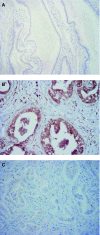
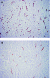
Midkine (MK) is a heparin-binding growth factor and a product of a retinoic acid-responsive gene. Midkine is overexpressed in many carcinomas and thought to play an important role in carcinogenesis. However, no studies have been focussed on the role of MK in pancreatic carcinoma. This study sought to evaluate the clinical significance of MK expression in pancreatic head carcinoma, including the relationship between immunohistochemical expression and clinicopathologic factors such as prognosis. Immunohistochemical expression of MK and CD34 was evaluated in pancreatic head carcinoma specimens from 75 patients who underwent surgical resection. Midkine was expressed in 53.3% of patients. Midkine expression was significantly correlated with venous invasion, microvessel density, and liver metastasis (P=0.0063, 0.0025, and 0.0153, respectively). The 5-year survival rate was significantly lower for patients positive for MK vs patients negative for MK (P=0.0073). Multivariate analysis revealed that MK expression was an independent prognostic factor (P=0.0033). This is the first report of an association between MK expression and pancreatic head carcinoma. Midkine may play an important role in the progression of pancreatic head carcinoma, and evaluation of MK expression is useful for predicting malignant properties of pancreatic head carcinoma.
Figures


Similar articles
-
Expression of pigment epithelium-derived factor decreases liver metastasis and correlates with favorable prognosis for patients with ductal pancreatic adenocarcinoma.Cancer Res. 2004 May 15;64(10):3533-7. doi: 10.1158/0008-5472.CAN-03-3725. Cancer Res. 2004. PMID: 15150108
-
Midkine promotes perineural invasion in human pancreatic cancer.World J Gastroenterol. 2014 Mar 21;20(11):3018-24. doi: 10.3748/wjg.v20.i11.3018. World J Gastroenterol. 2014. PMID: 24659893 Free PMC article.
-
Increased midkine gene expression in human gastrointestinal cancers.Jpn J Cancer Res. 1995 Jul;86(7):655-61. doi: 10.1111/j.1349-7006.1995.tb02449.x. Jpn J Cancer Res. 1995. PMID: 7559083 Free PMC article.
-
Midkine translocated to nucleoli and involved in carcinogenesis.World J Gastroenterol. 2009 Jan 28;15(4):412-6. doi: 10.3748/wjg.15.412. World J Gastroenterol. 2009. PMID: 19152444 Free PMC article. Review.
-
Measuring midkine: the utility of midkine as a biomarker in cancer and other diseases.Br J Pharmacol. 2014 Jun;171(12):2925-39. doi: 10.1111/bph.12601. Br J Pharmacol. 2014. PMID: 24460734 Free PMC article. Review.
Cited by
-
ZNF488 Promotes the Invasion and Migration of Pancreatic Carcinoma Cells through the Akt/mTOR Pathway.Comput Math Methods Med. 2022 Jan 24;2022:4622877. doi: 10.1155/2022/4622877. eCollection 2022. Comput Math Methods Med. 2022. Retraction in: Comput Math Methods Med. 2023 Sep 27;2023:9840346. doi: 10.1155/2023/9840346 PMID: 35111235 Free PMC article. Retracted.
-
Farnesoid X receptor, overexpressed in pancreatic cancer with lymph node metastasis promotes cell migration and invasion.Br J Cancer. 2011 Mar 15;104(6):1027-37. doi: 10.1038/bjc.2011.37. Epub 2011 Mar 1. Br J Cancer. 2011. PMID: 21364590 Free PMC article.
-
CD44v/CD44s expression patterns are associated with the survival of pancreatic carcinoma patients.Diagn Pathol. 2014 Apr 8;9:79. doi: 10.1186/1746-1596-9-79. Diagn Pathol. 2014. PMID: 24708709 Free PMC article.
-
Midkine and NANOG Have Similar Immunohistochemical Expression Patterns and Contribute Equally to an Adverse Prognosis of Oral Squamous Cell Carcinoma.Int J Mol Sci. 2017 Nov 6;18(11):2339. doi: 10.3390/ijms18112339. Int J Mol Sci. 2017. PMID: 29113102 Free PMC article.
-
Midkine: a novel prognostic biomarker for cancer.Cancers (Basel). 2010 Apr 20;2(2):624-41. doi: 10.3390/cancers2020624. Cancers (Basel). 2010. PMID: 24281085 Free PMC article.
References
-
- Choudhuri R, Zhang HT, Donnini S, Ziche M, Bicknell R (1997) An angiogenic role for the neurokines midkine and pleiotrophin in tumorigenesis. Cancer Res 57: 1814–1819 - PubMed
-
- Hsu SM, Raine L, Fanger H (1981) Use of avidin–biotin–peroxidase complex (ABC) in immunoperoxidase techniques: a comparison between ABC and unlabeled antibody (PAP) procedures. J Histochem Cytochem 29: 577–580 - PubMed
-
- Ikematsu S, Okamoto K, Yoshida Y, Oda M, Sugano-Nagano H, Ashida K, Kumai H, Kadomatsu K, Muramatsu H, Takashi M, Sakuma S (2003) High levels of urinary midkine in various cancer patients. Biochem Biophys Res Commun 306: 329–332 - PubMed
Publication types
MeSH terms
Substances
LinkOut - more resources
Full Text Sources
Other Literature Sources
Medical

