Comparative Study
. 2007 Nov 1;110(9):3192-201.
doi: 10.1182/blood-2007-06-094615. Epub 2007 Jul 6.

IL-2 immunotoxin denileukin diftitox reduces regulatory T cells and enhances vaccine-mediated T-cell immunity

Affiliations
Comparative Study

IL-2 immunotoxin denileukin diftitox reduces regulatory T cells and enhances vaccine-mediated T-cell immunity

Mary T Litzinger et al. Blood. .

Abstract

CD4+CD25+Foxp3+ regulatory T (Treg) cells have been implicated in the lack of effective antitumor immunity. Denileukin diftitox (DAB(389)IL-2), a fusion protein of interleukin 2 (IL-2) and diphtheria toxin, provides a means of targeting Treg cells. In this study, we examined (1) the effect of denileukin diftitox on the deletion of Treg cells in various lymphoid compartments and (2) the dose scheduling of denileukin diftitox in combination with a recombinant poxviral vaccine to enhance antigen-specific immune responses. Treg cells in spleen, peripheral blood, and bone marrow of normal C57BL/6 mice were variously reduced after a single intraperitoneal injection of denileukin diftitox; the reduction was evident within 24 hours and lasted approximately 10 days. Injection of denileukin diftitox 1 day before vaccination enhanced antigen-specific T-cell responses above levels induced by vaccination alone. These studies show for the first time in a murine model (1) the differential effects of denileukin diftitox on Treg cells in different cellular compartments, (2) the advantage of combining denileukin diftitox with a vaccine to enhance antigen-specific T-cell immune responses, (3) the lack of inhibition by denileukin diftitox of host immune responses directed against a live viral vector, and (4) the importance of dose scheduling of denileukin diftitox when used in combination with a vaccine.


Figures

Figure 1
Figure 1
Effect of denileukin diftitox on murine and human Treg cells in vitro. (A) Whole murine splenocytes from C57BL/6 mice were treated with the indicated doses of denileukin diftitox for 2 hours, then washed, and incubated at 37°C for 72 hours. Splenocytes were stained for CD4+CD25+Foxp3+ Treg cells. The plots shown were gated on CD4+ T cells. Percentage of CD25+Foxp3+ cells is indicated in the upper right quadrant of each plot. (B-D) PBMCs from healthy human donors were treated with 290 ng/mL (5 nM) denileukin diftitox for 72 hours. Cells were stained for CD4 and CD25. CD4+CD25+ cells were gated into CD25high and CD25low subpopulations based on CD25 mean fluorescent intensity. The low threshold for the CD25high gate was set above the population of CD4CD25+ cells; the low threshold for the CD25low gate was set above the isotype control. (B) Percentage CD25high, CD25low, and CD25+ of total CD4+ T cells is shown in the left, center, and right graphs, respectively, for untreated and denileukin diftitox-treated samples from 6 donors. (C) Representative plots for untreated and denileukin diftitox-treated samples from one donor are shown. Percentage CD25high of total CD4+ T cells is indicated on each plot. (D) CD4+CD25+ cells were isolated from untreated and denileukin diftitox-treated PBMC samples by FACS. Purified CD4+CD25 effector T cells (2.5 × 10⁴) from untreated PBMCs were then cultured in the presence or absence of the isolated CD4+CD25+ cells at a 1:1 ratio (with irradiated untreated PBMCs as antigen-presenting cells [APCs; 1.25 × 10⁵]) on an anti–CD3-coated 96-well plate. Mean proliferation (± SD) was measured by 3H-thymidine incorporation of triplicate wells.
Figure 2
Figure 2
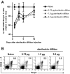
Dosing of denileukin diftitox for deletion of splenic Treg cells in vivo. A single intraperitoneal injection of denileukin diftitox (0.75, 1.5, or 3.0 μg) was administered to C57BL/6 mice. (A) Spleen cells from individual mice were stained for CD4+CD25+Foxp3+ Treg cells on days 1, 3, 5, and 10 after the injection. Mean values for percentage of Treg cells (± SD) are shown on the graph as percentage relative to naive mice; actual values for percentage Treg cells of total CD4+ T cells averaged 9.7% (range, 7.3%-12.0%) for the naive group. (B) One representative plot for each group is shown for day 1 after denileukin diftitox injection. The plots shown were gated on CD4+ T cells. Percentage of CD25+Foxp3+ cells is indicated in the upper right quadrant of each plot.
Figure 3
Figure 3
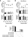
Specificity of denileukin diftitox for deletion of Treg cells versus activated T cells. A single intraperitoneal injection of 0.75 μg denileukin diftitox was administered to C57BL/6 mice (n ≥ 10/group). Flow cytometry analysis was performed on tissues on days 1, 3, 5, and 10 after injection. (A) Spleen, bone marrow, and peripheral blood cells from individual mice were stained for CD4+CD25+Foxp3+ Treg cells. Individual values and means for percentage of Treg cells are represented as percentage relative to naive mice; actual values for percentage Treg cells of total CD4+ T cells in the naive group averaged 8.9% for spleen (range, 5.6%-12%), 8.4% for bone marrow (range, 3.7%-11.1%), and 5.0% for peripheral blood (range, 2.2%-7.8%). Statistical evaluation was performed by one-way ANOVA using GraphPad Prism (GraphPad Software, San Diego, CA). (B) Spleen cells from individual mice were stained for CD4+CD25+Foxp3 and CD8+CD25+ activated T cells. Mean values (± SD) are shown on the graph as percentage relative to naive mice. Actual values for percentage activated CD4+ of total CD4+ T cells and activated CD8+ of total CD8+ T cells averaged 1.4% (range, 0.6%-2.9%) and 0.9% (range, 0.2%-1.6%), respectively, for the naive group.
Figure 4
Figure 4
Effect of denileukin diftitox on proliferation, apoptosis, and functionality of Treg cells. (A,B) A single intraperitoneal injection of 0.75 μg denileukin diftitox was administered to C57BL/6 mice. Flow cytometry analysis was performed on tissues on days 1, 3, 5, and 10 after injection. Animals were fed BrdU water for 5 days before being killed. At time of being killed, spleen (A) and bone marrow (B) cells from individual mice were stained for CD4+CD25+Foxp3+ Treg cells (individual values and means represented as percentage relative to naive mice, top), CD4+CD25+BrdU+ cells (mean values ± SD, middle), and CD4+CD25+AnnexinV+ cells (mean values ± SD, bottom). (C) Splenic CD4+CD25+ cells were isolated from naive mice, as well as from mice that were treated with 0.75 μg denileukin diftitox on day −7 or day −1 before being killed on day 0. Purified CD4+CD25 effector T cells (5 × 10⁴) from naive mice were then cultured in the presence or absence of the various isolated Treg cells (CD4+CD25+) at a 1:1 ratio (with 105 irradiated naive T-cell–depleted splenocytes as APCs) on an anti–CD3-coated 96-well plate. Mean proliferation (± SD) was measured by 3H-thymidine incorporation of triplicate wells.
Figure 5
Figure 5
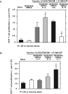
Scheduling of denileukin diftitox treatment for optimization of CD4+ T-cell proliferation to rV-CEA/TRICOM (+rF-GM-CSF) vaccination in a self-antigen system. CEA transgenic mice were subcutaneously vaccinated on day 0 with 1 × 10⁸ pfu rV-CEA/TRICOM (admixed with 1 × 10⁷ pfu rF-GM-CSF). On day 28, CD4+ T cells were added to proliferation assays with 50 μg/mL CEA protein. Mean proliferation (± SD) was measured by 3H-thymidine incorporation of triplicate wells; nonspecific proliferation to β-Gal protein was subtracted from CEA-specific proliferation. A single intraperitoneal injection of 0.75 μg denileukin diftitox was administered to indicated groups (A) before (day −4), concurrent with (day 0), or after (day 3) vaccination or (B) on day −7, −3, or −1 before vaccination. Statistical evaluation was performed by repeated-measures one-way ANOVA using GraphPad Prism.
Figure 6
Figure 6
Immune responses to Flu antigen after pretreatment with denileukin diftitox and vaccination with rV-Inf(A)NP34/TRICOM (+ rF-GM-CSF) in a foreign antigen system. C57BL/6 mice were subcutaneously vaccinated on day 0 with 5 × 10⁷ pfu rV-Inf(A)NP34/TRICOM (admixed with 1 × 10⁷ pfu rF-GM-CSF) and boosted on day 14 with 5 × 10⁷ pfu rF-Inf(A)NP34/TRICOM (admixed with 1 × 10⁷ pfu rF-GM-CSF) (vaccine alone). One group additionally received an intraperitoneal injection of 0.75 μg denileukin diftitox 1 day before the vaccine prime (vaccine + denileukin diftitox, day −1). Immune assays were performed on day 35, 3 weeks after the boost. Background-subtracted values are shown. (A) CD4+ T-cell proliferation to the indicated concentrations of inactivated Influenza A virus was measured by 3H-thymidine incorporation. Values are represented as mean proliferation (± SD) of triplicate wells. Statistical evaluation was performed by repeated-measures one-way ANOVA using GraphPad Prism. All values for the group pretreated with denileukin diftitox on day −1 relative to vaccine prime were statistically significant (P < .01) compared with vaccine alone. (B) CD4+ T cells were stimulated with 32.5 μg/mL inactivated Influenza A virus for 72 hours. Mean cytokine concentration in supernatants (± SD) was measured by Cytometric Bead Array analysis of triplicate samples. Statistical evaluation was performed by repeated-measures one-way ANOVA using GraphPad Prism. (C) CD8+ T cells were stimulated with the indicated concentrations of NP34 peptide for 48 hours. IFN-γ concentration in supernatants was measured by ELISA. (D) Mean serum levels of anti-Flu IgG (± SD) were measured by ELISA.
Figure 7
Figure 7
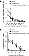
Immune responses to vaccinia virus after pretreatment with denileukin diftitox and vaccination with rV-Inf(A)NP34/TRICOM (+ rF-GM-CSF) in a foreign antigen system. C57BL/6 mice were subcutaneously vaccinated on day 0 with 5 × 10⁷ pfu rV-Inf(A)NP34/TRICOM (admixed with 107 pfu rF-GM-CSF) and boosted on day 14 with 5 × 10⁷ pfu rF-Inf(A)NP34/TRICOM (admixed with 107 pfu rF-GM-CSF) (vaccine alone). One group additionally received an intraperitoneal injection of 0.75 μg denileukin diftitox 1 day before the vaccine prime (vaccine + denileukin diftitox, day −1). Immune assays were performed on day 35, 3 weeks after the boost. Background-subtracted values are shown. (A) Mean CD4+ T-cell proliferation to the indicated concentrations of inactivated vaccinia virus (± SD) was assessed by 3H-thymidine uptake of triplicate wells. (B) Mean serum levels of antivaccinia IgG (± SD) were measured by ELISA.

Similar articles

Cited by

References

    1. Sakaguchi S. Naturally arising Foxp3-expressing CD25+CD4+ regulatory T cells in immunological tolerance to self and non-self. Nat Immunol. 2005;6:345–352. - PubMed
    1. Gavin M, Rudensky A. Control of immune homeostasis by naturally arising regulatory CD4+ T cells. Curr Opin Immunol. 2003;15:690–696. - PubMed
    1. Woo EY, Chu CS, Goletz TJ, et al. Regulatory CD4(+)CD25(+) T cells in tumors from patients with early-stage non-small cell lung cancer and late-stage ovarian cancer. Cancer Res. 2001;61:4766–4772. - PubMed
    1. Liyanage UK, Moore TT, Joo HG, et al. Prevalence of regulatory T cells is increased in peripheral blood and tumor microenvironment of patients with pancreas or breast adenocarcinoma. J Immunol. 2002;169:2756–2761. - PubMed
    1. Ichihara F, Kono K, Takahashi A, Kawaida H, Sugai H, Fujii H. Increased populations of regulatory T cells in peripheral blood and tumor-infiltrating lymphocytes in patients with gastric and esophageal cancers. Clin Cancer Res. 2003;9:4404–4408. - PubMed

Publication types

MeSH terms