. 2007 Jul;117(7):1794-804.
doi: 10.1172/JCI31731.

Wnt/beta-catenin signaling promotes expansion of Isl-1-positive cardiac progenitor cells through regulation of FGF signaling

Affiliations

Wnt/beta-catenin signaling promotes expansion of Isl-1-positive cardiac progenitor cells through regulation of FGF signaling

Ethan David Cohen et al. J Clin Invest. 2007 Jul.

Abstract

The anterior heart field (AHF), which contributes to the outflow tract and right ventricle of the heart, is defined in part by expression of the LIM homeobox transcription factor Isl-1. The importance of Isl-1-positive cells in cardiac development and homeostasis is underscored by the finding that these cells are required for cardiac development and act as cardiac stem/progenitor cells within the postnatal heart. However, the molecular pathways regulating these cells' expansion and differentiation are poorly understood. We show that Isl-1-positive AHF progenitor cells in mice were responsive to Wnt/beta-catenin signaling, and these responsive cells contributed to the outflow tract and right ventricle of the heart. Loss of Wnt/beta-catenin signaling in the AHF caused defective outflow tract and right ventricular development with a decrease in Isl-1-positive progenitors and loss of FGF signaling. Conversely, Wnt gain of function in these cells led to expansion of Isl-1-positive progenitors with a concomitant increase in FGF signaling through activation of a specific set of FGF ligands including FGF3, FGF10, FGF16, and FGF20. These data reveal what we believe to be a novel Wnt-FGF signaling axis required for expansion of Isl-1-positive AHF progenitors and suggest future therapies to increase the number and function of these cells for cardiac regeneration.


Figures

Figure 1
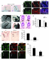
Figure 1. Expression of Wnt/β-catenin signaling components during cardiac development.
Expression of TCF3 was observed in the aortic sac (as) and atria (a) at E9.5 (A) and in the developing outflow tract (OFT) at E12.5 (B) and E14.5 (C). v, ventricle. TCF4 expression was also observed in the aortic sac and atria at E9.5 (D) and at much lower levels in the developing outflow tract at E12.5 (E) and E14.5 (F). Arrowheads indicate compact zone of the myocardium. (G) BATGAL lacZ expression was not observed in the heart proper at E10.5 or any other time tested. (H and I) However, extensive lacZ expression was observed in the outflow tract and pharyngeal mesoderm (PM) at E10.5 (arrowheads). (J) Wnt signaling was fate-mapped using the TOP–cre-ERT2 transgenic line, which consists of 3 reiterated LEF/TCF DNA binding sites upstream of a minimal c-fos promoter driving the tamoxifen-inducible cre-ERT2 cDNA (14). (K) Strong lacZ expression was observed in the outflow tract and right ventricle of the heart but little contribution was observed in the left ventricle. (LN) LacZ expression driven from the BATGAL transgene colocalized with Isl-1 expression in the outflow tract of the developing heart (brackets) using immunostaining with anti–β-galactosidase and anti–Isl-1 antibodies. Scale bars: 125 μm (A, D, H, I, and LN); 500 μm (B, C, E, and F).
Figure 2
Figure 2. Loss of Wnt/β-catenin signaling in the AHF leads to decreased right heart development and loss of Isl-1 progenitors.
(A and B) SM22α-cre is active in the AHF, demonstrated by lacZ expression throughout the outflow tract (brackets) and in the pharyngeal mesodermal apex (dotted lines) of SM22α-cre × R26R mice at E9.5 and E10.5. (CH) Immunofluorescent staining for Isl-1 and β-galactosidase expression shows extensive overlap within the outflow tract (arrowheads) and AHF (dotted lines) at E9.5. Loss of β-catenin using the SM22α-cre transgenic line caused hypoplastic right ventricle as assessed by scanning electron microscopy (I and J) and ink injections of wild-type (K) and SM22cre/Catnbflox/flox (SM/Catfl/fl) embryos (L) at E9.5. Histological sectioning at multiple levels showed the reduction in right ventricle size (arrows) at E9.5 in SM22cre/Catnbflox/flox (O and P) compared with wild-type embryos (M and N). (Q) Right ventricular diameter in SM22cre/Catnbflox/flox compared with wild-type embryos. (RT) To assess changes in Isl-1 AHF progenitors, wild-type and SM22cre/Catnbflox/flox embryos were immunostained for Isl-1 protein expression. SM22cre/Catnbflox/flox mutants have severely reduced numbers of Isl-1 AHF progenitors in the outflow tract at E9.5. (UAA) Isl-1 and Ki-67 double immunofluorescence was performed to determine changes in proliferation in AHF progenitors. Reduced Ki-67 staining in Isl-1–positive cells within the outflow tract was observed in SM22cre/Catnbflox/flox mutant embryos (arrowheads). (AA) Quantitation showed an almost 50% reduction in Isl-1 AHF progenitor proliferation. **P < 0.005. Scale bars: 100 μm (AJ); 500 μm (MP); 125 μm (R and S); 75 μm (UZ).
Figure 3
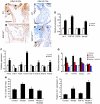
Figure 3. Regulation of AHF marker genes and progenitor number by Wnt signaling.
(A and B) FGF10 and Hand2 expression was reduced specifically in the hearts of SM22cre/Catnbflox/flox mutants (arrowheads). (C) Q-PCR was used to quantitate expression changes in the hearts of E9.5 SM22cre/Catnbflox/flox mutants. Expression of FGF10 and Hand2 was significantly downregulated, while expression of cTnI and Nkx2.5 was not appreciably affected by loss of β-catenin. (DF) To activate canonical Wnt signaling in vivo, developing embryos were treated with LiCl as described in Methods. LiCl treatment of embryos increased outflow tract (brackets) length. (G and H) This increased outflow tract length was associated with an increase in the number of Isl-1–positive AHF progenitors migrating into the outflow tract (brackets) and increased Isl-1 staining in the pharyngeal mesoderm harboring the Isl-1–positive AHF progenitor pool (arrows). (I) The number of Isl-1–positive cells in the outflow tract/right ventricle increased approximately 50% in Isl-1–positive AHF progenitors after LiCl treatment. (J and K) FGF10 and Hand2 expression was upregulated in the outflow tract and right ventricle after LiCl treatment (arrows). ***P < 0.001. Scale bars: 250 μm (D and E); 125 μm (G and H).
Figure 4
Figure 4. Activation of canonical Wnt signaling by β-catenin stabilization increases AHF and right heart development and proliferation.
(A and B) SM22α-cre mice were crossed to Catnbflox(ex3)/+ mice to generate SM22cre/Catnbflox(ex3)/+ mutant embryos. SM22cre/Catnbflox(ex3)/+ mutants had enlarged hearts, in particular the right ventricles (arrows), at E9.5. (C and D) Increased cell mass was also observed in the anterior foregut mesoderm surrounding the trachea, where a pool of Isl-1–positive AHF progenitors resides, at E10.5. (E and F) Isl-1 immunostaining was increased in the AHF (brackets) and outflow tracts (arrowheads) of SM22cre/Catnbflox(ex3)/+ mutants at E10.5. (G) Quantitation of the increase in Isl-1 immunostaining revealed a greater than 50% increase in Catnbflox(ex3)/+ mutants. (HM) Isl-1 and Ki-67 double immunofluorescent staining reveal increased proliferation in Isl-1–positive cells within the outflow tracts of SM22cre/Catnbflox(ex3)/+ mutant embryos at E10.5 (arrowheads). (N) Ki-67 staining in Isl-1–positive cells increased approximately 40% within SM22cre/Catnbflox(ex3)/+ embryos. *P < 0.02. Scale bars: 500 μm (A and B); 75 μm (CF); 100 μm (HM).
Figure 5
Figure 5. Wnt signaling expands the number of Isl-1–positive progenitors in AHF explants.
(AC) The AHF was explanted as shown (A) and cultured for 2 days in the absence (B) or presence (C) of Wnt3a. Isl-1 immunostaining increased upon treatment with Wnt3a. (D) Isl-1–positive cells increased approximately 50% in Wnt3a-treated explants. (E) Isl-1 mRNA expression also increased, as determined by Q-PCR. To determine whether this increase was due to an increase in Isl-1 expression per cell or due primarily to an increase in Isl-1–positive cells, explants were treated with HUA to inhibit cell proliferation. HUA treatment inhibited the increase in Isl-1 mRNA expression, which suggests that the majority of increased Isl-1 immunostaining and mRNA expression was the result of expansion of Isl-1–positive cells after Wnt3a treatment. **P < 0.005.
Figure 6
Figure 6. FGF signaling is activated by Wnt signaling in the AHF.
(A and B) ERK1/2 phosphorylation was used to assess the activity of FGF signaling in the AHF. SM22cre/Catnbflox/flox mutants expressed less phosphorylated ERK1/2 in the AHF and outflow tract than did wild-type littermates at E9.5 (arrowheads). PO4, phosphorylation. (C and D) ERK1/2 phosphorylation increased in the outflow tract and right ventricular myocardium in SM22cre/Catnbflox(ex3)/+ embryos at E10.5 (arrowheads). (E) FGF10 and FGF8 expression was assessed by Q-PCR in AHF explants treated with Wnt3a. Surprisingly, FGF10 expression was significantly upregulated, while expression of FGF8, which is also expressed in the AHF, was unchanged. Expression of both Isl-1 and Hand2 was upregulated as expected. (F) Expression of additional FGF ligands was determined by Q-PCR, and FGF3, FGF10, FGF16, and FGF20 were all significantly upregulated by Wnt3a treatment, whereas FGF4 was downregulated. (G and H) Activation of AHF gene expression (G) and Isl-1–positive AHF progenitor number (H) by Wnt3a was attenuated by the FGF receptor inhibitor SU5402, indicating that these pathways act cooperatively in regulating AHF development. (I) Conversely, Wnt3a and FGF10 cooperatively increased Isl-1 expression in AHF explants, further supporting interaction between Wnt and FGF signaling in AHF development. **P < 0.005. Scale bars: 100 μm.
Figure 7
Figure 7. FGF10 is a direct target of Wnt/β-catenin signaling in the AHF.
(A) The FGF10-AHF enhancer from the FGF10 gene contains 2 cross-species–conserved LEF/TCF DNA binding sites. (B and C) The FGF10-AHF enhancer is activated and repressed by an activated form of β-catenin (β-catenin41A/45A) and dominant-negative TCF3 (dnTCF2), respectively. (D and E) ChIP assays showed that β-catenin formed a complex on the FGF10-AHF enhancer in vivo (D), and LiCl treatment of 293 T cells increased the association of β-catenin with this enhancer element (E). (F) Model of Wnt/FGF signaling promoting expansion of Isl-1–positive AHF progenitors through activation of specific FGF ligands, leading to proliferation and proper development of the outflow tract and right ventricle of the heart.

Similar articles

Cited by

References

    1. Kelly R.G. Molecular inroads into the anterior heart field. Trends Cardiovasc. Med. 2005;15:51–56. - PubMed
    1. Cai C.L., et al. Isl1 identifies a cardiac progenitor population that proliferates prior to differentiation and contributes a majority of cells to the heart. Dev. Cell. 2003;5:877–889. - PMC - PubMed
    1. Moretti A., et al. Multipotent embryonic isl1+ progenitor cells lead to cardiac, smooth muscle, and endothelial cell diversification. Cell. 2006;127:1151–1165. - PubMed
    1. Wu S.M., et al. Developmental origin of a bipotential myocardial and smooth muscle cell precursor in the mammalian heart. Cell. 2006;127:1137–1150. - PubMed
    1. Laugwitz K.L., et al. Postnatal isl1+ cardioblasts enter fully differentiated cardiomyocyte lineages. Nature. 2005;433:647–653. - PMC - PubMed

Publication types

MeSH terms