. 2007 Jul 25;26(14):3451-62.
doi: 10.1038/sj.emboj.7601773. Epub 2007 Jun 28.

Involvement of the ubiquitin-like domain of TBK1/IKK-i kinases in regulation of IFN-inducible genes

Affiliations

Involvement of the ubiquitin-like domain of TBK1/IKK-i kinases in regulation of IFN-inducible genes

Fumiyo Ikeda et al. EMBO J. .

Abstract

TANK-binding kinase 1 (TBK1/NAK/T2K) and I-kappaB Kinase (IKK-i/IKK-epsilon) play important roles in the regulation of interferon (IFN)-inducible genes during the immune response to bacterial and viral infections. Cell stimulation with ssRNA virus, dsDNA virus or gram-negative bacteria leads to activation of TBK1 or IKK-i, which in turn phosphorylates the transcription factors, IFN-regulatory factor (IRF) 3 and IRF7, promoting their translocation in the nucleus. To understand the molecular basis of activation of TBK1, we analyzed the sequence of TBK1 and IKK-i and identified a ubiquitin-like domain (ULD) adjacent to their kinase domains. Deletion or mutations of the ULD in TBK1 or IKK-i impaired activation of respective kinases, failed to induce IRF3 phosphorylation and nuclear localization and to activate IFN-beta or RANTES promoters. The importance of the ULD of TBK1 in LPS- or poly(I:C)-stimulated IFN-beta production was demonstrated by reconstitution experiments in TBK1-IKK-i-deficient cells. We propose that the ULD is a regulatory component of the TBK1/IKK-i kinases involved in the control of the kinase activation, substrate presentation and downstream signaling pathways.


Figures

Figure 1
Figure 1
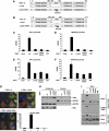
Identification and structural characterization of the ULD in TBK1 (A) An alignment of Fat10, ubiquitin and ULD domains of IKKβ, IKKα, IKK-i and TBK1. Conserved residues are shown on black or gray background. The hydrophobic patch at isoleucine 44 in ubiquitin (arrowhead) is conserved in ULD domain in TBK1. (dm: Drosophila, hs: Homo sapiens) (B) Primary and secondary structure alignment of mouse TBK1-ULD and human ubiquitin. Secondary structure elements (arrows for β-strands, boxes for helical regions) for TBK1-ULD represent the consensus of the chemical shifts analyses with the programs TALOS and CSI. Secondary structure elements as well as atomic coordinates for ubiquitin are taken from PDB entry 1UBI. (C) Dynamic behavior of TBK1-ULD. The ribbon diagram of the structure of human ubiquitin is colored according to the broadening and splitting of the TBK1-ULD resonances (red: significant, yellow: intermediate, green: no line broadening).
Figure 2
Figure 2
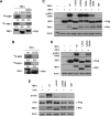
The ULD plays an important role in the regulation of downstream signaling. (A, B) Deletion-ULD (Δ-ULD) of TBK1 and IKK-i, and mutants with mutations at the hydrophobic patch of TBK1-L352A I353A and IKK-i-L353AF354A were generated as described previously. (C, D) The effects of TBK1-wt, Δ-ULD, L352A I353A and KM on promoter activities of IFN-inducible genes were examined. Luciferase construct driven by IFN-β promoter or RANTES promoter was co-expressed in HEK293T cells with TBK1-wt or mutants. After 48 h of transfection, cells were harvested for the luciferase assay. The internal control was measured by β-GAL activity. (E, F) The effects of IKK-i wt, Δ-ULD, L353A F354A and KM on promoter activities of IFN-inducible genes were examined. (G) Effects of TBK1 mutants on IRF3-nuclear localization were examined. Cellular localization of IRF3 was examined by immunofluorescence. Myc-IRF3 was co-expressed with GFP-TBK1-wt (upper left), GFP-TBK1-Δ-ULD (upper right) or GFP-TBK1-L352AI353A mutant (lower left). Nuclear was stained by DAPI (blue). GFP-TBK1-wt, GFP-TBK1-Δ-ULD and GFP-TBK1-L352AI353A mutants were localized in the cytoplasm (green). Nuclear translocation of Myc-IRF3 (red) was only induced by TBK1-wt (upper left). More than 100 cells in which both of GFP-TBK1 (wt, Δ-ULD or L352A I353A) and Myc-IRF3 were expressed were counted and percentage of the cells with nuclear IRF3 was shown (lower right). All of the panels were under the magnification of × 400. (H) HeLa cells were co-transfected with plasmids encoding Flag-IRF3 and Myc-TBK1. Nuclear and cytoplasmic fractions were prepared and analyzed by immunoblot. NUP62 and GAPDH were used for fractionation markers of nuclear and cytoplasm, respectively. (I) Dimer formation of IRF3 was examined by native PAGE. Flag-IRF3 was transfected with Myc-TBK1 wt or mutants into HEK293T cells. Total cell lysates were used for native PAGE and the membrane transferred was blotted with α-IRF3 or α-pIRF3 S386 antibody. Expressions of Flag-IRF3, Myc-TBK1 and loading control were determined by SDS–PAGE.
Figure 3
Figure 3
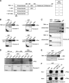
The ULD is required for the activation of downstream molecules and kinase activity of TBK1. (A) Various Myc-tagged TBK1 constructs were transfected into HEK293T cells and total cell lysates were subjected to immunoprecipitation by α-Myc antibody. Immunoprecipitated samples were subjected to in vitro kinase assay using MBP as substrate. The phosphorylation of MBP is shown in the upper panel, phosphorylation of TBK1 is shown in the middle panel and the expression level of TBK1 is shown in the lower panel. Densitometric analysis was performed using Scion Image software (Scion Corporations) and the numbers of the density are indicated. (B) Myc-IKK-i constructs were transfected into HEK293T cells and total cell lysates were subjected to immunoprecipitation by α-Myc antibody. Immunoprecipitated samples were subjected to in vitro kinase assay. (C, D) The effects of TBK1 and IKK-i-wt and mutants on phosphorylation of IRF3 were examined. Flag-IRF3 was co-expressed with Myc-TBK1 and IKK-i-wt or mutants in HEK293T cells. Total cell lysates were collected and examined by immunoblotting using α-phospho-IRF3 (Ser396) for the determination of IRF3-phosphorylation. The expression levels of Flag-IRF3, Myc-TBK1 and Myc-IKK-i were examined and are shown in the lower panels. Loading control was determined by using α-actin antibody. (E) The effects of TBK1-wt and mutants on phosphorylation of IκBα. Flag-IκBα was co-expressed with Myc-TBK1-wt or mutants in HEK293T cells. Total cell lysates were examined by immunoblotting using α-phospho-IκBα antibody for the determination of IκBα-phosphorylation. The expression levels of Flag-IκBα and Myc-TBK1 were examined and are shown in the lower panels. Loading control was determined by using α-actin antibody.
Figure 4
Figure 4
The ULD binds to kinase domain of TBK1 via hydrophobic patch L352 I353. (A) Deletion mutants of TBK1 were created as described. TBK1 only containing kinase domain with the ULD (1–383), kinase domain (1–301), kinase domain with mutations at ATP-binding site (1–301 KM) were generated as shown. (B) Myc-tagged TBK1-wt, 1–383, 1–301 (wt) and 1–301 (KM) were introduced into HEK293T cells and total cell lysates were used for GST-pull-down assay. After incubation of total cell lysates with GST-control or GST-ULD proteins, the binding was determined by immunoblotting using α-Myc antibody. (C) Myc-tagged TBK1-wt, L352A I353A and Δ-ULD were introduced into HEK293T cells and total cell lysates were used for GST-pull-down assay. (D) Myc-tagged IKK-i-wt, Δ-ULD and L353F F354A were introduced into HEK293T cells and total cell lysates were used for GST-pull-down assay. (E) GST-ULD and GST-ULD with L352A I353A-mutation (ULD-L352A I353A) were purified and used to examine the binding to Myc-tagged TBK1-wt. The amount of GST-fusion proteins was determined by Ponceau S staining, shown in the lower panels (B–E). (F) Sts-1 and -2, which have a UBA domain, do not bind the ULD of TBK1. Flag-Sts-1 and -2 were transfected into HEK293T cells and cell were harvested for the GST-pull-down assay. Total cell lysates were incubated with GST-fusion proteins of ubiquitin, TBK1-ULD or Parkin-UBL and immunoblotting using α-Flag antibody was performed. The amount of GST-fusion proteins was checked by Ponceau S staining as shown in the lower panels.
Figure 5
Figure 5
The ULD binds to IRF3. (A) Myc-IRF3 was introduced into HEK293T cells and total cell lysates were used to examine the binding to GST-ULD, GST-ULD-L352A I353A and GST-Ub. (B) The deletion mutants were generated for the experiments as indicated: IRF3 lacking serine-rich region (SRR, 1–384), lacking SRR and IAD (1–189) containing IAD domain and SRR (190–427) and containing IAD domain (190–384) were created. The positive binding between each mutant and GST-ULD is indicated as +, and negative binding is indicated as – on the right panel. (C) Each of the Myc-tagged IRF3 mutants was introduced into the HEK293T cells and total cell lysates were subjected to GST-pull-down assay. The binding between GST-ULD and Myc-IRF3 was examined by immunoblotting. (D) Flag-IRF3 with or without Myc-TBK1-wt or KM was transfected into HEK293T cells. Total cell lysates were incubated with GST-ULD. The precipitates were subjected to immunoblotting using α-Flag antibody or α-Myc antibody.
Figure 6
Figure 6
The ULD of TBK1 plays a role in LPS- and poly(I:C)-induced signaling pathway. (A) Mock, TBK1-wt, Δ-ULD, L352A I353A or KM retrovirus was infected into control (TBK1+/+IKK-i+/−) and TBK1−/−IKK-i−/− MEF cells. After 48 h of infection, cells were harvested and expression of TBK1 and IRF3 in total cell lysates was analyzed by immunoblotting using α-TBK1 antibody or α-IRF3 antibody. The endogenous TBK1 is indicated with *. The expression of IRF3 was not affected by retrovirus infection. The loading control was examined using α-actin antibody. (B, C) TBK1-wt, TBK1-Δ-ULD, TBK1-L352A I353A and TBK1-KM were introduced in TBK1−/−IKK-i−/− MEF cells by retrovirus. IFN-β promoter-driven luciferase construct was transfected together with β-GAL construct. The cells were treated or untreated with LPS (10 μg/ml) or poly(I:C) (6 μg/ml) for 16 h and harvested for the luciferase assay. The internal control was measured by β-GAL. (D) IFN-β production of infected TBK1−/−IKK-i−/− MEF cells after 24 h of poly(I:C) (6 μg/ml) treatment was examined by ELISA. (nd, not detected) (*P<0.05, **P<0.01).
Figure 7
Figure 7
Active and inactive form of TBK1. (A) Under the nonstimulated condition, the domain interfaces of the ULD and kinase domain of TBK1 are not in the close proximity, failing to be fully functional. (B) TBK1 is active when the ULD interacts with the kinase domain. LPS- or poly(I:C)-induced active form of TBK1 interacts with IAD of IRF3, at least partially, via the ULD and phosphorylates it. Phosphorylated IRF3 dissociates from TBK1 and translocates into the nucleus, binds to the ISRE motif in the promoter region of the target genes and regulates their transcription. In the scheme, we draw interactions between single TBK1 and IRF molecules due to the clarity of presentation. It is, however, likely that these interactions are taking place in larger oligomeric TBK1/IRF complexes in vivo.

Similar articles

Cited by

References

    1. Akira S, Uematsu S, Takeuchi O (2006) Pathogen recognition and innate immunity. Cell 124: 783–801 - PubMed
    1. Andrejeva J, Childs KS, Young DF, Carlos TS, Stock N, Goodbourn S, Randall RE (2004) The V proteins of paramyxoviruses bind the IFN-inducible RNA helicase, mda-5, and inhibit its activation of the IFN-beta promoter. Proc Natl Acad Sci USA 101: 17264–17269 - PMC - PubMed
    1. Bayliss R, Sardon T, Vernos I, Conti E (2003) Structural basis of Aurora-A activation by TPX2 at the mitotic spindle. Mol Cell 12: 851–862 - PubMed
    1. Bienko M, Green CM, Crosetto N, Rudolf F, Zapart G, Coull B, Kannouche P, Wider G, Peter M, Lehmann AR, Hofmann K, Dikic I (2005) Ubiquitin-binding domains in Y-family polymerases regulate translesion synthesis. Science 310: 1821–1824 - PubMed
    1. Cornilescu G, Delaglio F, Bax A (1999) Protein backbone angle restraints from searching a database for chemical shift and sequence homology. J Biomol NMR 13: 289–302 - PubMed

Publication types

MeSH terms