. 2007 Jul 25;26(14):3296-307.
doi: 10.1038/sj.emboj.7601775. Epub 2007 Jun 28.

The Caenorhabditis elegans septin complex is nonpolar

Affiliations

The Caenorhabditis elegans septin complex is nonpolar

Corinne M John et al. EMBO J. .

Abstract

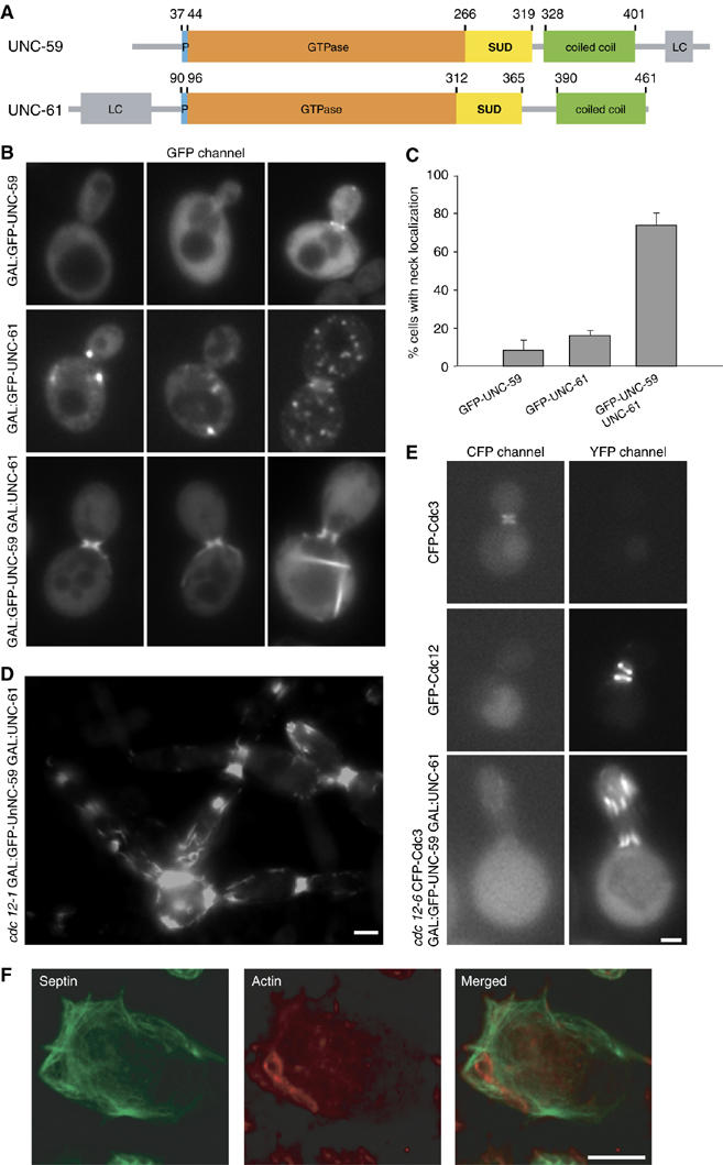
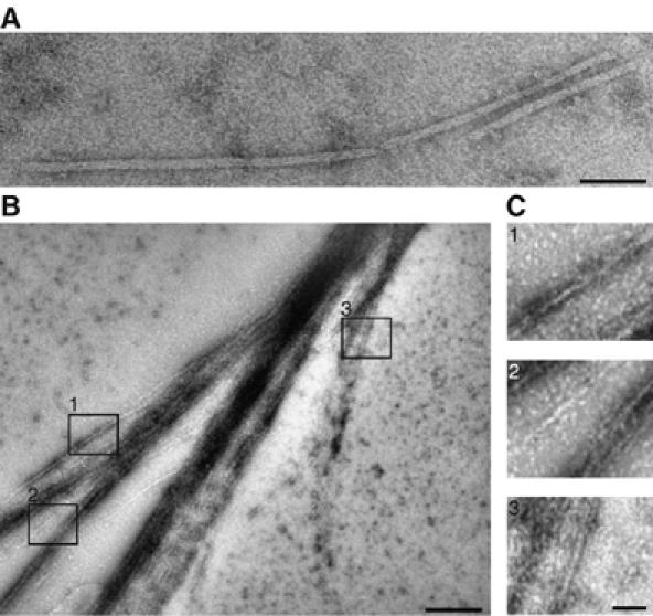
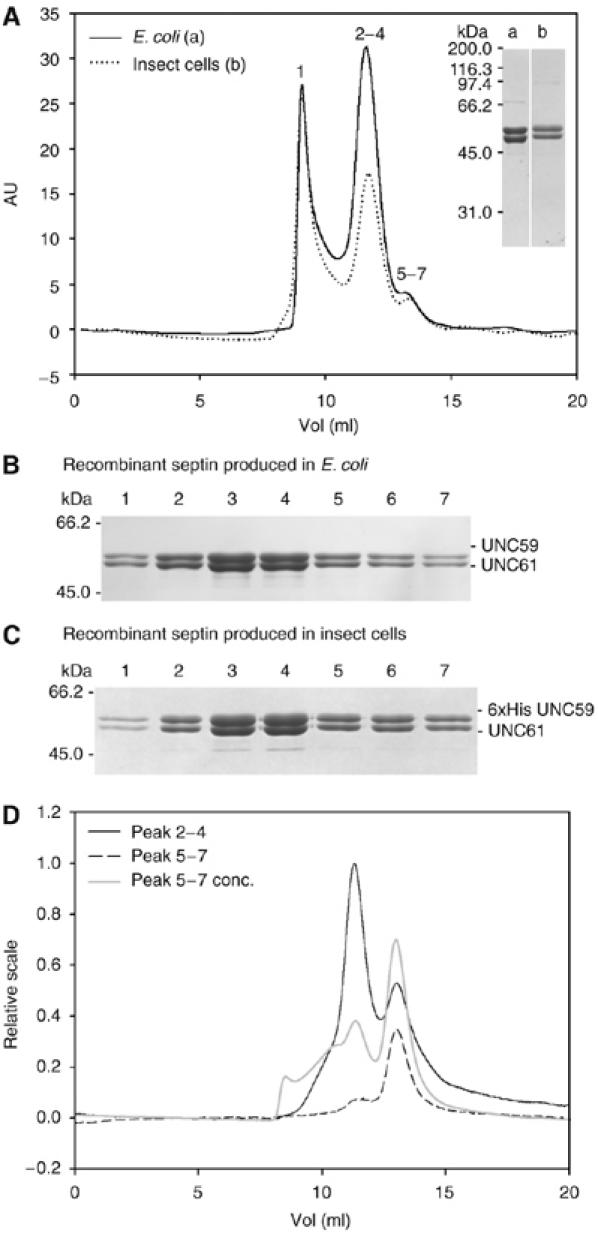
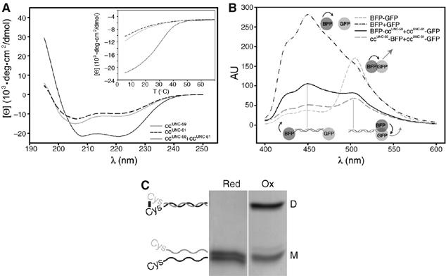
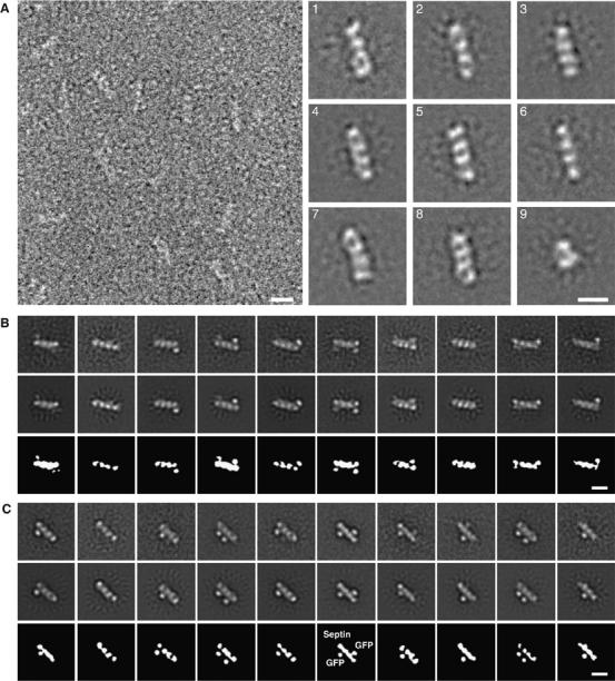
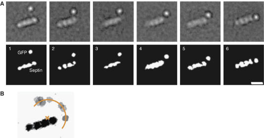
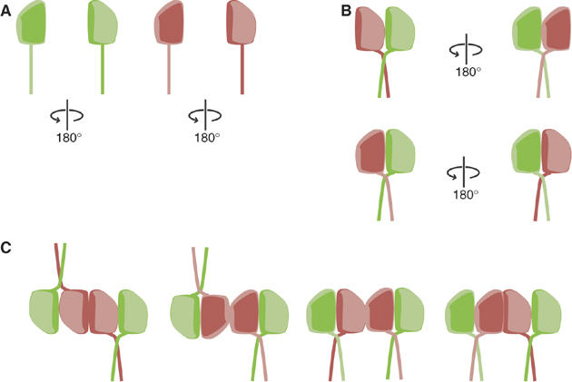
Septins are conserved GTPases that form heteromultimeric complexes and assemble into filaments that play a critical role in cell division and polarity. Results from budding and fission yeast indicate that septin complexes form around a tetrameric core. However, the molecular structure of the core and its influence on the polarity of septin complexes and filaments is poorly defined. The septin complex of the nematode Caenorhabditis elegans is formed entirely by the core septins UNC-59 and UNC-61. We show that UNC-59 and UNC-61 form a dimer of coiled-coil-mediated heterodimers. By electron microscopy, this heterotetramer appears as a linear arrangement of four densities representing the four septin subunits. Fusion of GFP to the N termini of UNC-59 and UNC-61 and subsequent electron microscopic visualization suggests that the sequence of septin subunits is UNC-59/UNC-61/UNC-61/UNC-59. Visualization of GFP extensions fused to the extremity of the C-terminal coiled coils indicates that these extend laterally from the heterotetrameric core. Together, our study establishes that the septin core complex is symmetric, and suggests that septins form nonpolar filaments.


Figures

Figure 1
Figure 1
Localization of C. elegans septins expressed in yeast and insect cells. (A) Schematic representation of the domain organization of the C. elegans septins UNC-59 and UNC-61. Both septins contain a polybasic region (P; blue), a GTPase domain (orange), a septin specific domain (SUD; yellow) and a predicted coiled-coil domain (green). The N- and the C-termini of UNC-61 and UNC-59, respectively, contain low complexity regions (LC; gray). Corresponding domain boundaries are indicated by residue positions. (B–E) Localization of C. elegans septins in S. cerevisiae as visualized by fluorescence microscopy. (B) WT yeast cells expressing the indicated combinations of GFP-tagged or untagged UNC-59 and UNC-61. (C) Quantification of budded cells showing septin localization at the bud neck where the indicated septin is expressed. (D) Typical localization of GFP-UNC-59 coexpressed with UNC-61 in the cdc12–1 septin mutant upon overnight incubation at restrictive temperature. (E) Localization of CFP-Cdc3 and GFP-UNC-59 coexpressed with UNC-61 in cdc12–6 cells after growth at 25°C overnight, followed by a 1 h shift to 37°C. As controls, WT yeast expressing either CFP-Cdc3 or GFP-Cdc12 was visualized in both YFP and CFP channels. (F) Visualization of C. elegans septins in SF21 insect cells by indirect immunofluorescence. SF21 cells expressing 6 × His-UNC-59 and UNC-61 were stained with anti-penta-His antibodies (green) and filamentous actin was stained with rhodamin phalloidin (red). The scale bars represent 2 μm in (B, D, E) and 10 μm in (F).
Figure 2
Figure 2
C. elegans septins form filaments in vitro. Electron micrographs of negatively stained septin filaments produced in E. coli (A) and insect cells (B, C) after dialysis from 500 mM NaCl into 20 mM NaCl buffer. (B) Overview of a septin filament sheet. (C) Enlargement of the indicated areas 1, 2, 3 from (B). The scale bar represents 50 nm in (A, C) and 250 nm in (B).
Figure 3
Figure 3
Analysis of the UNC-59/UNC-61 complex by size-exclusion chromatography. (A) Affinity-purified septin complex expressed in E. coli (solid line; 100 μl of 3 mg/ml injected) or in insect cells (dotted line; 100 μl of 1 mg/ml injected) was analyzed on a Superdex 200 column. Numbers on top of the peaks correspond to the lanes in the SDS–PAGE analysis (Coomassie stained) of septins produced in E. coli (B) or in insect cells (C). For SDS–PAGE comparison of septins (6 × His-UNC-59/UNC-61) produced in bacteria (a) or insect cells (b) see inset in (A). (D) Analysis of tetramer and dimer fractions. The tetramer fractions (2–4) (black line; 100 μl of 0.5 mg/ml injected), the dimer fractions (5–7) (black dashed line; 100 μl of 0.05 mg/ml injected) and a fourfold concentrated dimer fraction (gray line; 100 μl of 0.2 mg/ml injected) were rechromatographed on a Superdex 200 column.
Figure 4
Figure 4
Analysis of the coiled-coil domains of UNC-59 and UNC-61. (A) Far-UV CD spectra of ccUNC−59 (gray line), ccUNC−61 (dashed line), and ccUNC−59 and ccUNC−61 mixed in equimolar ratio (solid line). (B, inset) Thermal unfolding profile recorded by CD at 222 nm. (B, C) Analysis of septin coiled-coil orientation. (B) FRET efficiency of soluble fractions of cells expressing ccUNC−59BFP and ccUNC−61GFP (solid line) or BFPccUNC−59 and ccUNC−61GFP (large dashed line), respectively. Positive control: BFP-GFP (small dashed line); negative control: BFP and GFP (dashed/dotted line). (C) Coomassie blue stained SDS–PAGE of CGGccUNC−59 mixed in equimolar ratio with CGGccUNC−61 under reducing (red) or oxidizing (ox) conditions. The migration positions of monomers (M) and dimers (D) are indicated schematically.
Figure 5
Figure 5
EM of septin complexes. (A) Electron micrograph and projection averages of negatively stained 6 × His-UNC-59/UNC-61 heterotetramers expressed in E. coli. The septin complexes stain poorly, making it difficult to see the linear septin molecules. The first eight panels show averages obtained from representative classes of septin heterotetramers containing four linearly arranged densities. The last panel is an average of heterodimers, which are composed of only two densities. (B) Projection averages of negatively stained GFP-UNC-59 (23–459)/6 × His-UNC-61 heterotetramers. The top panels show ten representative class averages from the multi-reference alignment using randomly selected raw images as initial references. These averages were used as input references for the second round of multi-reference alignment, whose outputs are shown in the middle row. Schematic diagrams of the averages from the second alignment are shown in the bottom row. (C) Projection averages of negatively stained 6 × His-UNC-59/GFP-UNC-61 (77–461) heterotetramers. The top panels show ten representative class averages from the multireference alignment using random references. These averages were used as input references for the second round of multireference alignment, whose outputs are shown in the middle row. Schematic diagrams of the averages from the second alignment are shown in the bottom row. All scale bars represent 10 nm.
Figure 6
Figure 6
Flexibility of the coiled-coil domain. (A) Six representative class averages of UNC-59/UNC-61-GFP complexes showing one resolved GFP. The GFP is located at various positions relative to the heterotetramer core, demonstrating the flexibility of the coiled-coil/septin core junction. The scale bars represent 10 nm. (B) Schematic representation obtained by overlaying nine class averages of negatively stained 6 × His-UNC-59/UNC-61-GFP. Averages with the one resolved GFP moiety at the maximum distance from the four aligned globular domains were chosen. The coiled-coil domains occupy a semicircle around a heterodimer. The center lies at the interface between the UNC-59 and UNC-61 globular domains. The radius is about 10 nm.
Figure 7
Figure 7
Molecular organization of the heterotetrameric septin core complex. (A) Schematic representations of UNC-59 (green) and UNC-61 (red), showing their GTPase domains and coiled-coil domains (rod). The GTPase domains are not internally symmetric, as represented by the ‘top' face of the protein (arbitrary) shown as pale green or pink, and the ‘bottom' side as dark green or red. (B) The septin heterodimer interacts via its globular domains and its parallel coiled coils. Diagrams show two possible modes of interaction between UNC-59 and UNC-61: ‘top' to ‘top', in which UNC-59 and UNC-61 are in the same orientation (top), and ‘top' to ‘bottom', in which they face opposite directions (bottom). (C) All models for heterotetramer formation require that two of three axes are nonpolar. Two of UNC-59/UNC-61 heterodimers dimerize along their longitudinal axis (X-axis). One two-fold symmetry axis between the two UNC-59/UNC-61 heterodimers is perpendicular to the drawing plane (Z-axis). Thus, the tetrameric septin subunit is nonpolar along X. In the first two models, the tetramer is also symmetric along the Y-axis, but asymmetric along the Z-axis. In the second two models, the tetramer is asymmetric along the y-axis, but symmetric along the Z-axis.

Similar articles

Cited by

References

    1. An H, Morrell JL, Jennings JL, Link AJ, Gould KL (2004) Requirements of fission yeast septins for complex formation, localization, and function. Mol Biol Cell 15: 5551–5564 - PMC - PubMed
    1. Ausubel FM, Brent R, Kingston RE, Moore DD, Seidman JG, Smth JA, Struhl K (eds). (1987) Current Protocols in Molecular Biology. Harvard Medical School, Boston: John Wiley and Sons
    1. Barral Y, Mermall V, Mooseker MS, Snyder M (2000) Compartmentalization of the cell cortex by septins is required for maintenance of cell polarity in yeast. Mol Cell 5: 841–851 - PubMed
    1. Barral Y, Parra M, Bidlingmaier S, Snyder M (1999) Nim1-related kinases coordinate cell cycle progression with the organization of the peripheral cytoskeleton in yeast. Genes Dev 13: 176–187 - PMC - PubMed
    1. Beites CL, Xie H, Bowser R, Trimble WS (1999) The septin CDCrel-1 binds syntaxin and inhibits exocytosis. Nat Neurosci 2: 434–439 - PubMed

Publication types

MeSH terms

LinkOut - more resources