. 2007 Jul;5(7):e170.
doi: 10.1371/journal.pbio.0050170. Epub 2007 Jun 19.

The spindle pole bodies facilitate nuclear envelope division during closed mitosis in fission yeast

Affiliations

The spindle pole bodies facilitate nuclear envelope division during closed mitosis in fission yeast

Liling Zheng et al. PLoS Biol. 2007 Jul.

Abstract

Many organisms divide chromosomes within the confines of the nuclear envelope (NE) in a process known as closed mitosis. Thus, they must ensure coordination between segregation of the genetic material and division of the NE itself. Although many years of work have led to a reasonably clear understanding of mitotic spindle function in chromosome segregation, the NE division mechanism remains obscure. Here, we show that fission yeast cells overexpressing the transforming acid coiled coil (TACC)-related protein, Mia1p/Alp7p, failed to separate the spindle pole bodies (SPBs) at the onset of mitosis, but could assemble acentrosomal bipolar and antiparallel spindle structures. Most of these cells arrested in anaphase with fully extended spindles and nonsegregated chromosomes. Spindle poles that lacked the SPBs did not lead the division of the NE during spindle elongation, but deformed it, trapping the chromosomes within. When the SPBs were severed by laser microsurgery in wild-type cells, we observed analogous deformations of the NE by elongating spindle remnants, resulting in NE division failure. Analysis of dis1Delta cells that elongate spindles despite unattached kinetochores indicated that the SPBs were required for maintaining nuclear shape at anaphase onset. Strikingly, when the NE was disassembled by utilizing a temperature-sensitive allele of the Ran GEF, Pim1p, the abnormal spindles induced by Mia1p overexpression were capable of segregating sister chromatids to daughter cells, suggesting that the failure to divide the NE prevents chromosome partitioning. Our results imply that the SPBs preclude deformation of the NE during spindle elongation and thus serve as specialized structures enabling nuclear division during closed mitosis in fission yeast.


Conflict of interest statement

Competing interests. The authors have declared that no competing interests exist.

Figures

Figure 1
Figure 1. Fission Yeast Cells Overexpressing Mia1p Arrest in Mitosis with Fully Extended Spindles
(A) The centromere markers, Cen1-GFP, Clp1-GFP, and Rlc1p-GFP, exhibit late mitotic localization in cells overexpressing Mia1p. Shown are single maximum-intensity reconstructions of live cells. (B) Graph quantifying the proportion of cells exhibiting mitotic localization of marker proteins (n = 100). (C) Immunofluorescence images of wild-type and Mia1p-overexpressing cells using anti–α-tubulin antibodies and the DNA dye, DAPI. (D) Graph quantifying the proportion of mitotic cells exhibiting wild-type or abnormal spindle architecture (n = 300). MT, microtubule.
Figure 2
Figure 2. The SPBs Are Displaced from the Spindle Poles, and NE Division Fails in Cells Overexpressing Mia1p
(A) Immunofluorescence images of wild-type and Mia1p-overexpressing cells using anti–α-tubulin antibodies, Pcp1p-GFP, and the DNA dye, DAPI. (B) Graph quantifying the proportion of mitotic Mia1p-overexpressing cells exhibiting normal or aberrant spindle architecture (n = 100). Figures over each bar schematically represent position of the SPBs (black dots) with respect to the spindle (grey line). (C) Mia1p overexpression induces panhandle-shaped deformations of the NE. Shown are single maximum-intensity reconstructions of live wild-type and Mia1p-overexpressing cells containing the NE marker proteins Cut11p-GFP and Uch2p-GFP. (D) Graph quantifying the proportion of cells exhibiting the panhandle-shaped NE deformations (n = 100). (E) Time-lapse sequence of Cut11p-GFP–expressing wild-type cells undergoing mitosis. Numbers refer to the time, in minutes. (F) Time-lapse sequence of Mia1p-overexpressing Cut11p-GFP cells undergoing mitosis. Note an elongating protrusion of the NE and the SPB pair on the opposite side of the nucleus. Numbers refer to the time, in minutes. (G) The SPBs largely localize away from the NE protrusion tips in cells overexpressing Mia1p. Note that in wild-type cells, the SPBs seem to lead the NE division. Shown are single maximum-intensity reconstructions of live wild-type and Mia1p-overexpressing cells containing the NE marker protein, Uch2p-GFP, and the SPB protein, Sad1p-DSRed.
Figure 3
Figure 3. The SPBs Are Duplicated but Not Separated in Cells Overexpressing Mia1p
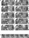
(A) Electron microscopy images of serial sections of a Mia1p-overexpressing cell showing the abnormal panhandle-shaped protrusion of the NE. Note that microtubules extend throughout the protrusion (indicated by indented arrowheads, panel 4). The SPB pair is located at the base of protrusion. The NE protrusion and position of the SPBs are indicated on panel 4. Scale bar represents 1 μm. (B) Higher magnification image of the duplicated SPB pair. Mitochondrion is labeled as M, nucleus as N. Scale bar represents 0.2 μm.
Figure 4
Figure 4. Spindle Structures in Mia1p-Overexpressing Cells Are Bipolar and Antiparallel
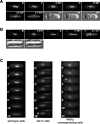
(A) Time-lapse sequence of Cut7p-GFP–expressing wild-type cells undergoing spindle elongation. Numbers refer to the time, in minutes. (B) Time-lapse sequence of Mia1p-overexpressing Cut7p-GFP cells undergoing mitosis. Note that Cut7p-GFP clearly localizes to both poles early in anaphase and is progressively depleted from one of them as the cell progresses through mitosis. The midzone localization is similar to wild type. Shown are single maximum-intensity reconstructions of z-stacks. Numbers refer to the time, in minutes. (C) Cut7p-mCherry is enriched on acentrosomal spindle poles as judged by double fluorescence microscopy analysis with the SPB core marker, Pcp1p-GFP. Shown are single maximum-intensity reconstructions of z-stacks. (D) The γ-TuRC marker, Alp4p-GFP, is present on acentrosomal poles in Mia1p-overexpressing cells, where it co-localizes with Cut7p-mCherry. Shown are single maximum-intensity reconstructions of z-stacks. (E) The chromosomal passenger complex protein, Ark1p-GFP, defines the spindle midzone in acentrosomal spindles. Shown are single maximum-intensity reconstructions of z-stacks. (F) Ase1p-GFP localizes to the spindle poles and spindle midzone in wild-type and Mia1p-overexpressing cells. Shown are single maximum-intensity reconstructions of z-stacks. (G) Graph quantifying the proportion of cells exhibiting monopolar spindles upon Mia1p overexpressing in wild-type and ase1Δ genetic backgrounds (n = 300). Inset, immunofluorescence image of the monopolar spindle in a Mia1p-overexpressing ase1Δ cell using anti–α-tubulin antibodies and the DNA dye, DAPI. MTs, microtubules.
Figure 5
Figure 5. NE Division Fails When the SPBs Are Not Positioned at Elongating Spindle Poles
(A) Time-lapse sequence of a Cut11p-GFP α-tubulin-GFP–expressing wild-type cell when both SPBs were severed from the elongating anaphase spindle by laser microsurgery. (B) Time-lapse sequence of a Cut11p-GFP α-tubulin-GFP–expressing wild-type cell when one SPB was cut off the spindle. Note that the NE is forming the panhandle-shaped protrusions at the sites of contact with the elongating spindle remnants. DIC images at the end of the sequence show normally proceeding septation. Shown are single maximum-intensity reconstructions of z-stacks. Numbers refer to the time, in minutes and seconds. (C) Time-lapse fluorescence microscopy analyses of the NE and spindle dynamics in Uch2p-GFP α-tubulin-GFP–expressing wild-type, dis1Δ, and Mia1p-overexpressing cells. Formation of membrane tethers in dis1Δ and Mia1p-overexpressing cells is indicated by arrowheads. Shown are single maximum-intensity reconstructions of z-stacks. Numbers refer to the time, in minutes.
Figure 6
Figure 6. DNA Segregation Occurs in Mia1p-Overexpressing Cells When the NE Is Fragmented in Mitotic Cells Harboring the pim1–1 Mutation
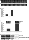
(A) Time-lapse sequence of an Uch2p-GFP α-tubulin-GFP–expressing pim1–1 cell undergoing anaphase at the permissive temperature of 24 °C. (B) Time-lapse sequence of an Uch2p-GFP α-tubulin-GFP–expressing pim1–1 ts cell undergoing anaphase at the restrictive temperature of 36 °C. Cells were shifted to the restrictive temperature 3 h prior to imaging to allow inactivation of Pim1p protein. Note that the NE is fragmented shortly after anaphase B onset. Shown are single maximum-intensity reconstructions of z-stacks. Numbers refer to the time, in minutes. (C) Graph quantifying the proportion of binucleate Pcp1p-GFP Uch2p-GFP pim1–1 cells overexpressing Mia1p at 24 °C and 36 °C (n = 300). (D) Graph quantifying the proportion of uninucleate and binucleate Mia1p-overexpressing Pcp1p-GFP Uch2p-GFP pim1–1 cells that do not separate the SPBs at 36 °C (n = 100). Insets, examples of the scored phenotypes. Epifluorescence images of fixed Mia1p-overexpressing pim1–1 cells containing the core SPB marker, Pcp1p-GFP, and the outer NE marker, Uch2p-GFP, at 36 °C. DNA is stained with DAPI. The NE is fragmented. Shown are single maximum-intensity reconstructions of z-stacks. (E) Epifluorescence images of Cen1-GFP Sad1-DSRed–containing pim1–1 cells, with and without Mia1p overexpression. Shown are single maximum-intensity reconstructions of z-stacks. Cartoons outline the phenotypes scored. Circle indicates the SPB; star indicates the centromere I.

Similar articles

Cited by

References

    1. Ding R, West RR, Morphew DM, Oakley BR, McIntosh JR. The spindle pole body of Schizosaccharomyces pombe enters and leaves the nuclear envelope as the cell cycle proceeds. Mol Biol Cell. 1997;8:1461–1479. - PMC - PubMed
    1. Uzawa S, Li F, Jin Y, McDonald KL, Braunfeld MB, et al. Spindle pole body duplication in fission yeast occurs at the G1/S boundary but maturation is blocked until exit from S by an event downstream of cdc10+ Mol Biol Cell. 2004;15:5219–5230. - PMC - PubMed
    1. Tran PT, Marsh L, Doye V, Inoue S, Chang F. A mechanism for nuclear positioning in fission yeast based on microtubule pushing. J Cell Biol. 2001;153:397–411. - PMC - PubMed
    1. Zheng L, Schwartz C, Wee L, Oliferenko S. The fission yeast transforming acidic coiled coil-related protein Mia1p/Alp7p is required for formation and maintenance of persistent microtubule-organizing centers at the nuclear envelope. Mol Biol Cell. 2006;17:2212–2222. - PMC - PubMed
    1. McCully EK, Robinow CF. Mitosis in the fission yeast Schizosaccharomyces pombe: A comparative study with light and electron microscopy. J Cell Sci. 1971;9:475–507. - PubMed

Publication types

MeSH terms

Substances

LinkOut - more resources