. 2007 Aug;293(2):L417-28.
doi: 10.1152/ajplung.00489.2006. Epub 2007 Jun 8.

PGE(2) inhibition of TGF-beta1-induced myofibroblast differentiation is Smad-independent but involves cell shape and adhesion-dependent signaling

Affiliations

PGE(2) inhibition of TGF-beta1-induced myofibroblast differentiation is Smad-independent but involves cell shape and adhesion-dependent signaling

Peedikayil E Thomas et al. Am J Physiol Lung Cell Mol Physiol. 2007 Aug.

Abstract

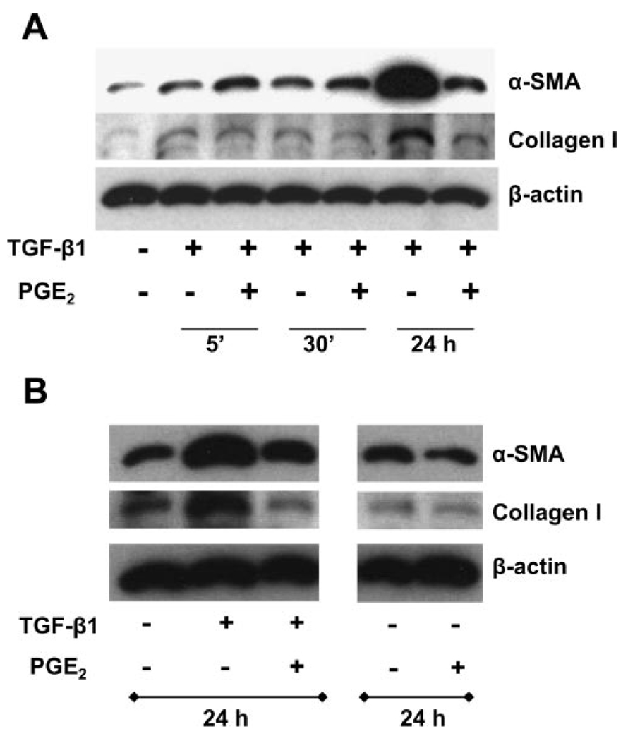
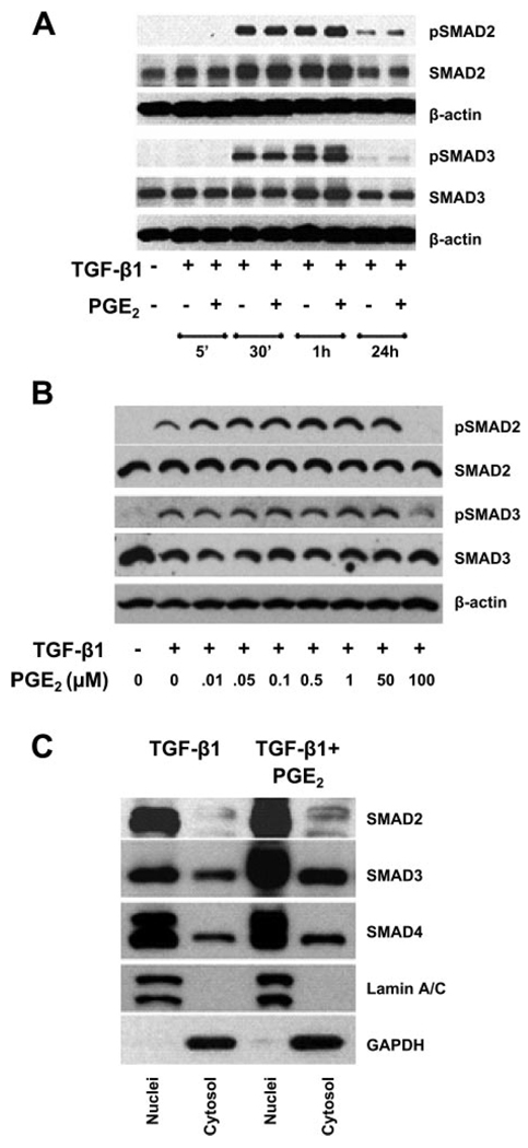
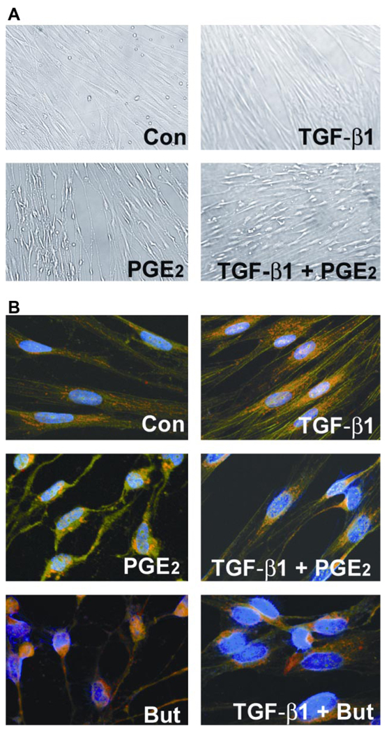
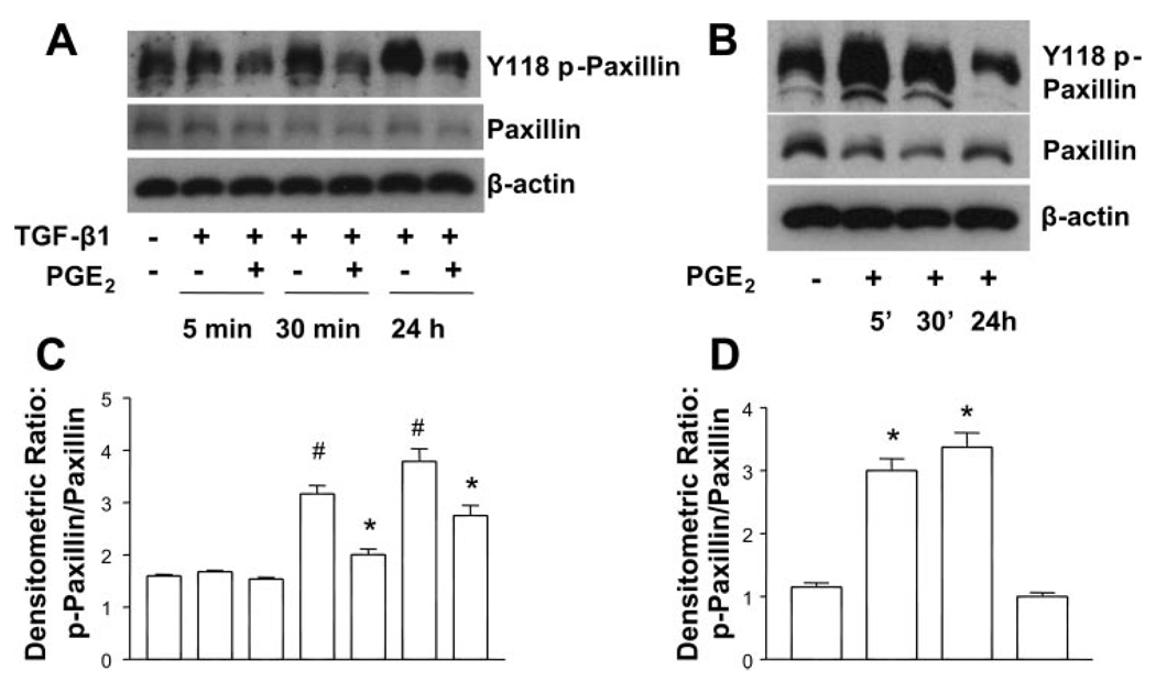
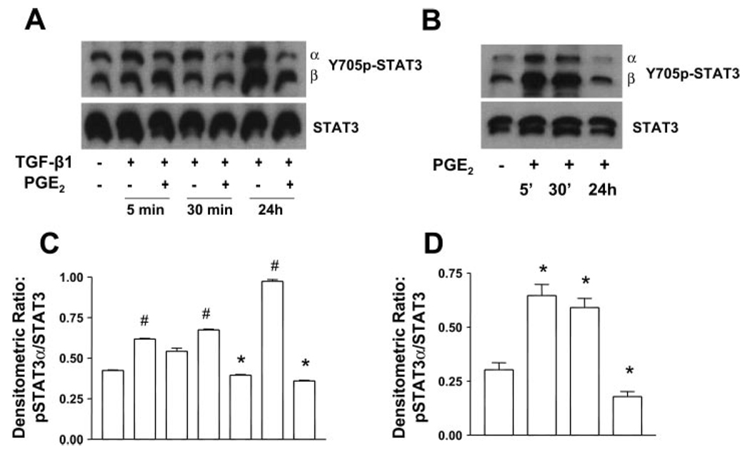
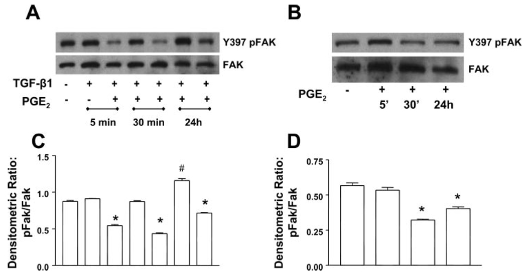
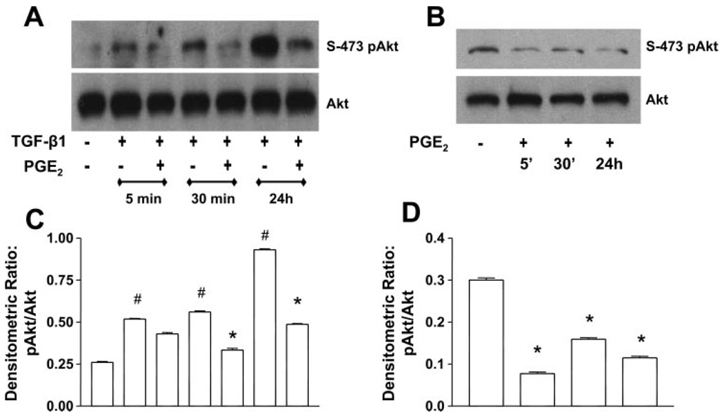
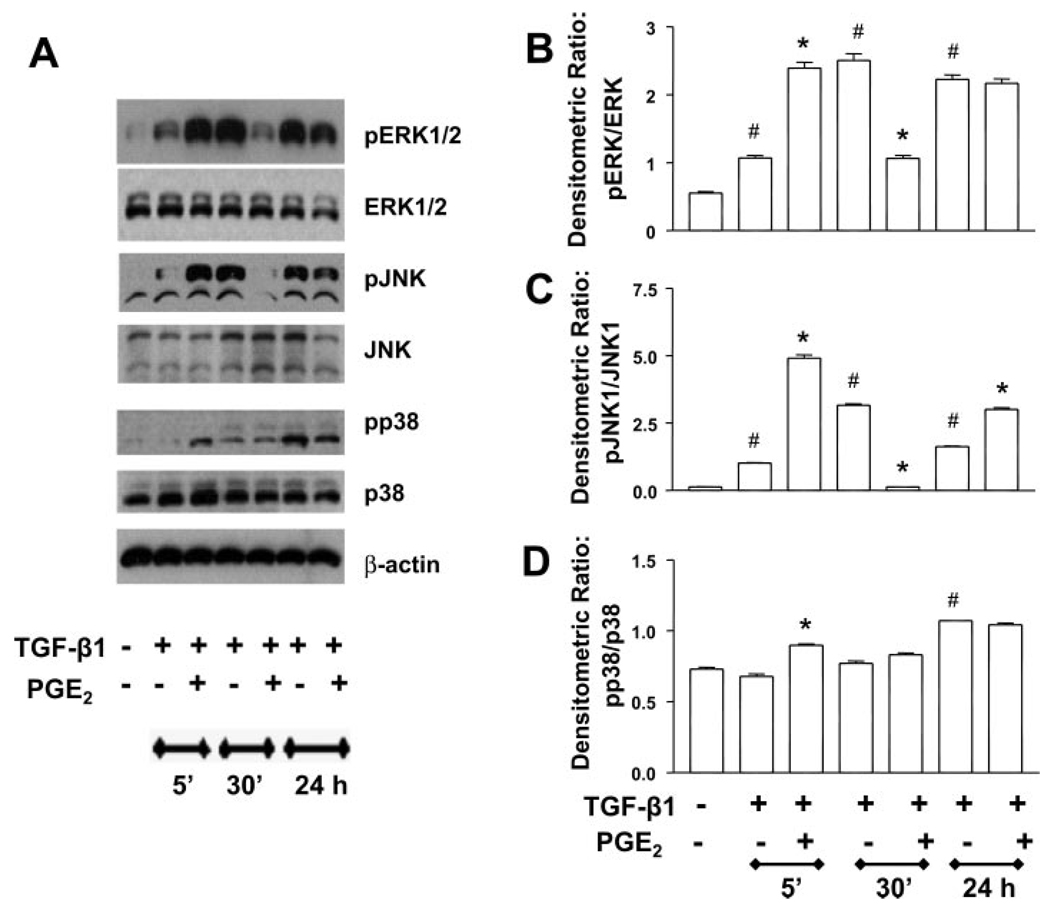
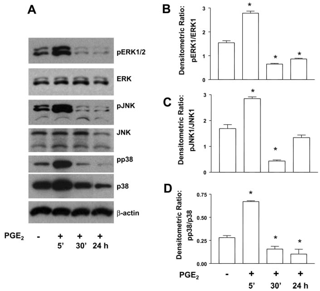
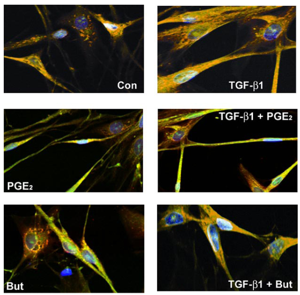
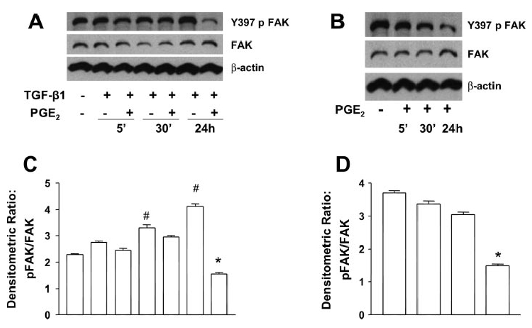
Myofibroblasts are pathogenic in pulmonary fibrotic disease due to their exuberant production of matrix rich in collagen that interferes with gas exchange and the ability of these cells to contract and distort the alveolar space. Transforming growth factor-beta1 (TGF-beta1) is a well-known inducer of myofibroblast differentiation. TGF-beta1-induced transformation of fibroblasts to apoptosis-resistant myofibroblasts is adhesion-dependent and focal adhesion kinase (FAK)-mediated. Prostaglandin E(2) (PGE(2)) inhibits this differentiation via E prostanoid receptor 2 (EP2) signaling and cAMP elevation, but whether PGE(2) does so by interfering with TGF-beta1 signaling is unknown. Thus we examined the effects of PGE(2) in the presence and absence of TGF-beta1 stimulation on candidate signaling pathways in human lung fibroblasts. We now demonstrate that PGE(2) does not interfere with TGF-beta1-induced Smad phosphorylation or its translocation to the nucleus. Rather, PGE(2) has dramatic effects on cell shape and cytoskeletal architecture and disrupts the formation of appropriate focal adhesions. PGE(2) treatment diminishes TGF-beta1-induced phosphorylation of paxillin, STAT-3, and FAK and, in turn, limits activation of the protein kinase B (PKB/Akt) pathway. These alterations do not, however, result in increased apoptosis within the first 24 h of treatment. Interestingly, the effects of PGE(2) stimulation alone do not always mirror the effects of PGE(2) in the presence of TGF-beta1, indicating that the context for EP2 signaling is different in the presence of TGF-beta1. Taken together, our results demonstrate that PGE(2) has the potential to limit TGF-beta1-induced myofibroblast differentiation via adhesion-dependent, but Smad-independent, pathways.


Figures

Fig. 1
Fig. 1
PGE2 suppresses the expression of collagen and smooth muscle actin-α (α-SMA). A: quiescent human lung fibroblasts (IMR-90) were treated with TGF-β1 (2 ng/ml), or TGF-β1 (2 ng/ml) and PGE2 (10 nM), under serum-free conditions for indicated times, and cell lysates were prepared. Cell lysates were subjected to SDS-PAGE followed by immunoblotting with antibodies against collagen 1, α-SMA, and β-actin. B: IMR-90 cells were treated with TGF-β1 alone, PGE2 alone (10 nM), or TGF-β1 + PGE2, and cell lysates were analyzed as above after 24 h. Data are representative of 3 independent experiments.
Fig. 2
Fig. 2
PGE2 did not affect the TGF-β1-stimulated phosphorylation (p) of Smad2/3. A: quiescent human lung fibroblasts (IMR-90) were treated with TGF-β1 (2 ng/ml), or TGF-β1 (2 ng/ml) and PGE2 (10 nM), under serum-free conditions, and cell lysates were prepared at the times indicated. Cell lysates were subjected to SDS-PAGE followed by immunoblotting with antibodies against pS465/467 Smad2 and pS422/424 Smad3, Smad2, and Smad3. Blots were probed first with phospho-specific Smad antibodies, stripped, and reblotted with antibodies for respective nonphosphorylated Smad proteins or β-actin. B: IMR-90 cells were treated as above with TGF-β1 in the presence or absence of increasing concentrations of PGE2 ranging from 0.01 to 100 µM, and phosphorylation of Smad2 and Smad3 was assessed as in A. C: PGE2 did not abrogate the translocation of Smad2/3/4 complex to the nucleus in human lung fibroblasts. Quiescent human lung fibroblasts (IMR-90) were treated with TGF-β1 (2 ng/ml), or TGF-β1 (2 ng/ml) and PGE2 (10 nM), under serum-free conditions for 30 min. Cells were lysed, and nuclei and cytosol were fractionated. Reduced nuclear and cytosolic fractions were used for Western blot analysis with antibodies against Smad2, Smad3, and Smad4. GAPDH and lamin A/C were used as markers for cytosol and nuclear fractions, respectively. Data are representative of 3 independent experiments.
Fig. 3
Fig. 3
PGE2 induces sudden and sustained morphological changes. A: IMR-90 cells were growth-arrested at 85% confluency for 48 h. Medium was replaced with serum-free media or that containing TGF-β1 (2 ng/ml) or PGE2 (10 nM) or both TGF-β1 and PGE2. Cells were photographed after 5 min of treatment. Control (Con) cells show characteristic flattened spindle shape. TGF-β1-treated cells show spindle shape with a more 3-dimensional appearance. PGE2-treated cells are no longer spindle shaped and appear shrunken longitudinally with decrease in area of surface fibrillar and focal adhesion. The appearance of the cells treated with both TGF-β1 and PGE2 is intermediate between those treated with TGF-β1 alone and those treated with PGE2 alone. B: PGE2 alters cytoskeletal structure and focal adhesions in human lung fibroblasts. Shown are FITC-phalloidin staining (green), immunocytochemistry with paxillin (red), and 4,6-diamidino-2-phenylindole (DAPI) staining of nuclei (blue). IMR-90 cells were serum-deprived for 48 h. Medium was replaced with serum-free media or that containing TGF-β1, PGE2, butaprost (But), or combinations of both TGF-β1 and PGE2 or butaprost. After 24 h, cells were washed, fixed, and stained. Cells were then analyzed using laser-scanning confocal microscopy with appropriate wavelengths using a ×60 water immersion objective. Merged images are shown, and focal adhesions appear as yellow/orange.
Fig. 4
Fig. 4
PGE2 inhibits the TGF-β1-stimulated phosphorylation of paxillin. A: quiescent human lung fibroblasts (IMR-90) were treated with TGF-β1 (2 ng/ml), or TGF-β1 (2 ng/ml) and PGE2 (10 nM), under serum-free conditions, and cell lysates were prepared at the times indicated. B: quiescent human lung fibroblasts (IMR-90) were treated with PGE2 (10 nM) under serum-free conditions, and cell lysates were prepared at the times indicated. Cell lysates were subjected to SDS-PAGE followed by immunoblotting with antibodies against p-paxillin. Blots probed with antibody against p-paxillin were stripped and blotted with antibodies against paxillin and β-actin sequentially. C: densitometry data for n = 3 experiments as in A. #P < 0.05 for TGF-β1-treated samples compared with untreated control; *P < 0.05 for TGF-β1 + PGE2-treated samples compared with TGF-β1 treatment alone. D: densitometry data for n = 3 experiments as in B. *P < 0.05 compared with untreated control.
Fig. 5
Fig. 5
PGE2 inhibits TGF-β1-stimulated phosphorylation of STAT-3 (Y705p-STAT-3). A: quiescent human lung fibroblasts (IMR-90) were treated with TGF-β1 (2 ng/ml), or TGF-β1 (2 ng/ml) and PGE2 (10 nM), under serum-free conditions, and cell lysates were prepared at the times indicated. B: quiescent human lung fibroblasts (IMR-90) were treated with PGE2 (10 nM) under serum-free conditions, and cell lysates were prepared at the times indicated. Cell lysates were subjected to SDS-PAGE followed by immunoblotting with antibodies against Y705p-STAT-3, which can detect both the alpha (α) and beta (β) splice forms. Blots probed with antibody against Y705p-STAT-3 were stripped and blotted with antibodies against STAT-3 sequentially. C: densitometry data for n = 3 experiments as in A. D: densitometry data for n = 3 experiments as in B. Significance is indicated with # and * symbols as in Fig 4.
Fig. 6
Fig. 6
PGE2 inhibits the TGF-β1-stimulated Y-397 focal adhesion kinase (FAK) autophosphorylation (Y397pFAK). A: quiescent human lung fibroblasts (IMR-90) were treated with TGF-β1 (2 ng/ml), or TGF-β1 (2 ng/ml) and PGE2 (10 nM), under serum-free conditions, and cell lysates were prepared at the times indicated. B: quiescent human lung fibroblasts (IMR-90) were treated with PGE2 (10 nM) under serum-free conditions, and cell lysates were prepared at the times indicated. Cell lysates were subjected to SDS-PAGE followed by immunoblotting with antibodies against Y397pFAK. Blots probed with antibody against Y397pFAK were stripped and blotted with antibodies against FAK sequentially. C: densitometry data for n = 3 experiments as in A. D: densitometry data for n = 3 experiments as in B. Significance is indicated with # and * symbols as in Fig 4.
Fig. 7
Fig. 7
PGE2 inhibits the TGF-β1-stimulated PKB/Akt phosphorylation. A: quiescent human lung fibroblasts (IMR-90) were treated with TGF-β1 (2 ng/ml), or TGF-β1 (2 ng/ml) and PGE2 (10 nM), under serum-free conditions, and cell lysates were prepared at the times indicated. B: quiescent human lung fibroblasts (IMR-90) were treated with PGE2 (10 nM) under serum-free conditions, and cell lysates were prepared at the times indicated. Cell lysates were subjected to SDS-PAGE followed by immunoblotting with antibodies against phosphorylated Akt (S-473p-Akt). Blots probed with antibody against S-473p-Akt were stripped and blotted with antibodies against Akt sequentially. C: densitometry data for n = 3 experiments as in A. D: densitometry data for n = 3 experiments as in B. Significance is indicated with # and * symbols as in Fig 4.
Fig. 8
Fig. 8
PGE2 alters the kinetics of TGF-β1-stimulated MAP kinase activation. A: quiescent human lung fibroblasts (IMR-90) were treated with TGF-β1 (2 ng/ml), or TGF-β1 (2 ng/ml) and PGE2 (10 nM), under serum-free conditions, and cell lysates were prepared at the times indicated. Cell lysates were subjected to SDS-PAGE followed by immunoblotting with antibodies against pERK1/2, pJNK, or pp38. Blots were then stripped and blotted with antibodies against ERK1/2, JNK, or p38 sequentially. B: densitometry data for n = 3 experiments looking at ERK phosphorylation. C: densitometry data for n = 3 experiments looking at JNK phosphorylation. D: densitometry data for n = 3 experiments looking at p38 phosphorylation. Significance is indicated with # and * symbols as in Fig 4.
Fig. 9
Fig. 9
PGE2 stimulates rapid transient MAP kinase activation but diminished late activation. A: quiescent human lung fibroblasts (IMR-90) were treated with PGE2 (10 nM) under serum-free conditions, and cell lysates were prepared at the times indicated. Cell lysates were subjected to SDS-PAGE followed by immunoblotting with antibodies against pERK1/2, pJNK, or pp38. Blots were then stripped and blotted with antibodies against ERK1/2, JNK, or p38 sequentially. B: densitometry data for n = 3 experiments looking at ERK phosphorylation. C: densitometry data for n = 3 experiments looking at JNK phosphorylation. D: densitometry data for n = 3 experiments looking at p38 phosphorylation. *P < 0.05 compared with untreated controls.
Fig. 10
Fig. 10
PGE2 alters cell morphology and inhibits the formation of TGF-β1-induced focal adhesions in adult lung fibroblasts. Normal adult lung fibroblasts were serum-starved for 48 h before culture with serum-free media alone, TGF-β1 alone (2 ng/ml), PGE2 alone (100 nM), butaprost alone (5 µM), or TGF-β1 in combination with PGE2 or butaprost. Cells were then fixed and stained with FITC-phalloidin (green), paxillin (red), and DAPI staining of nuclei (blue). Cells were then analyzed using laser-scanning confocal microscopy with appropriate wavelengths using a ×60 water immersion objective. Merged images are shown, and focal adhesions appear as orange/yellow. Z-stack analysis confirmed the colocalization of the FITC and indocarbocyanine (Cy3) staining.
Fig. 11
Fig. 11
PGE2 inhibits the TGF-β1-stimulated Y-397 FAK autophosphorylation in adult lung fibroblasts. A: quiescent adult lung fibroblasts were treated with TGF-β1 (2 ng/ml), or TGF-β1 (2 ng/ml) and PGE2 (100 nM), under serum-free conditions, and cell lysates were prepared at the times indicated. B: quiescent adult lung fibroblasts were treated with PGE2 (100 nM) under serum-free conditions, and cell lysates were prepared at the times indicated. Cell lysates were subjected to SDS-PAGE followed by immunoblotting with antibodies against Y397pFAK. Blots probed with antibody against Y397pFAK were stripped and blotted with antibodies against total FAK sequentially. C: densitometry data for n = 3 experiments as in A. D: densitometry data for n = 3 experiments as in B. Significance is indicated with # and * symbols as in Fig 4.

Similar articles

Cited by

References

    1. Bhattacharyya S, Ghosh AK, Pannu J, Mori Y, Takagawa S, Chen G, Trojanowska M, Gilliam AC, Varga J. Fibroblast expression of the coactivator p300 governs the intensity of profibrotic response to transforming growth factor beta. Arthritis Rheum. 2005;52:1248–1258. - PubMed
    1. Border WA, Noble NA. Transforming growth factor beta in tissue fibrosis. N Engl J Med. 1994;331:1286–1292. - PubMed
    1. Border WA, Ruoslahti E. Transforming growth factor-beta in disease: the dark side of tissue repair. J Clin Invest. 1992;90:1–7. - PMC - PubMed
    1. Bulin C, Albrecht U, Bode JG, Weber AA, Schror K, Levkau B, Fischer JW. Differential effects of vasodilatory prostaglandins on focal adhesions, cytoskeletal architecture, and migration in human aortic smooth muscle cells. Arterioscler Thromb Vasc Biol. 2005;25:84–89. - PubMed
    1. Charbeneau RP, Christensen PJ, Chrisman CJ, Paine R, Toews GB, Peters-Golden M, Moore BB. Impaired synthesis of prostaglandin E2 by lung fibroblasts and alveolar epithelial cells from GM-CSF−/−mice: implications for fibroproliferation. Am J Physiol Lung Cell Mol Physiol. 2003;284:L1103–L1111. - PubMed

Publication types

MeSH terms