. 2007 Jul 11;26(13):3086-97.
doi: 10.1038/sj.emboj.7601746. Epub 2007 Jun 7.

Selective redox regulation of cytokine receptor signaling by extracellular thioredoxin-1

Affiliations

Selective redox regulation of cytokine receptor signaling by extracellular thioredoxin-1

Ulla Schwertassek et al. EMBO J. .

Abstract

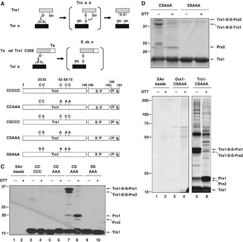
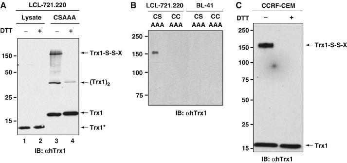
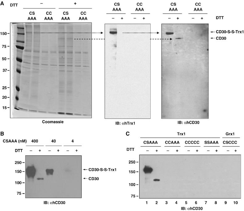
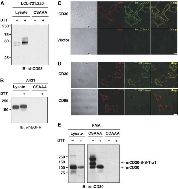
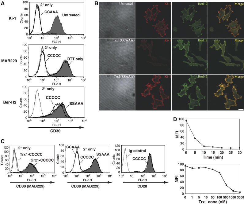
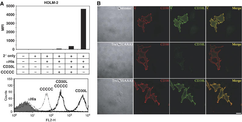
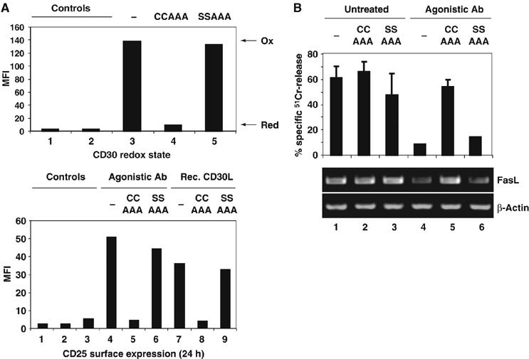
The thiol-disulfide oxidoreductase thioredoxin-1 (Trx1) is known to be secreted by leukocytes and to exhibit cytokine-like properties. Extracellular effects of Trx1 require a functional active site, suggesting a redox-based mechanism of action. However, specific cell surface proteins and pathways coupling extracellular Trx1 redox activity to cellular responses have not been identified so far. Using a mechanism-based kinetic trapping technique to identify disulfide exchange interactions on the intact surface of living lymphocytes, we found that Trx1 catalytically interacts with a single principal target protein. This target protein was identified as the tumor necrosis factor receptor superfamily member 8 (TNFRSF8/CD30). We demonstrate that the redox interaction is highly specific for both Trx1 and CD30 and that the redox state of CD30 determines its ability to engage the cognate ligand and transduce signals. Furthermore, we confirm that Trx1 affects CD30-dependent changes in lymphocyte effector function. Thus, we conclude that receptor-ligand signaling interactions can be selectively regulated by an extracellular redox catalyst.


Figures

Figure 1
Figure 1
Mechanism-based kinetic trapping identifies target proteins of human Trx1. (A) Catalytic mechanism of wild-type Trx1 (upper panel) and the principle of substrate trapping (lower panel). The mixed disulfide intermediate is normally resolved by cysteine-35. Replacement of cysteine-35 by serine (C35S) stabilizes the covalent intermediate. The affinity tag enables purification of resulting enzyme–substrate complexes. (B) Schematic representation of recombinant Trx1 constructs. Catalytic (C32, C35) and structural cysteines (C62, C69, C73) are indicated. His6: hexahistidine tag; SBP: streptavidin-binding peptide. (C) Trapping of cytosolic Trx1 target proteins. Cytosolic proteins released from digitonin-permeabilized Jurkat cells were incubated with different recombinant Trx1 proteins. Disulfide-linked Trx1 complexes were analyzed by silver staining under non-reducing and reducing conditions. Trx1 complexes involving Prx1 and Prx2 are indicated; SAv streptavidin. (D) Trapping of extracellular Trx1 target proteins. Trapping (CSAAA) and non-trapping (SSAAA) mutants of Trx1 were incubated with a <30 kDa fraction prepared from fresh human serum. Disulfide-linked complexes were analyzed by colloidal Coomassie staining under non-reducing and reducing conditions. The Trx1–Prx2 conjugate and the Trx1 dimer as well as monomeric Prx2 and Trx1 are indicated. (E) Kinetic trapping is mediated by specific protein–protein interactions. Cytosolic proteins from digitonin-permeabilized Jurkat cells were incubated with SAv sepharose, Grx1(CSAAA) or Trx1(CSAAA). Disulfide-linked complexes were analyzed by silver staining under non-reducing and reducing conditions. Trx1 conjugates formed with Prx1 and Prx2 are indicated. Other bands correspond to additional cytosolic proteins interacting with Trx1.
Figure 2
Figure 2
Trx1 targets a single principal interaction partner on the surface of lymphocytic cell lines. (A) LCL-721.220 cells were incubated with Trx1(CSAAA) and disulfide-linked complexes analyzed by anti-Trx1 immunoblotting under non-reducing and reducing conditions. Cellular lysate was included as an immunoblotting control (Trx1*: endogenous Trx1). Dimerization of exogenously added Trx1 is indicated. (B) LCL-721.220 or BL-41 B cells were treated as in (A). Trx1(CCAAA) was included as a control. Disulfide-linked complexes were analyzed as in (A). (C) CCRF-CEM T cells were treated as in (A) and complexes analyzed by anti-Trx1 immunoblotting under non-reducing and reducing conditions. Conjugation of the unknown cell surface protein is indicated (Trx1-S-S-X).
Figure 3
Figure 3
TNFRSF8/CD30 is the main Trx1 interaction partner on the surface of lymphoid cell lines. (A) A total of 5 × 10⁹ LCL-721.220 cells were incubated with Trx1(CSAAA) or Trx1(CCAAA) as control. Disulfide-linked Trx1 complexes were purified by SAv affinity purification and analyzed by colloidal Coomassie staining under non-reducing and reducing conditions (left panel). Indicated bands were subjected to tryptic digestion and LC-MS/MS analysis. In parallel, part of the same sample was analyzed by anti-Trx1 immunoblotting (middle panel). The blot was stripped and reprobed with anti-human CD30 antibody (right panel). (B) CCRF-CEM T cells were incubated with different amounts of Trx1(CSAAA) and disulfide-linked Trx1 complexes were analyzed by anti-CD30 immunoblotting under non-reducing and reducing conditions. (C) CCRF-CEM T cells were incubated with recombinant Trx1 constructs or the Grx1(C25S) trapping mutant as indicated and complexes were analyzed as described in (B). The difference in signal intensity between non-reduced and reduced form of CD30 is due to less efficient recognition of the reduced form by the CD30-specific antibody.
Figure 4
Figure 4
Trx1 discriminates between CD30 and other receptors with CRDs. (A) LCL-721.220 B cells were incubated with Trx1(CSAAA) and disulfide-linked Trx1 complexes were analyzed by anti-CD95 immunoblotting. (B) A431 epithelial carcinoma cells were incubated with Trx1(CSAAA) and complexes were analyzed by anti-EGFR immunoblotting. Cellular lysate was included as control. (C) HeLa cells were transiently transfected with an expression construct for human CD30 or empty vector incubated with Trx1(CSAAA) and analyzed by immunofluorescence microscopy using CD30- and Trx1-specific antibodies (scale bar, 20 μm). (D) HeLa cells were transiently transfected with expression constructs for human CD30 or CD95, treated as described in (C) and analyzed by immunofluorescence microscopy using CD30-, Trx1- and CD95-specific antibodies (scale bar, 20 μm). (E) RMA mouse lymphoma cells were incubated with Trx1(CSAAA) or Trx1(CCAAA). Disulfide-linked Trx1 complexes were analyzed by anti-mouse CD30 (mCD30) immunoblotting under non-reducing and reducing conditions. The disulfide-linked Trx1-mCD30 complex and monomeric mCD30 are indicated. The additional band of higher molecular weight has not been further characterized but might represent a conjugate between Trx1 and a dimer of mCD30.
Figure 5
Figure 5
Trx1 catalyzes disulfide bond reduction within the CD30 ectodomain. (A) CCRF-CEM T cells were left untreated or treated with active (CCAAA/CCCCC) or inactive Trx1 (SSAAA) in the presence of DTT as a regenerating system or with DTT only. Cell surface CD30 was analyzed by flow cytometry using three different anti-CD30 antibodies (Ki-1: upper panel; MAB229: middle panel; Ber-H2: lower panel). PE-labeled secondary antibody only was included as control. Untreated cells are shown in gray. (B) HeLa cells were transiently transfected with a CD30 expression vector and either left untreated or treated with active (CCAAA) or inactive Trx1 (SSAAA) in the presence of DTT. Cells were analyzed by immunofluorescence microscopy using CD30-specific antibodies Ki-1 and Ber-H2 (scale bar, 20 μm). (C) CCRF-CEM cells were left untreated or treated with different Trx1 constructs or wild-type Grx1 as indicated. Cell surface CD30 was analyzed by anti-CD30 antibody MAB229 (left and middle panel). Alternatively, cells treated with wild-type Trx1 were analyzed by PE-conjugated anti-CD28 antibody (right panel). Untreated cells are shown in gray. (D) CCRF-CEM cells were treated with 5 μM active Trx1 for different time points (upper graph) or with different Trx1 concentrations (lower graph) for 30 min. Cells were analyzed as described in (C).
Figure 6
Figure 6
Trx1-mediated disulfide bond reduction in CD30 prevents binding of the CD30L. (A) HDLM-2 Hodgkin cells were either left untreated or treated with wild-type Trx1 in the presence of DTT as a regenerating system. After washing, cells were incubated with recombinant CD30L-His10 as indicated. Bound CD30L was detected by flow cytometry using anti-polyHis antibody. Cells incubated with anti-polyHis antibody only were used as control. Unstained cells are shown in gray. (B) HeLa cells were transiently transfected with an expression construct for CD30 and either left untreated or treated with active (CCAAA) or inactive Trx1 (SSAAA) in the presence of DTT. After washing, cells were incubated with recombinant CD30L-His10. CD30L bound to the cell surface was analyzed by immunofluorescence microscopy using anti-CD30L antibody (scale bar, 20 μm).
Figure 7
Figure 7
Trx1 controls CD30-mediated functional changes in lymphoma cells. (A) Trx1 inhibits CD30-mediated changes in gene expression. YT large granular lymphoma cells were left untreated or treated with active (CCAAA) or inactive Trx1 (SSAAA) in the presence of TrxR/NADPH as a regenerating system. After treatment, the change in CD30 redox state was verified by flow cytometry using antibody MAB229 (upper panel). Cells were then stimulated with agonistic anti-CD30 antibody (MAB229) or recombinant CD30L, or left unstimulated (column 3). After 24 h, cell surface expression of the IL-2Rα chain (CD25) was analyzed by flow cytometry using anti-CD25 antibody. As control, unstained cells and cells stained with 2° antibody only (columns 1 and 2) were used. (B) Trx1 inhibits CD30-mediated changes in effector function. YT cells were treated as described in (A). After 24 h, YT cells were harvested and analyzed for cytotoxicity on 51Cr-labeled Raji cells (E:T ratio of 25:1). The experiment was performed in triplicates. Expression levels of FasL-mRNA were analyzed by RT–PCR. β-Actin levels are shown as control.

Similar articles

Cited by

References

    1. Angelini G, Gardella S, Ardy M, Ciriolo MR, Filomeni G, Di Trapani G, Clarke F, Sitia R, Rubartelli A (2002) Antigen-presenting dendritic cells provide the reducing extracellular microenvironment required for T lymphocyte activation. Proc Natl Acad Sci USA 99: 1491–1496 - PMC - PubMed
    1. Bertini R, Howard OM, Dong HF, Oppenheim JJ, Bizzarri C, Sergi R, Caselli G, Pagliei S, Romines B, Wilshire JA, Mengozzi M, Nakamura H, Yodoi J, Pekkari K, Gurunath R, Holmgren A, Herzenberg LA, Ghezzi P (1999) Thioredoxin, a redox enzyme released in infection and inflammation, is a unique chemoattractant for neutrophils, monocytes, and T cells. J Exp Med 189: 1783–1789 - PMC - PubMed
    1. Bodmer JL, Schneider P, Tschopp J (2002) The molecular architecture of the TNF superfamily. Trends Biochem Sci 27: 19–26 - PubMed
    1. Bowen MA, Olsen KJ, Cheng L, Avila D, Podack ER (1993) Functional effects of CD30 on a large granular lymphoma cell line, YT. Inhibition of cytotoxicity, regulation of CD28 and IL-2R, and induction of homotypic aggregation. J Immunol 151: 5896–5906 - PubMed
    1. Burgess SC, Young JR, Baaten BJ, Hunt L, Ross LN, Parcells MS, Kumar PM, Tregaskes CA, Lee LF, Davison TF (2004) Marek's disease is a natural model for lymphomas overexpressing Hodgkin's disease antigen (CD30). Proc Natl Acad Sci USA 101: 13879–13884 - PMC - PubMed

Publication types

MeSH terms