

Semliki Forest virus nonstructural protein 2 is involved in suppression of the type I interferon response
- PMID: 17553895
- PMCID: PMC1951358
- DOI: 10.1128/JVI.02411-06
Semliki Forest virus nonstructural protein 2 is involved in suppression of the type I interferon response
Abstract
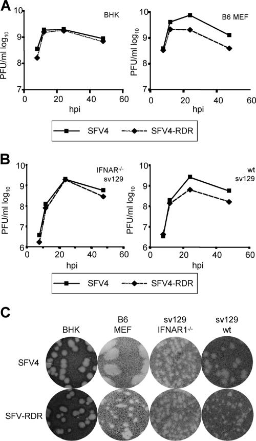
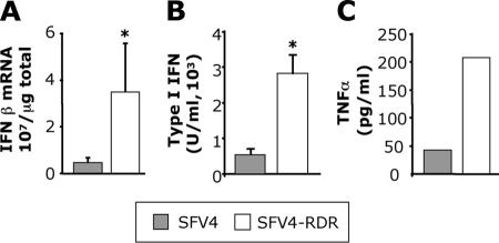
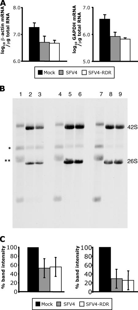
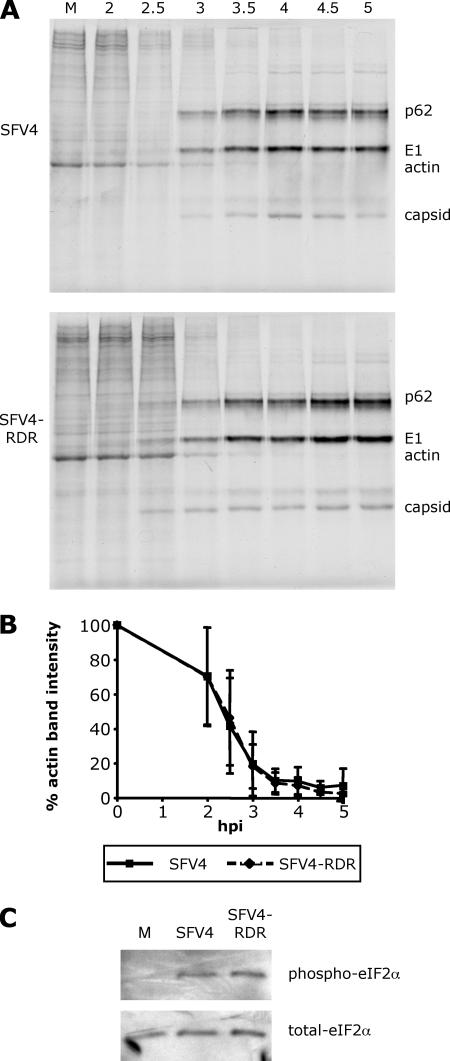
The type I interferons (IFNs) are potent mediators of antiviral immunity, and many viruses have developed means to block their expression or their effects. Semliki Forest virus (SFV) infection induces rapid and profound silencing of host cell gene expression, a process believed to be important for the inhibition of the IFN response. In SFV-infected cells, a large proportion of the nonstructural protein nsp2 is found in the nucleus, but a role for this localization has not been described. In this work we demonstrate that a viral mutant, SFV4-RDR, in which the nuclear localization sequence of nsp2 has been rendered inactive, induces a significantly more robust IFN response in infected cells. This mutant virus replicates at a rate similar to that of the parental SFV4 strain and also shuts off host cell gene expression to similar levels, indicating that the general cellular shutoff is not responsible for the inhibition of IFN expression. Further, the rate of virus-induced nuclear translocation of early IFN transcription factors was not found to differ between the wild-type and mutant viruses, indicating that the effect of nsp2 is at a later stage. These results provide novel information about the mode of action of this viral IFN antagonist.
Figures
Similar articles
-
Functional significance of the nuclear-targeting and NTP-binding motifs of Semliki Forest virus nonstructural protein nsP2.Virology. 1996 Apr 15;218(2):352-61. doi: 10.1006/viro.1996.0204. Virology. 1996. PMID: 8610462
-
A single amino acid change in the nuclear localization sequence of the nsP2 protein affects the neurovirulence of Semliki Forest virus.J Virol. 2002 Jan;76(1):392-6. doi: 10.1128/jvi.76.1.392-396.2002. J Virol. 2002. PMID: 11739703 Free PMC article.
-
MicroRNA-Attenuated Clone of Virulent Semliki Forest Virus Overcomes Antiviral Type I Interferon in Resistant Mouse CT-2A Glioma.J Virol. 2015 Oct;89(20):10637-47. doi: 10.1128/JVI.01868-15. Epub 2015 Aug 12. J Virol. 2015. PMID: 26269187 Free PMC article.
-
The Methyltransferase-Like Domain of Chikungunya Virus nsP2 Inhibits the Interferon Response by Promoting the Nuclear Export of STAT1.J Virol. 2018 Aug 16;92(17):e01008-18. doi: 10.1128/JVI.01008-18. Print 2018 Sep 1. J Virol. 2018. PMID: 29925658 Free PMC article.
-
Regulation of Semliki Forest virus RNA replication: a model for the control of alphavirus pathogenesis in invertebrate hosts.Virology. 2004 May 20;323(1):153-63. doi: 10.1016/j.virol.2004.03.009. Virology. 2004. PMID: 15165827
Cited by
-
Deciphering the differential response of two human fibroblast cell lines following Chikungunya virus infection.Virol J. 2012 Sep 20;9:213. doi: 10.1186/1743-422X-9-213. Virol J. 2012. PMID: 22992396 Free PMC article.
-
The C-terminal domain of chikungunya virus nsP2 independently governs viral RNA replication, cytopathicity, and inhibition of interferon signaling.J Virol. 2013 Sep;87(18):10394-400. doi: 10.1128/JVI.00884-13. Epub 2013 Jul 17. J Virol. 2013. PMID: 23864632 Free PMC article.
-
Evasion of the innate immune response: the Old World alphavirus nsP2 protein induces rapid degradation of Rpb1, a catalytic subunit of RNA polymerase II.J Virol. 2012 Jul;86(13):7180-91. doi: 10.1128/JVI.00541-12. Epub 2012 Apr 18. J Virol. 2012. PMID: 22514352 Free PMC article.
-
ViralORFeome: an integrated database to generate a versatile collection of viral ORFs.Nucleic Acids Res. 2010 Jan;38(Database issue):D371-8. doi: 10.1093/nar/gkp1000. Epub 2009 Dec 8. Nucleic Acids Res. 2010. PMID: 20007148 Free PMC article.
-
Chikungunya virus nonstructural protein 2 inhibits type I/II interferon-stimulated JAK-STAT signaling.J Virol. 2010 Oct;84(20):10877-87. doi: 10.1128/JVI.00949-10. Epub 2010 Aug 4. J Virol. 2010. PMID: 20686047 Free PMC article.
References
-
- Ahmed, M., M. O. McKenzie, S. Puckett, M. Hojnacki, L. Poliquin, and D. S. Lyles. 2003. Ability of the matrix protein of vesicular stomatitis virus to suppress beta interferon gene expression is genetically correlated with the inhibition of host RNA and protein synthesis. J. Virol. 77:4646-4657. - PMC - PubMed
-
- Amineva, S. P., A. G. Aminev, A. C. Palmenberg, and J. E. Gern. 2004. Rhinovirus 3C protease precursors 3CD and 3CD′ localize to the nuclei of infected cells. J. Gen. Virol. 85:2969-2979. - PubMed
Publication types
MeSH terms
Substances
LinkOut - more resources
Full Text Sources
Other Literature Sources
