. 2007 Jun;4(6):e186.
doi: 10.1371/journal.pmed.0040186.

Vascular endothelial growth factor mediates intracrine survival in human breast carcinoma cells through internally expressed VEGFR1/FLT1

Affiliations

Vascular endothelial growth factor mediates intracrine survival in human breast carcinoma cells through internally expressed VEGFR1/FLT1

Tae-Hee Lee et al. PLoS Med. 2007 Jun.

Abstract

Background: While vascular endothelial growth factor (VEGF) expression in breast tumors has been correlated with a poor outcome in the pathogenesis of breast cancer, the expression, localization, and function of VEGF receptors VEGFR1 (also known as FLT1) and VEGFR2 (also known as KDR or FLK1), as well as neuropilin 1 (NRP1), in breast cancer are controversial.

Methods and findings: We investigated the expression and function of VEGF and VEGF receptors in breast cancer cells. We observed that VEGFR1 expression was abundant, VEGFR2 expression was low, and NRP1 expression was variable. MDA-MB-231 and MCF-7 breast cancer cells, transfected with antisense VEGF cDNA or with siVEGF (VEGF-targeted small interfering RNA), showed a significant reduction in VEGF expression and increased apoptosis as compared to the control cells. Additionally, specifically targeted knockdown of VEGFR1 expression by siRNA (siVEGFR1) significantly decreased the survival of breast cancer cells through down-regulation of protein kinase B (AKT) phosphorylation, while targeted knockdown of VEGFR2 or NRP1 expression had no effect on the survival of these cancer cells. Since a VEGFR1-specific ligand, placenta growth factor (PGF), did not, as expected, inhibit the breast cancer cell apoptosis induced by siVEGF, and since VEGFR1 antibody also had no effects on the survival of these cells, we examined VEGFR1 localization. VEGFR1 was predominantly expressed internally in MDA-MB-231 and MCF-7 breast cancer cells. Specifically, VEGFR1 was found to be colocalized with lamin A/C and was expressed mainly in the nuclear envelope in breast cancer cell lines and primary breast cancer tumors. Breast cancer cells treated with siVEGFR1 showed significantly decreased VEGFR1 expression levels and a lack of VEGFR1 expression in the nuclear envelope.

Conclusions: This study provides, to our knowledge for the first time, evidence of a unique survival system in breast cancer cells by which VEGF can act as an internal autocrine (intracrine) survival factor through its binding to VEGFR1. These results may lead to an improved strategy for tumor therapy based on the inhibition of angiogenesis.


Conflict of interest statement

Competing Interests: The authors have declared that no competing interests exist.

Figures

Figure 1
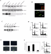
Figure 1. Expression of VEGF Receptors in Breast Cancer Cells and Apoptosis of MDA-MB-231 Cells by Down-Regulation of VEGF
(A) VEGF receptor expression was examined by using immunoprecipitation (IP) and Western blot (WB) analyses in several breast cancer cell lines. (B) VEGFR2 expression in MDA-MB-231 cells was analyzed by confocal microscopy. Cells were stained with anti-VEGFR2 antibody or IgG (inset). The asterisk indicates cells transduced with adenovirus encoding soluble human VEGFR2, which served as a positive control. (C) MDA-MB-231 cells were transfected with antisense VEGF vector and selected in the presence of Zeocin (1 mg/ml). VEGF expression in the MDA-MB-231 transfectants (pZeoSV, AS-C1, and AS-C2) was assessed by Western blotting (WB) using a polyclonal anti-VEGF antibody. Total protein extracts were analyzed by Western blotting using anti-Csk antibody as an internal control. (D and E) MDA-MB-231 cells were stably transfected with antisense VEGF vector and the survival of these cells was measured by using cell cycle analysis (D) and TUNEL assay (E). (F) MDA-MB-231 cells were transiently transfected with siCTL or siVEGF oligonucleotides, and VEGF was quantitated in the supernatants (left graph) using an ELISA kit as directed by the manufacturer. The apoptosis of these cells was measured by using cell cycle analysis. The data are representative of three individual studies (right graphs).
Figure 2
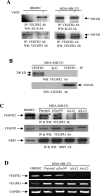
Figure 2. VEGF Receptor Expression in VEGF-Reduced MDA-MB-231 Cells
(A) MDA-MB-231 cells and HBMECs were treated with VEGF (30 ng/ml) to examine its effects on the phosphorylation (top blots) and expression (bottom blots) of VEGFR2. Total cell lysates were prepared and then subjected to immunoprecipitation (IP) and Western blotting (WB) using 4G10 or anti-VEGFR2 antibodies. The arrowhead indicates a nonspecific protein band. (B) Lysates (5 mg) from MDA-MB-231 cells were immunoprecipitated by using anti-VEGFR1 or anti-VEGFR2 antibodies. The immune complexes were analyzed by Western blotting using anti-VEGFR2 antibodies. After stripping, the membrane was subjected to Western blotting using anti- VEGFR1 antibodies. (C) Lysates (2 mg) from variously transfected MDA-MB-231 cells were subjected to immunoprecipitation (IP) and Western blot (WB) analyses for VEGF and NRP1 receptor expression, as indicated. Total protein lysates (0.4 mg) from HBMECs were used as a positive control. (D) Total RNA (5 μg) from variously transfected MDA-MB-231 cells was subjected to RT-PCR analysis for VEGFR1 or VEGFR2 mRNA expression. GAPDH mRNA is shown as an internal control.
Figure 3
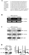
Figure 3. Effects of siVEGFR2 or siNRP1 Oligonucleotides on the Survival of MDA-MB-231 Cells
(A) The siRNA oligonucleotides (and their sequences) used in this study were purchased from Dharmacon. (B) MDA-MB-231 cells were infected with retrovirus encoding wild-type (w) or mutant (m) siVEGFR2 or siLuc and then selected with puromycin (5 μg/ml). Total RNA (5 μg) was subjected to RT-PCR analysis for VEGFR2 mRNA expression. GAPDH mRNA is shown as an internal control. (C) Total protein extracts (5 mg) from puromycin-selected cells were subjected to immunoprecipitation (IP) and Western blot (WB) analyses for VEGF and NRP1 receptor expression, as indicated. (D) MDA-MB-231 cells were transiently transfected with siCTL or siNRP1 oligonucleotides. After 5 d of incubation, total cell lysates were prepared and subjected to Western blotting (WB) using anti-NRP1 antibodies (left blots). Total protein extracts were analyzed by Western blotting using anti-Csk antibody as an internal control. The survival of siNRP1-treated cells was measured by using cell cycle analysis. The data are representative of three individual studies (right graphs).
Figure 4
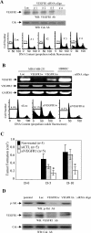
Figure 4. Effects of VEGFR1 siRNA Oligonucleotides on the Survival of MDA-MB-231 Cells
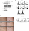
(A) Effects of siVEGFR1 oligonucleotides on the apoptosis of MDA-MB-231 cells. MDA-MB-231 cells were transiently transfected with siLuc or with various siVEGFR1 oligonucleotides (#1 through #4). After 5 d of incubation, the cells were lysed and subjected to Western blotting (WB) using anti-VEGFR1 antibodies. Total protein extracts were analyzed by Western blotting using anti-Csk antibody as an internal control. The apoptosis of siVEGFR1-treated cells was analyzed by using cell cycle analysis, as indicated. (B) MDA-MB-231 cells were transiently transfected with wild-type (w) or mutated (m) siVEGFR1 oligonucleotides for 5 d. Total RNA (5 μg) was subjected to RT-PCR analysis for VEGFR1 and VEGFR2 mRNA expression; GAPDH mRNA is shown as an internal control (top gels). The survival of siVEGFR1-treated cells was measured by using cell cycle analysis (lower graphs). The data are representative of four individual studies. (C) MDA-MB-231 cells (5 × 10⁶), transfected with either siVEGFR1 or siCTL, were subcutaneously injected into the flank of athymic nude mice. Tumors were measured at days 0, 5, and 10 (D-0, D-5, D10, respectively) and expressed in cm3. Number of mice per group is indicated by n-value. *p < 0.05. (D) MDA-MB-231 cells were transiently transfected with siLuc or with wild-type (w) or mutated (m) siVEGFR1 oligonucleotides. After 5 d of incubation, total cell lysates were prepared and subjected to Western blotting (WB) by using phospho-AKT (p-AKT) or VEGFR1 antibodies. Total protein extracts were analyzed by Western blotting using anti-Csk antibody as an internal control for loading.
Figure 5
Figure 5. Effect of VEGF and VEGF Receptor siRNA Oligonucleotides on the Survival of MCF-7 Cells
(A) MCF-7 cells were transiently transfected with various VEGF or VEGF receptor siRNA oligonucleotides, as indicated. Transfection procedures were performed with DharmaFECT-1 reagent according to the manufacturer's protocol. After 5 d of incubation, the cells were lysed and subjected to Western blotting (WB) using anti-VEGFR1 or anti-NRP1 antibodies. Total protein extracts were analyzed by Western blotting using anti-Csk antibody as an internal control. Alternatively, after 5 d of transfection, total RNA (5 μg) was isolated and subjected to RT-PCR analysis for VEGF and VEGFR2 mRNA expression. GAPDH mRNA is shown as an internal control. (B) MCF-7 cells were transiently transfected with siLuc, siNRP1, siVEGF, or various VEGF receptor siRNA oligonucleotides in the absence or presence of PGF (20 ng/ml) or VEGF (20 ng/ml), as indicated. After 5 d of incubation, the cells were harvested and subjected to cell cycle analysis. The data are representative of three individual studies. m, mutant; w, wild type. (C) MCF-7 cells were transiently transfected with siLuc, siNRP1, siVEGF, or various VEGF receptor siRNA oligonucleotides as shown. After 5 d of incubation, the cells were observed under a light microscope. (D) Effect of VEGF on the cell cycle in MDA-MB-231 cells. MDA-MB-231 cells were transiently transfected with siLuc or with siVEGF oligonucleotides in the presence or absence of VEGF (20 ng/ml). After 5 d of incubation, cells were harvested and subjected to cell cycle analysis. The data are representative of three individual studies.
Figure 6
Figure 6. Internal Expression of VEGFR1 in Breast Cancer Cells
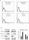
(A) VEGFR1 expression in MDA-MB-231 and MCF-7 cells was analyzed using flow cytometry, under permeable and nonpermeable conditions. VEGFR1 expression (grey tracing) is detected mostly internally under permeable conditions. Staining with control antibody is indicated by the black tracing. (B) Cytoplasmic and nuclear fractions extracted from MDA-MB-231 cells and HUVEC were subjected to Western blotting (WB) using anti-VEGFR1, anti-lamin B1, anti-GAPDH, and anti-actin antibodies. The figure shows the relative expression levels of VEGFR1 in the cytoplasmic (CE) and nuclear (NE) extracts from MDA-MB-231 cells and HUVEC. *p < 0.05.
Figure 7
Figure 7. Localization of VEGFR1 in MCF-7 and MDA-MB-231 Cells
Cells were stained with anti-VEGFR1 and anti-LMNA (Lamin A/C; a nuclear envelope marker) antibodies. Localization of VEGFR1 was then examined using confocal microscopy. LMNA was specifically localized at the nuclear envelope. Strong VEGFR1 protein expression was observed in the nuclear envelopes of the MCF-7 and MDA-MB-231 cells. Merged staining shows colocalization of VEGFR1 and LMNA at the nuclear envelopes.
Figure 8
Figure 8. Localization of VEGFR1 in MDA-MB-231 Cells after siVEGFR1 Treatment
MDA-MB-231 cells were transfected with siCTL, mutant siVEGFR1 (siVEGFR1m), or wild-type siVEGFR1 (siVEGFR1w), and then stained with anti-VEGFR1 and anti-LMNA (Lamin A/C) antibodies. Strong nuclear envelope staining and weak nuclear and cytoplasmic staining were observed for the VEGFR1 protein in the control siCTL and siVEGFR1m-transfected cells, whereas VEGFR1 protein expression was significantly decreased in the siVEGFR1w-transfected cells, and no VEGFR1 expression was observed in the nuclear envelope. The merged staining indicates the colocalization of VEGFR1 and LMNA.
Figure 9
Figure 9. Localization of VEGFR1 Protein in Human Breast Tissues
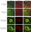
Tissue sections from human breast tumors and normal mammary glands were stained with anti-VEGFR1 and anti-LMNA (Lamin A/C) antibodies to determine their localization. (A and B) VEGFR1 was intensively colocalized with the nuclear envelope marker LMNA at the nuclear envelope and in the cytoplasm in human breast tumors. (C and D) VEGFR1 was colocalized with LMNA at the nuclear envelope in normal mammary glands.
Figure 10
Figure 10. Effects of VEGFR1 Antibody on Cell Growth
MDA-MB-231 and MCF-7 cells were grown subconfluently on six-well plates and treated with various concentrations of monoclonal antibody to VEGFR1. After 4 d of incubation, cells were harvested and counted on a hemocytometer. Data are presented as the means ± SD of three individual studies.

Similar articles

Cited by

References

    1. Hicklin DJ, Ellis LM. Role of the vascular endothelial growth factor pathway in tumor growth and angiogenesis. J Clin Oncol. 2004;23:1011–1027. - PubMed
    1. Cai J, Ahmad S, Jiang WG, Huang J, Kontos CD, et al. Activation of vascular endothelial growth factor receptor-1 sustains angiogenesis and Bcl-2 expression via the phosphatidylinositol 3-kinase pathway in endothelial cells. Diabetes. 2003;52:2959–2968. - PubMed
    1. Senger DR, Perruzzi CA, Feder J, Dvorak HF. A highly conserved vascular permeability factor secreted by a variety of human and rodent tumor cell lines. Cancer Res. 1986;46:5629–5632. - PubMed
    1. Berkman RA, Merrill MJ, Reinhold WC, Monacci WT, Saxena A, et al. Expression of the vascular permeability factor/vascular endothelial growth factor gene in central nervous system neoplasms. J Clin Invest. 1993;91:153–159. - PMC - PubMed
    1. Boocock CA, Charnock-Jones DS, Sharkey AM, McLaren J, Barker PJ, et al. Expression of vascular endothelial growth factor and its receptors flt and KDR in ovarian carcinoma. J Natl Cancer Inst. 1995;87:506–516. - PubMed

Publication types

MeSH terms

Substances