Preprotein-controlled catalysis in the helicase motor of SecA
- PMID: 17525736
- PMCID: PMC1894763
- DOI: 10.1038/sj.emboj.7601721
Preprotein-controlled catalysis in the helicase motor of SecA
Abstract
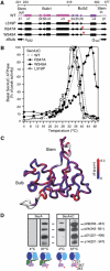
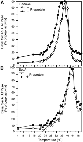
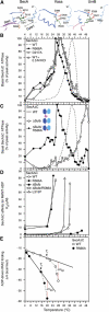
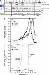
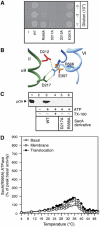
The cornerstone of the functionality of almost all motor proteins is the regulation of their activity by binding interactions with their respective substrates. In most cases, the underlying mechanism of this regulation remains unknown. Here, we reveal a novel mechanism used by secretory preproteins to control the catalytic cycle of the helicase 'DEAD' motor of SecA, the preprotein translocase ATPase. The central feature of this mechanism is a highly conserved salt-bridge, Gate1, that controls the opening/closure of the nucleotide cleft. Gate1 regulates the propagation of binding signal generated at the Preprotein Binding Domain to the nucleotide cleft, thus allowing the physical coupling of preprotein binding and release to the ATPase cycle. This relay mechanism is at play only after SecA has been previously 'primed' by binding to SecYEG, the transmembrane protein-conducting channel. The Gate1-controlled relay mechanism is essential for protein translocase catalysis and may be common in helicase motors.
Figures


Similar articles
-
Disorder-order folding transitions underlie catalysis in the helicase motor of SecA.Nat Struct Mol Biol. 2006 Jul;13(7):594-602. doi: 10.1038/nsmb1108. Epub 2006 Jun 18. Nat Struct Mol Biol. 2006. PMID: 16783375
-
Helicase Motif III in SecA is essential for coupling preprotein binding to translocation ATPase.EMBO Rep. 2004 Aug;5(8):807-11. doi: 10.1038/sj.embor.7400206. Epub 2004 Jul 23. EMBO Rep. 2004. PMID: 15272299 Free PMC article.
-
Quaternary dynamics of the SecA motor drive translocase catalysis.Mol Cell. 2013 Dec 12;52(5):655-66. doi: 10.1016/j.molcel.2013.10.036. Mol Cell. 2013. PMID: 24332176
-
SecA-mediated targeting and translocation of secretory proteins.Biochim Biophys Acta. 2014 Aug;1843(8):1466-74. doi: 10.1016/j.bbamcr.2014.02.014. Epub 2014 Feb 25. Biochim Biophys Acta. 2014. PMID: 24583121 Review.
-
Bacterial preprotein translocase: mechanism and conformational dynamics of a processive enzyme.Mol Microbiol. 1998 Feb;27(3):511-8. doi: 10.1046/j.1365-2958.1998.00713.x. Mol Microbiol. 1998. PMID: 9489663 Review.
Cited by
-
Signal peptides are allosteric activators of the protein translocase.Nature. 2009 Nov 19;462(7271):363-7. doi: 10.1038/nature08559. Nature. 2009. PMID: 19924216 Free PMC article.
-
Conformational transition of Sec machinery inferred from bacterial SecYE structures.Nature. 2008 Oct 16;455(7215):988-91. doi: 10.1038/nature07421. Nature. 2008. PMID: 18923527 Free PMC article.
-
The Dynamic SecYEG Translocon.Front Mol Biosci. 2021 Apr 15;8:664241. doi: 10.3389/fmolb.2021.664241. eCollection 2021. Front Mol Biosci. 2021. PMID: 33937339 Free PMC article. Review.
-
Molecular Mimicry of SecA and Signal Recognition Particle Binding to the Bacterial Ribosome.mBio. 2019 Aug 13;10(4):e01317-19. doi: 10.1128/mBio.01317-19. mBio. 2019. PMID: 31409676 Free PMC article.
-
Cotranslational folding of alkaline phosphatase in the periplasm of Escherichia coli.Protein Sci. 2020 Oct;29(10):2028-2037. doi: 10.1002/pro.3927. Epub 2020 Aug 24. Protein Sci. 2020. PMID: 32790204 Free PMC article.
References
-
- Baud C, Karamanou S, Sianidis G, Vrontou E, Politou AS, Economou A (2002) Allosteric communication between signal peptides and the SecA protein DEAD motor ATPase domain. J Biol Chem 277: 13724–13731 - PubMed
-
- Baud C, Papanikou E, Karamanou S, Sianidis G, Kuhn A, Economou A (2005) Purification of a functional mature region from a SecA-dependent preprotein. Protein Expr Purif 40: 336–339 - PubMed
-
- Benach J, Chou YT, Fak JJ, Itkin A, Nicolae DD, Smith PC, Wittrock G, Floyd DL, Golsaz CM, Gierasch LM, Hunt JF (2003) Phospholipid-induced monomerization and signal-peptide-induced oligomerization of SecA. J Biol Chem 278: 3628–3638 - PubMed
-
- Caruthers JM, McKay DB (2002) Helicase structure and mechanism. Curr Opin Struct Biol 12: 123–133 - PubMed
-
- Cavanaugh LF, Palmer AG III, Gierasch LM, Hunt JF (2006) Disorder breathes life into a DEAD motor. Nat Struct Mol Biol 13: 566–569 - PubMed
Publication types
MeSH terms
Substances
Grants and funding
LinkOut - more resources
Full Text Sources
Other Literature Sources
Molecular Biology Databases

