

Sequential structures provide insights into the fidelity of RNA replication
- PMID: 17517631
- PMCID: PMC1890517
- DOI: 10.1073/pnas.0700518104
Sequential structures provide insights into the fidelity of RNA replication
Abstract
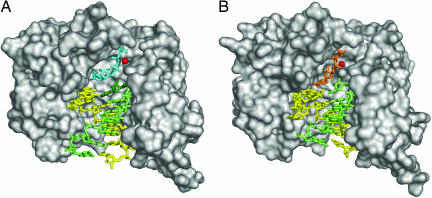
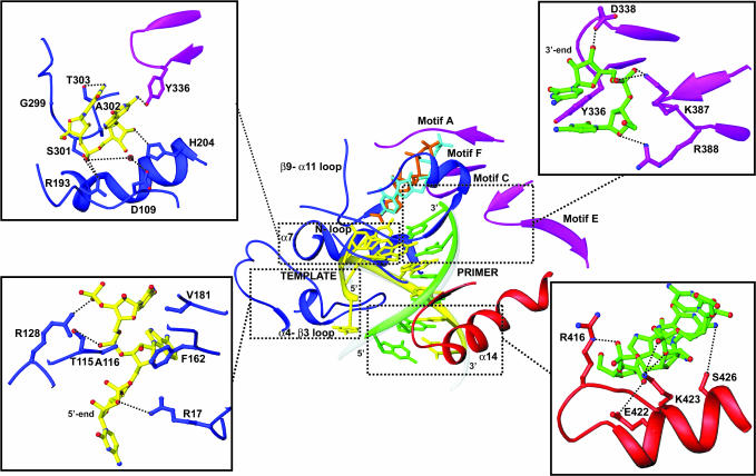
RNA virus replication is an error-prone event caused by the low fidelity of viral RNA-dependent RNA polymerases. Replication fidelity can be decreased further by the use of mutagenic ribonucleoside analogs to a point where viral genetic information can no longer be maintained. For foot-and-mouth disease virus, the antiviral analogs ribavirin and 5-fluorouracil have been shown to be mutagenic, contributing to virus extinction through lethal mutagenesis. Here, we report the x-ray structure of four elongation complexes of foot-and-mouth disease virus polymerase 3D obtained in presence of natural substrates, ATP and UTP, or mutagenic nucleotides, ribavirin triphosphate and 5-fluorouridine triphosphate with different RNAs as template-primer molecules. The ability of these complexes to synthesize RNA in crystals allowed us to capture different successive replication events and to define the critical amino acids involved in (i) the recognition and positioning of the incoming nucleotide or analog; (ii) the positioning of the acceptor base of the template strand; and (iii) the positioning of the 3'-OH group of the primer nucleotide during RNA replication. The structures identify key interactions involved in viral RNA replication and provide insights into the molecular basis of the low fidelity of viral RNA polymerases.
Conflict of interest statement
The authors declare no conflict of interest.
Figures

Similar articles
-
Molecular characterization of a dual inhibitory and mutagenic activity of 5-fluorouridine triphosphate on viral RNA synthesis. Implications for lethal mutagenesis.J Mol Biol. 2008 Oct 10;382(3):652-66. doi: 10.1016/j.jmb.2008.07.033. Epub 2008 Jul 17. J Mol Biol. 2008. PMID: 18662697
-
Residues within the Foot-and-Mouth Disease Virus 3Dpol Nuclear Localization Signal Affect Polymerase Fidelity.J Virol. 2020 Aug 17;94(17):e00833-20. doi: 10.1128/JVI.00833-20. Print 2020 Aug 17. J Virol. 2020. PMID: 32581111 Free PMC article.
-
Foot-and-mouth disease virus low-fidelity polymerase mutants are attenuated.Arch Virol. 2014 Oct;159(10):2641-50. doi: 10.1007/s00705-014-2126-z. Epub 2014 Jun 3. Arch Virol. 2014. PMID: 24888311
-
Structural insights into replication initiation and elongation processes by the FMDV RNA-dependent RNA polymerase.Curr Opin Struct Biol. 2009 Dec;19(6):752-8. doi: 10.1016/j.sbi.2009.10.016. Epub 2009 Nov 13. Curr Opin Struct Biol. 2009. PMID: 19914060 Review.
-
5-fluorouracil in lethal mutagenesis of foot-and-mouth disease virus.Future Med Chem. 2009 Jun;1(3):529-39. doi: 10.4155/fmc.09.26. Future Med Chem. 2009. PMID: 21426129 Review.
Cited by
-
Homology-Based Identification of a Mutation in the Coronavirus RNA-Dependent RNA Polymerase That Confers Resistance to Multiple Mutagens.J Virol. 2016 Jul 27;90(16):7415-7428. doi: 10.1128/JVI.00080-16. Print 2016 Aug 15. J Virol. 2016. PMID: 27279608 Free PMC article.
-
RNA-Dependent RNA Polymerases of Picornaviruses: From the Structure to Regulatory Mechanisms.Viruses. 2015 Aug 6;7(8):4438-60. doi: 10.3390/v7082829. Viruses. 2015. PMID: 26258787 Free PMC article. Review.
-
Motif D of viral RNA-dependent RNA polymerases determines efficiency and fidelity of nucleotide addition.Structure. 2012 Sep 5;20(9):1519-27. doi: 10.1016/j.str.2012.06.012. Epub 2012 Jul 19. Structure. 2012. PMID: 22819218 Free PMC article.
-
A multi-step process of viral adaptation to a mutagenic nucleoside analogue by modulation of transition types leads to extinction-escape.PLoS Pathog. 2010 Aug 26;6(8):e1001072. doi: 10.1371/journal.ppat.1001072. PLoS Pathog. 2010. PMID: 20865120 Free PMC article.
-
Distinct conformations of a putative translocation element in poliovirus polymerase.J Mol Biol. 2014 Apr 3;426(7):1407-19. doi: 10.1016/j.jmb.2013.12.031. Epub 2014 Jan 12. J Mol Biol. 2014. PMID: 24424421 Free PMC article.
References
Publication types
MeSH terms
Substances
Associated data
- Actions
- Actions
- Actions
- Actions
LinkOut - more resources
Full Text Sources
Other Literature Sources
