

Bacteria of the genus Asaia stably associate with Anopheles stephensi, an Asian malarial mosquito vector
- PMID: 17502606
- PMCID: PMC1885625
- DOI: 10.1073/pnas.0610451104
Bacteria of the genus Asaia stably associate with Anopheles stephensi, an Asian malarial mosquito vector
Abstract
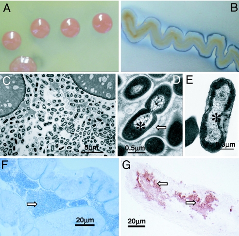
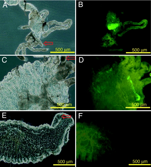
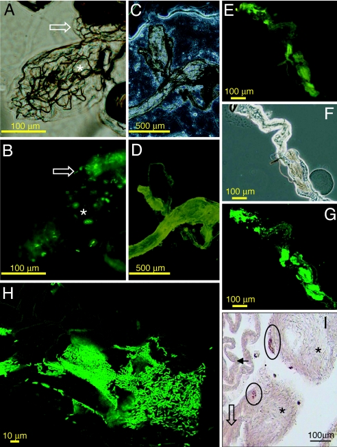
Here, we show that an alpha-proteobacterium of the genus Asaia is stably associated with larvae and adults of Anopheles stephensi, an important mosquito vector of Plasmodium vivax, a main malaria agent in Asia. Asaia bacteria dominate mosquito-associated microbiota, as shown by 16S rRNA gene abundance, quantitative PCR, transmission electron microscopy and in situ-hybridization of 16S rRNA genes. In adult mosquitoes, Asaia sp. is present in high population density in the female gut and in the male reproductive tract. Asaia sp. from An. stephensi has been cultured in cell-free media and then transformed with foreign DNA. A green fluorescent protein-tagged Asaia sp. strain effectively lodged in the female gut and salivary glands, sites that are crucial for Plasmodium sp. development and transmission. The larval gut and the male reproductive system were also colonized by the transformed Asaia sp. strain. As an efficient inducible colonizer of mosquitoes that transmit Plasmodium sp., Asaia sp. may be a candidate for malaria control.
Conflict of interest statement
The authors declare no conflict of interest.
Figures
Similar articles
-
Bacteria of the genus Asaia: a potential paratransgenic weapon against malaria.Adv Exp Med Biol. 2008;627:49-59. doi: 10.1007/978-0-387-78225-6_4. Adv Exp Med Biol. 2008. PMID: 18510013 Review.
-
Isolation and identification of Asaia sp. in Anopheles spp. mosquitoes collected from Iranian malaria settings: steps toward applying paratransgenic tools against malaria.Parasit Vectors. 2018 Jun 28;11(1):367. doi: 10.1186/s13071-018-2955-9. Parasit Vectors. 2018. PMID: 29950179 Free PMC article.
-
Interactions between Asaia, Plasmodium and Anopheles: new insights into mosquito symbiosis and implications in malaria symbiotic control.Parasit Vectors. 2013 Jun 18;6(1):182. doi: 10.1186/1756-3305-6-182. Parasit Vectors. 2013. PMID: 23777746 Free PMC article.
-
Mosquito-bacteria symbiosis: the case of Anopheles gambiae and Asaia.Microb Ecol. 2010 Oct;60(3):644-54. doi: 10.1007/s00248-010-9704-8. Epub 2010 Jun 23. Microb Ecol. 2010. PMID: 20571792
-
Using bacteria to express and display anti-parasite molecules in mosquitoes: current and future strategies.Insect Biochem Mol Biol. 2005 Jul;35(7):699-707. doi: 10.1016/j.ibmb.2005.02.008. Epub 2005 Mar 25. Insect Biochem Mol Biol. 2005. PMID: 15894187 Review.
Cited by
-
Identification of the midgut microbiota of An. stephensi and An. maculipennis for their application as a paratransgenic tool against malaria.PLoS One. 2011;6(12):e28484. doi: 10.1371/journal.pone.0028484. Epub 2011 Dec 6. PLoS One. 2011. PMID: 22163022 Free PMC article.
-
Use of the checkerboard DNA-DNA hybridization technique for bacteria detection in Aedes aegypti (Diptera:Culicidae) (L.).Parasit Vectors. 2011 Dec 20;4:237. doi: 10.1186/1756-3305-4-237. Parasit Vectors. 2011. PMID: 22185193 Free PMC article.
-
Delivery of a Genetically Marked Serratia AS1 to Medically Important Arthropods for Use in RNAi and Paratransgenic Control Strategies.Microb Ecol. 2019 Jul;78(1):185-194. doi: 10.1007/s00248-018-1289-7. Epub 2018 Nov 20. Microb Ecol. 2019. PMID: 30460544
-
Bacterial community composition in the salivary glands of triatomines (Hemiptera: Reduviidae).PLoS Negl Trop Dis. 2018 Sep 13;12(9):e0006739. doi: 10.1371/journal.pntd.0006739. eCollection 2018 Sep. PLoS Negl Trop Dis. 2018. PMID: 30212460 Free PMC article.
-
Mosquito-Borne Diseases Emergence/Resurgence and How to Effectively Control It Biologically.Pathogens. 2020 Apr 23;9(4):310. doi: 10.3390/pathogens9040310. Pathogens. 2020. PMID: 32340230 Free PMC article. Review.
References
-
- Atkinson PW, Michel K. Genesis. 2002;32:42–48. - PubMed
-
- Grossman GL, Rafferty CS, Clayton JR, Stevens TK, Mukabayire O, Benedict MQ. Insect Mol Biol. 2001;10:597–604. - PubMed
-
- Catteruccia F, Nolan T, Loukeris TG, Blass C, Savakis C, Kafatos FC, Crisanti A. Nature. 2000;405:959–962. - PubMed
-
- Ito J, Ghosh A, Moreira AL, Wimmer EA, Jacobs-Lorena M. Nature. 2002;417:452–455. - PubMed
-
- Catteruccia F, Godfray HC, Crisanti A. Science. 2003;299:1225–1227. - PubMed
MeSH terms
Substances
Associated data
- Actions
- Actions
- Actions
LinkOut - more resources
Full Text Sources
Other Literature Sources
Molecular Biology Databases
Research Materials
