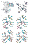
Insights into the influence of nucleotides on actin family proteins from seven structures of Arp2/3 complex
- PMID: 17499050
- PMCID: PMC1997283
- DOI: 10.1016/j.molcel.2007.04.017
Insights into the influence of nucleotides on actin family proteins from seven structures of Arp2/3 complex
Abstract
ATP is required for nucleation of actin filament branches by Arp2/3 complex, but the influence of ATP binding and hydrolysis are poorly understood. We determined crystal structures of bovine Arp2/3 complex cocrystallized with various bound adenine nucleotides and cations. Nucleotide binding favors closure of the nucleotide-binding cleft of Arp3, but no large-scale conformational changes in the complex. Thus, ATP binding does not directly activate Arp2/3 complex but is part of a network of interactions that contribute to nucleation. We compared nucleotide-induced conformational changes of residues lining the cleft in Arp3 and actin structures to construct a movie depicting the proposed ATPase cycle for the actin family. Chemical crosslinking stabilized subdomain 1 of Arp2, revealing new electron density for 69 residues in this subdomain. Steric clashes with Arp3 appear to be responsible for intrinsic disorder of subdomains 1 and 2 of Arp2 in inactive Arp2/3 complex.
Figures


Similar articles
-
Visualizing Arp2/3 complex activation mediated by binding of ATP and WASp using structural mass spectrometry.Proc Natl Acad Sci U S A. 2007 Jan 30;104(5):1552-7. doi: 10.1073/pnas.0605380104. Epub 2007 Jan 24. Proc Natl Acad Sci U S A. 2007. PMID: 17251352 Free PMC article.
-
Crystal structures of actin-related protein 2/3 complex with bound ATP or ADP.Proc Natl Acad Sci U S A. 2004 Nov 2;101(44):15627-32. doi: 10.1073/pnas.0407149101. Epub 2004 Oct 25. Proc Natl Acad Sci U S A. 2004. PMID: 15505213 Free PMC article.
-
Molecular dynamics simulation and coarse-grained analysis of the Arp2/3 complex.Biophys J. 2008 Dec;95(11):5324-33. doi: 10.1529/biophysj.108.143313. Epub 2008 Sep 19. Biophys J. 2008. PMID: 18805923 Free PMC article.
-
Regulation of actin filament assembly by Arp2/3 complex and formins.Annu Rev Biophys Biomol Struct. 2007;36:451-77. doi: 10.1146/annurev.biophys.35.040405.101936. Annu Rev Biophys Biomol Struct. 2007. PMID: 17477841 Review.
-
Structure and function of the Arp2/3 complex.Curr Opin Struct Biol. 1999 Apr;9(2):244-9. doi: 10.1016/s0959-440x(99)80034-7. Curr Opin Struct Biol. 1999. PMID: 10322212 Review.
Cited by
-
Nucleotide-dependent conformational states of actin.Proc Natl Acad Sci U S A. 2009 Aug 4;106(31):12723-8. doi: 10.1073/pnas.0902092106. Epub 2009 Jul 20. Proc Natl Acad Sci U S A. 2009. PMID: 19620726 Free PMC article.
-
Nucleotide- and activator-dependent structural and dynamic changes of arp2/3 complex monitored by hydrogen/deuterium exchange and mass spectrometry.J Mol Biol. 2009 Jul 17;390(3):414-27. doi: 10.1016/j.jmb.2009.03.028. Epub 2009 Mar 17. J Mol Biol. 2009. PMID: 19298826 Free PMC article.
-
Development of free-energy-based models for chaperonin containing TCP-1 mediated folding of actin.J R Soc Interface. 2008 Dec 6;5(29):1391-408. doi: 10.1098/rsif.2008.0185. J R Soc Interface. 2008. PMID: 18708324 Free PMC article. Review.
-
Small molecules CK-666 and CK-869 inhibit actin-related protein 2/3 complex by blocking an activating conformational change.Chem Biol. 2013 May 23;20(5):701-12. doi: 10.1016/j.chembiol.2013.03.019. Epub 2013 Apr 25. Chem Biol. 2013. PMID: 23623350 Free PMC article.
-
Structural basis for regulation of Arp2/3 complex by GMF.Nat Struct Mol Biol. 2013 Sep;20(9):1062-8. doi: 10.1038/nsmb.2628. Epub 2013 Jul 28. Nat Struct Mol Biol. 2013. PMID: 23893131 Free PMC article.
References
-
- Beltzner CC, Pollard TD. Identification of functionally important residues of Arp2/3 complex by analysis of homology models from diverse species. J Mol Biol. 2004;336:551–565. - PubMed
-
- Blanchoin L, Pollard TD. Interaction of actin monomers with Acanthamoeba actophorin (ADF/cofilin) and profilin. J Biol Chem. 1998;273:25106–25111. - PubMed
-
- Blanchoin L, Pollard TD. Mechanism of interaction of Acanthamoeba actophorin (ADF/Cofilin) with actin filaments. J Biol Chem. 1999;274:15538–15546. - PubMed
-
- Blanchoin L, Pollard TD. Hydrolysis of ATP by polymerized actin depends on the bound divalent cation but not profilin. Biochemistry. 2002;41:597–602. - PubMed
Publication types
MeSH terms
Substances
Grants and funding
LinkOut - more resources
Full Text Sources
Miscellaneous

