. 2007 Jul;81(14):7786-800.
doi: 10.1128/JVI.02780-06. Epub 2007 May 9.

Small interfering RNA profiling reveals key role of clathrin-mediated endocytosis and early endosome formation for infection by respiratory syncytial virus

Affiliations

Small interfering RNA profiling reveals key role of clathrin-mediated endocytosis and early endosome formation for infection by respiratory syncytial virus

Andrey A Kolokoltsov et al. J Virol. 2007 Jul.

Abstract

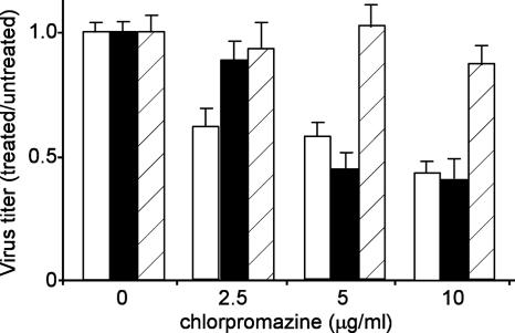
Respiratory syncytial virus (RSV) is a common cause of respiratory tract infections in infants and the elderly. Like many other pH-independent enveloped viruses, RSV is thought to enter at the cell surface, independently of common endocytic pathways. We have used a targeted small interfering RNA (siRNA) library to identify key cellular genes involved in cytoskeletal dynamics and endosome trafficking that are important for RSV infection. Surprisingly, RSV infection was potently inhibited by siRNAs targeting genes associated with clathrin-mediated endocytosis, including clathrin light chain. The important role of clathrin-mediated endocytosis was confirmed by the expression of well-characterized dominant-negative mutants of genes in this pathway and by using the clathrin endocytosis inhibitor chlorpromazine. We conclude that, while RSV may be competent to enter at the cell surface, clathrin function and endocytosis are a necessary and important part of a productive RSV infection, even though infection is strictly independent of pH. These findings raise the possibility that other pH-independent viruses may share a similar dependence on endocytosis for infection and provide a new potential avenue for treatment of infection.


Figures

FIG. 1.
FIG. 1.
RSV infection is insensitive to lysosomotropic agents. HeLa cells were treated with bafilomycin A1 (10 nM), ammonium chloride (Amm. Cl.; 10 mM), or chloroquine (10 μM) for 1 h prior to the addition of virus. RSV (open bars), VSV-G-pseudotyped MLV (solid bars) or ecotropic MLV envelope protein-pseudotyped MLV (diagonal striped bars) were then added (MOI, 0.1) to the cells and incubated for an additional 5 h in the presence of each drug. The medium was then replaced, and the infected cells were counted after 20 h for RSV or 36 h for ecotropic MLV envelope protein- or VSV-G-pseudotyped viruses. Each virus encoded GFP as an infection marker. The means ± SDs of the results of three independent experiments are shown.
FIG. 2.
FIG. 2.
The impacts of siRNAs targeting genes important in endocytosis and cytoskeleton rearrangement on RSV infection. HeLa cells were transfected with siRNAs and, after 2 days, the cells were infected with recombinant RSV encoding GFP at an MOI of 0.05. Twenty hours postinfection, the infected cells expressing GFP were counted. To establish a baseline of infection and transfection efficiency, cells were treated with a control set (top right) of siRNAs and/or transfection reagent, which included transfection lipids alone or together with “nontargeting” siGLO (fluorescently labeled) nonspecific siRNA or siRNAs targeting lamin A/C or glyceraldehyde-3-phosphate dehydrogenase. kif11 is required for cell survival, and siRNA targeting this gene rapidly kills cells. Together with the siGLO siRNA, the kif11 siRNA permitted monitoring of transfection efficiency. In all experiments, >90% of cells became fluorescent or died after treatment with siGLO or Kif11, respectively, indicating a high transfection efficiency and siRNA suppression of Kif11. The cells were then treated with siRNA pools targeting each of the indicated genes (total of 79). The cells were infected (as above), and the levels of infection monitored by GFP expression. The infection efficiencies were normalized to the average number of infected cells after treatment with the control set (except for Kif11). Dashed lines indicate the threshold used in the assay (two times the average SD of the data). The averages ± 1 SD of the results of three independent experiments are shown. Accession numbers and a brief description of the role of each gene are provided in Table 1.
FIG. 3.
FIG. 3.
Confirmation of siRNA suppression of protein expression. Where antibodies could be obtained and were of sufficient sensitivity to detect endogenous protein, Western blots were performed after treatment with siRNAs (as indicated above the blots). For each experiment, cell lysates were made from untreated cells (lane 1), cells treated with transfection reagent alone (lane 2), cells treated with transfection reagent together with nontargeting siRNA (lane 3), or transfection reagent with siRNA targeting the indicated gene (lane 4). The lysates were made 2 days after siRNA transfection in sodium dodecyl sulfate-polyacrylamide gel electrophoresis loading buffer with 2% β-mercaptoethanol. After samples were boiled for 5 min, each was loaded on 10 or 12% polyacrylamide gel electrophoresis gels as appropriate and transferred to nitrocellulose membranes and the blots were stained with each specific antibody (upper panels). The blots were then destained and reprobed with β-actin-specific antibody which served as a gel-loading control (lower panels).
FIG. 4.
FIG. 4.
Chlorpromazine inhibits infection by RSV. The clathrin endocytosis inhibitor, chlorpromazine, was used at the indicated dosages to confirm that RSV required clathrin-mediated endocytosis for infection. Cells were treated following the same regimen as described in the Fig. 1 legend, with 1 h of pretreatment of cells and removal of the drug 5 h after the addition of virus. The cells were infected with recombinant RSV encoding red fluorescent protein (open bars) or with a VSV-G (solid bars) or 10A1 MLV (bars with diagonal stripes) envelope protein-pseudotyped retrovirus encoding GFP. Infected cells were counted by microscopy and the results were normalized to those for the untreated control for each virus. The means ± 1 SD of the results of three replicate experiments are shown.
FIG. 5.
FIG. 5.
FACS analysis indicates that clathrin-mediated endocytosis is important for RSV infection. Cells were transfected with plasmids encoding β-galactosidase, GFP, Eps15 Δ95-295, wild-type or dominant-negative Rab5 (S34N), or constitutively active Rab5 (Q79L). The Eps15 and Rab5 proteins were each tagged with GFP. After 2 days to allow peak expression, the cells were challenged with wild-type RSV, VSV, or recombinant 10A1 MLV (encoding a truncated CD4 as an infection marker), each at an MOI of 0.2. (A) Cells were stained with RSV- or VSV-specific antibodies followed by Alexa Fluor647-labeled secondary antibody or with Alexa Fluor647-labeled anti-CD4. The cells were then analyzed by using a FACS. After gating for single cells, at least 5 × 10⁴ cells were analyzed. Cells transfected with β-galactosidase-encoding plasmids and stained prior to virus protein expression (3 h for VSV and 6 h for RSV or 20 h for 10A1 MLV) were used to define the autofluorescence levels. Cells expressing GFP alone were used to define the infection levels in normal cells. The gates for data analysis were uninfected cells expressing no GFP-tagged protein (indicated by −/− box), those not infected but expressing moderate to high levels of tagged protein (+/−), or those infected with virus and not expressing (−/+) or expressing tagged protein (+/+). The cells not expressing tagged protein served as important internal controls to monitor the levels of infection of normal cells compared to the levels in those expressing the recombinant protein. (B) The proportions of cells infected with VSV (open bars), RSV (solid bars), or 10A1 envelope protein-pseudotyped MLV (bars with diagonal lines) and expressing recombinant GFP-tagged protein were calculated by dividing the number of infected cells expressing tagged protein by the total number of cells expressing tagged protein. This value was then normalized to the proportion of infected cells not expressing GFP-tagged protein. The means ± 1 SD of the results of 3 independent experiments are given. wt, wild type.
FIG. 6.
FIG. 6.
Microscopy analysis confirms that RSV requires clathrin-mediated endocytosis for infection. Cells were transfected with plasmids carrying the indicated genes and challenged 2 days later with recombinant RSV encoding red fluorescent protein. After 30 h, the cells were photographed using a Leica DMIRB inverted fluorescence microscope detecting the green fluorescence of the GFP-tagged transfected gene and the red fluorescence of the RSV infection marker (upper panel). The impact of the expression of each gene was determined by counting the cells infected with RSV that were also expressing the GFP-tagged protein and dividing by the total number of infected cells (lower panel). The averages ± 1 SD of the results of three replicate experiments are shown. wt, wild type.
FIG. 7.
FIG. 7.
Expression of Eps15 or Rab5 proteins does not alter virus binding to cells. Cells were transfected with expression plasmids carrying the indicated genes. Each gene product was GFP tagged. (A) Virus was then added to the cells on ice and incubated for 2 h, after which excess virus was removed by washing and pelleting cells in ice-cold PBS. The cells were then fixed and stained as detailed in Materials and Methods. The cells were then photographed using a Zeiss LSM 510 confocal microscope, and the images analyzed using ImageJ software. Each image is a composite of the visible light phase-contrast image and the fluorescence images of GFP expression (green) and labeled virus (red). Midsections of cells expressing the GFP-tagged protein and untransfected cells in the same field are shown and virus is visible on the cell periphery. In some instances, the surface of the untransfected cell is in the focal plane and virus can be seen on the cell surface as well. wt, wild type. (B) For each cell population, the total number of labeled virus particles per cell was determined by counting using ImageJ software and was normalized to the cell area as described in Materials and Methods. At least five transfected and untransfected cells were used for each analysis. The average numbers of particles per cell ± 1 SD are shown.

Similar articles

Cited by

References

    1. Acton, S. L., D. H. Wong, P. Parham, F. M. Brodsky, and A. P. Jackson. 1993. Alteration of clathrin light chain expression by transfection and gene disruption. Mol. Biol. Cell 4:647-660. - PMC - PubMed
    1. Balachandran, S., P. C. Roberts, L. E. Brown, H. Truong, A. K. Pattnaik, D. R. Archer, and G. N. Barber. 2000. Essential role for the dsRNA-dependent protein kinase PKR in innate immunity to viral infection. Immunity 13:129-141. - PubMed
    1. Barnes, W. G., E. Reiter, J. D. Violin, X. R. Ren, G. Milligan, and R. J. Lefkowitz. 2005. β-Arrestin 1 and Gαq/11 coordinately activate RhoA and stress fiber formation following receptor stimulation. J. Biol. Chem. 280:8041-8050. - PubMed
    1. Benmerah, A., M. Bayrou, N. Cerf-Bensussan, and A. Dautry-Varsat. 1999. Inhibition of clathrin-coated pit assembly by an Eps15 mutant. J. Cell Sci. 112:1303-1311. - PubMed
    1. Benmerah, A., C. Lamaze, B. Begue, S. L. Schmid, A. Dautry-Varsat, and N. Cerf-Bensussan. 1998. AP-2/Eps15 interaction is required for receptor-mediated endocytosis. J. Cell Biol. 140:1055-1062. - PMC - PubMed

Publication types