. 2007 Jul;81(13):7178-88.
doi: 10.1128/JVI.00404-07. Epub 2007 May 2.

A novel Bcl-2-like inhibitor of apoptosis is encoded by the parapoxvirus ORF virus

Affiliations

A novel Bcl-2-like inhibitor of apoptosis is encoded by the parapoxvirus ORF virus

Dana Westphal et al. J Virol. 2007 Jul.

Abstract

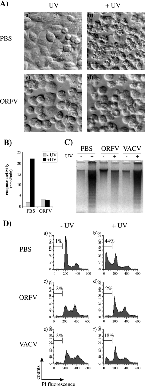
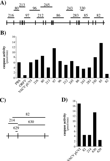
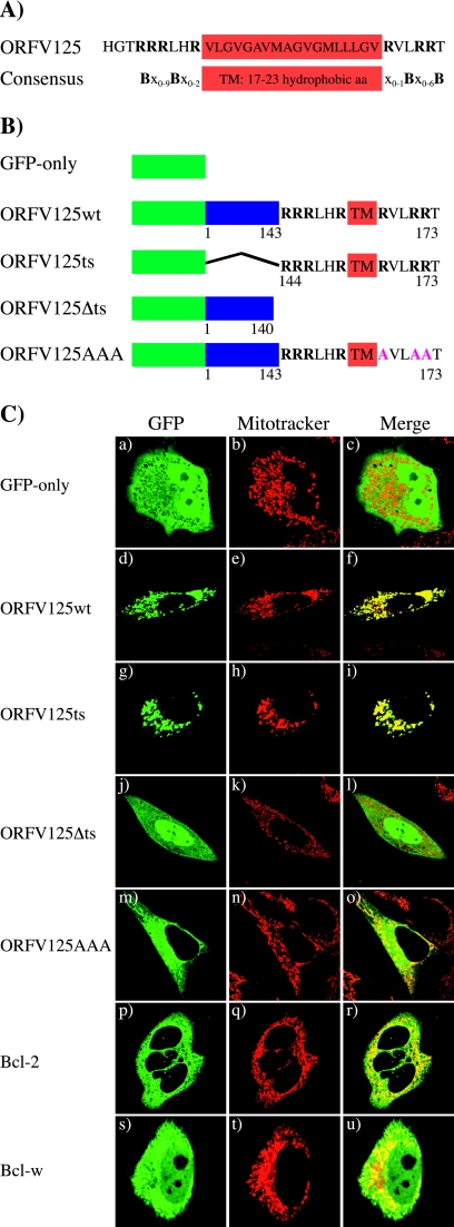
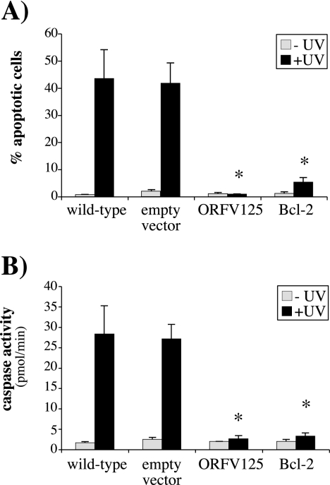
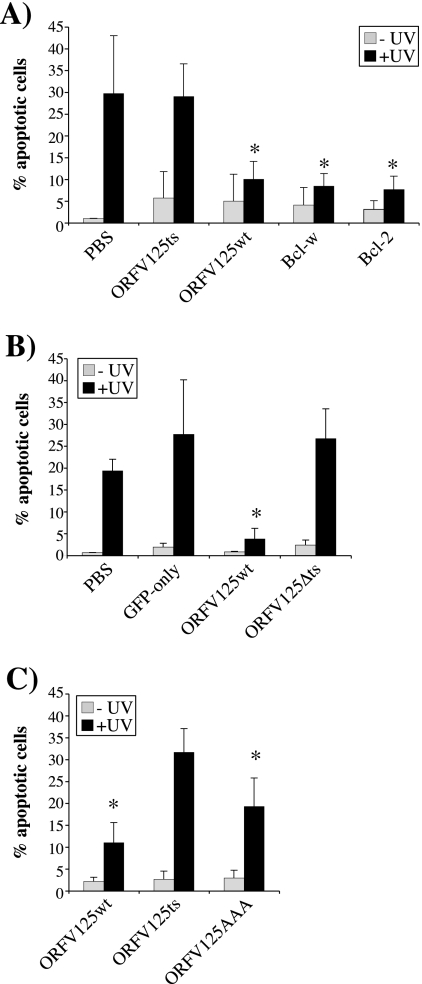
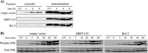
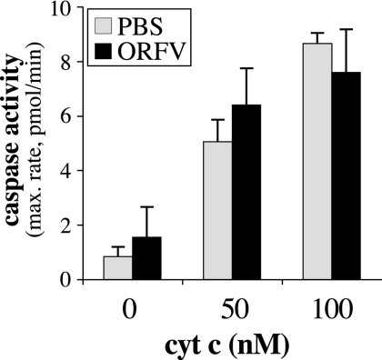
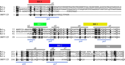
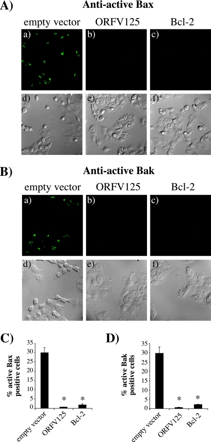
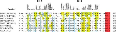
Apoptotic cell death forms part of the host defense against virus infection. We tested orf virus, a member of the poxvirus family, for the ability to inhibit apoptosis and found that orf virus-infected cells were fully resistant to UV-induced changes in cell morphology, caspase activation, and DNA fragmentation. By using a library of vaccinia virus-orf virus recombinants, we identified an orf virus gene (ORFV125) whose presence was linked with the inhibition of apoptosis. The 173-amino-acid predicted protein had no clear homologs in public databases other than those encoded by other parapoxviruses. However, ORFV125 possessed a distinctive C-terminal domain which was necessary and sufficient to direct the protein to the mitochondria. We determined that ORFV125 alone could fully inhibit UV-induced DNA fragmentation, caspase activation, and cytochrome c release and that its mitochondrial localization was required for its antiapoptotic function. In contrast, ORFV125 did not prevent UV-induced activation of c-Jun NH2-terminal kinase, an event occurring upstream of the mitochondria. These features are comparable to the antiapoptotic properties of the mitochondrial regulator Bcl-2. Furthermore, bioinformatic analyses revealed sequence and secondary-structure similarities to Bcl-2 family members, including characteristic residues of all four Bcl-2 homology domains. Consistent with this, the viral protein inhibited the UV-induced activation of the proapoptotic Bcl-2 family members Bax and Bak. ORFV125 is the first parapoxvirus apoptosis inhibitor to be identified, and we propose that it is a new antiapoptotic member of the Bcl-2 family.


Figures

FIG. 1.
FIG. 1.
ORFV inhibits UV-induced apoptosis. Cells were mock infected (PBS) or infected with ORFV or VACV, either strain MVA (C) or Lister (D), at a multiplicity of infection of 4 and UV irradiated (+UV) 7 h later. Cells not irradiated are indicated by −UV. (A) At 14 h after UV treatment, HeLa cells were examined with an inverted microscope (Olympus; Zeiss) equipped with (20×) Hoffmann modulation contrast optics. (B) Caspase activity in 143B cell lysates prepared 12 h after UV treatment. (C) DNA laddering was visualized by agarose gel electrophoresis of 143B cell lysates prepared 17 h after UV treatment. (D) At 17 h after UV irradiation, the DNA content of permeabilized, PI-stained 143B cells was analyzed by flow cytometry. The percentage of hypodiploid (apoptotic) cells in each population is indicated on each histogram.
FIG. 2.
FIG. 2.
Use of VACV-ORFV recombinants to map an ORFV inhibitor of UV-induced apoptosis. (A) The ORFV KpnI map is shown at the bottom, and the bars above indicate the locations of DNA fragments that were recombined into the VACV genome to create the VVOV recombinant library. (B) 143B cells were mock infected (PBS) or infected, at a multiplicity of infection of 4, with ORFV or the indicated VACV recombinants and treated with UV 7 h later. After 12 h, the samples were analyzed for caspase activity. The VACV recombinant pUVI is a control virus carrying no ORFV DNA (35). (C) An expanded view of the right end of the ORFV KpnI map with lines above showing the relative locations of the DNA fragments used to generate VVOV82, -214, 630, and -629. (D) Caspase activities in UV-irradiated 143B cells infected with the indicated VACV recombinants and prepared as described for panel B.
FIG. 3.
FIG. 3.
The mitochondrion-targeting motif of ORFV125 is necessary and sufficient to direct the protein to the mitochondria. (A) Comparison of the C-terminal region of ORFV125 with the consensus mitochondrion-targeting motif of tail-anchored proteins (26). The hydrophobic amino acids (aa) constituting the TM domain are boxed. The TM domain is flanked by positively charged amino acids, which are shown as B in the consensus sequence and are in bold in ORFV125. In the consensus sequence, x stands for any amino acid. (B) Schematic representations of GFP fusion constructs. ORFV125wt consists of the full-length 173-amino-acid protein. ORFV125Δts lacks the last 33 amino acids of the protein, which contain the TM domain flanked by the positively charged amino acids. The ORFV125ts construct possesses only the C-terminal 30 amino acids of the viral protein fused to GFP. In ORFV125AAA, the three arginines downstream of the TM domain were changed to alanines. (C) HeLa cells were transiently transfected with the indicated GFP fusion plasmids and visualized by confocal microscopy 24 h later. Mitochondria were labeled with Mitotracker.
FIG. 4.
FIG. 4.
ORFV125 inhibits UV-induced apoptosis in the absence of virus infection. (A) Stable 143B cell lines expressing ORFV125, Bcl-2, or the empty vector were UV irradiated. After 17 h, the DNA content of permeabilized, PI-stained cells was analyzed by flow cytometry and the percentage of hypodiploid (apoptotic) cells in each population was calculated. (B) Caspase activity in the indicated cell lines was quantified 7 h after UV treatment. The data shown in panels A and B are averages of three independent experiments with standard deviations indicated. UV-irradiated samples that showed significantly (P < 0.001) less apoptosis or caspase activity than the UV-irradiated wild-type cell line are indicated by asterisks.
FIG. 5.
FIG. 5.
The mitochondrial localization of ORFV125 is necessary for its antiapoptotic function. 143B cells were mock transfected (PBS) or transfected with the indicated GFP fusion constructs and UV irradiated either 30 h (A and C) or 12 h (B) later. After a further 17 h (A and C) or 12 h (B), the DNA content of permeabilized, PI-stained cells was examined by flow cytometry. The shortened time between transfection and harvesting in panel B was used to avoid degradation of the ORFV125Δts construct within the cell. The percentage of hypodiploid (apoptotic) cells in each population was calculated. The data presented are averages of three independent experiments with standard deviations indicated. Asterisks indicate UV-irradiated samples in which the apoptotic population is significantly reduced (A, P < 0.05 for ORFV125wt and P < 0.01 for Bcl-w and Bcl-2; B, P < 0.05; C, P < 0.001 for ORFV125wt and P < 0.01 for ORFV125AAA) compared with the corresponding UV-irradiated mock-transfected (A and B) or ORFV125ts-transfected (C) sample.
FIG. 6.
FIG. 6.
ORFV125 inhibits cytochrome c release, but not JNK phosphorylation, following UV treatment. Stable 143B cell lines expressing ORFV125, Bcl-2, or the empty vector were either mock treated (−UV) or UV irradiated. (A) Cells were fractionated into cytosolic and mitochondrial fractions after 4, 6, or 8 h. Release of cytochrome c from the mitochondria into the cytosol was determined by Western blot analysis with an anti-cytochrome c antibody. This blot is representative of three independent experiments. (B) Cell lysates were prepared after the indicated times. Cells harvested at 0 min were collected immediately after UV exposure. Immunoblot analysis was performed to compare the levels of two isozymes of phosphorylated JNK (p46-JNK1 and p54-JNK2) with total JNK. The latter also served as a loading control.
FIG. 7.
FIG. 7.
ORFV does not express a cytosolic protein able to inhibit caspase activation induced by cytosolic cytochrome c (cyt c). 143B cells were mock infected (PBS) or infected with ORFV NZ2 (ORFV) at a multiplicity of infection of 5 for 14 h. Cytosolic extracts were prepared and treated with 0, 50, or 100 nM cytochrome c and 1 mM dATP. Activation of caspases was measured in triplicate over 120 min, and the average maximum (max.) rate of AMC production was determined (standard deviations are indicated). The results shown are representative of three independent experiments.
FIG. 8.
FIG. 8.
Sequence and predicted secondary-structure similarities between ORFV125 and Bcl-2 family members. Amino acid sequence alignment of ORFV125 with three antiapoptotic Bcl-2 family members, Bcl-xL, Bcl-2, and Bcl-w. BH domains of Bcl-xL are shown as colored boxes, and the TM domain is shown as a gray box. Positions of the Bcl-xL α-helices (α1 to -6, -6′, and -7) (Jpred) are represented as black chains above the alignment, while the predicted α-helices for ORFV125 (pα1 to -7) are plotted below the alignment in blue. Amino acids conserved in all four sequences are marked with black boxes showing amino acids identical to ORFV125 and gray boxes indicating conserved substitutions (according to ClustalW).
FIG. 9.
FIG. 9.
ORFV125 inhibits the activation of Bax and Bak. 143B stable cell lines were incubated with 50 μM Z-VAD-FMK (Calbiochem), added 1 h before UV treatment. Eight hours after treatment, cells were stained with Bax (A) or Bak (B) conformation-specific antibodies and visualized by fluorescence (a to c) or phase-contrast (d to f) microscopy. The percentage of cells with active Bax (C) or Bak (D) in four different microscope fields was calculated and averaged. The results are presented as an average of three independent experiments (standard deviations are indicated). Samples that showed significantly (P < 0.001) less active Bax or Bak than the empty-vector control cell line are indicated by asterisks.
FIG. 10.
FIG. 10.
Representatives from seven of the eight vertebrate poxvirus genera encode proteins with Bcl-2-like features. Alignment of BH3-like and BH1-like domains of proteins from fowlpox virus (FWPV, FWPV039), ORFV (ORFV125), myxoma virus (MYXV, M11L), sheeppox virus (SPPV, SPPV014), swinepox virus (SWPV, SWPV012), Yaba monkey tumor virus (YMTV, 16L), deerpox virus (DPV, DPV022), and VACV (F1L). Deerpox virus is an unassigned member of the subfamily. Amino acids highlighted in yellow match the consensus sequence of the BH1 domain (Prosite accession no. PSO1080) or the BH3 domain (Prosite accession no. PSO1259). The Prosite consensus sequences are shown at the top, with X representing any amino acid. Amino acids boxed in blue are conserved in at least five of eight poxviral proteins. Predicted mitochondrion-targeting sequences (mTS) are marked as red boxes. xn indicates the numbers of intervening amino acids. The total number of amino acids in each protein is shown at the end of each sequence.

Similar articles

Cited by

References

    1. Afonso, C. L., E. R. Tulman, Z. Lu, L. Zsak, G. F. Kutish, and D. L. Rock. 2000. The genome of fowlpox virus. J. Virol. 74:3815-3831. - PMC - PubMed
    1. Antoine, G., F. Scheiflinger, F. Dorner, and F. G. Falkner. 1998. The complete genomic sequence of the modified vaccinia Ankara strain: comparison with other orthopoxviruses. Virology 244:365-396. - PubMed
    1. Breckenridge, D. G., and D. Xue. 2004. Regulation of mitochondrial membrane permeabilization by BCL-2 family proteins and caspases. Curr. Opin. Cell Biol. 16:647-652. - PubMed
    1. Brick, D. J., R. D. Burke, A. A. Minkley, and C. Upton. 2000. Ectromelia virus virulence factor p28 acts upstream of caspase-3 in response to UV light-induced apoptosis. J. Gen. Virol. 81:1087-1097. - PubMed
    1. Cartier, J. L., P. A. Hershberger, and P. D. Friesen. 1994. Suppression of apoptosis in insect cells stably transfected with baculovirus p35: dominant interference by N-terminal sequences p351-76. J. Virol. 68:7728-7737. - PMC - PubMed

Publication types

MeSH terms

Substances