. 2007 Jul;27(13):4815-24.
doi: 10.1128/MCB.02062-06. Epub 2007 Apr 23.

Potential interface between ribosomal protein production and pre-rRNA processing

Affiliations

Potential interface between ribosomal protein production and pre-rRNA processing

Dipayan Rudra et al. Mol Cell Biol. 2007 Jul.

Abstract

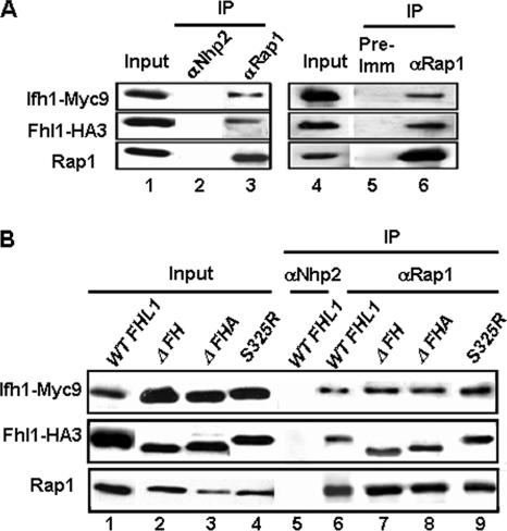
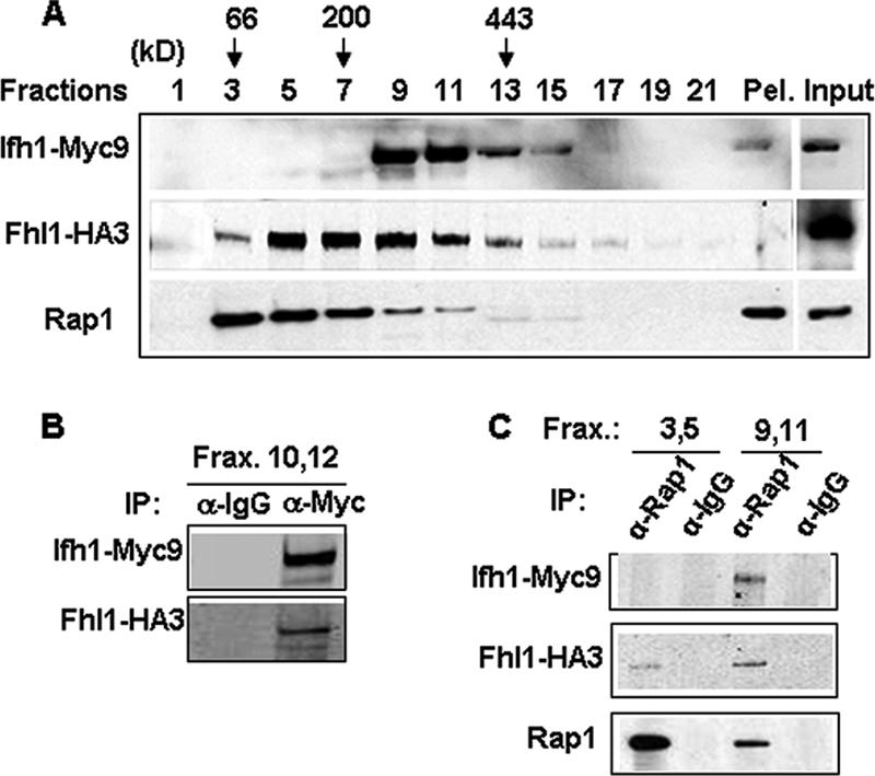
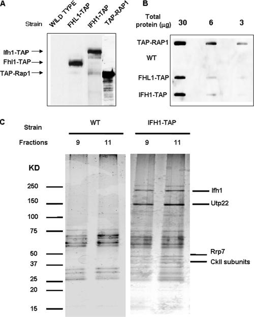
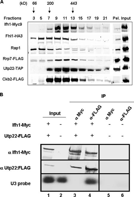
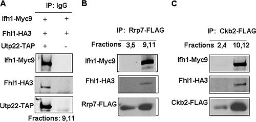
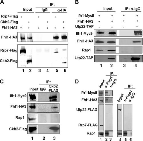
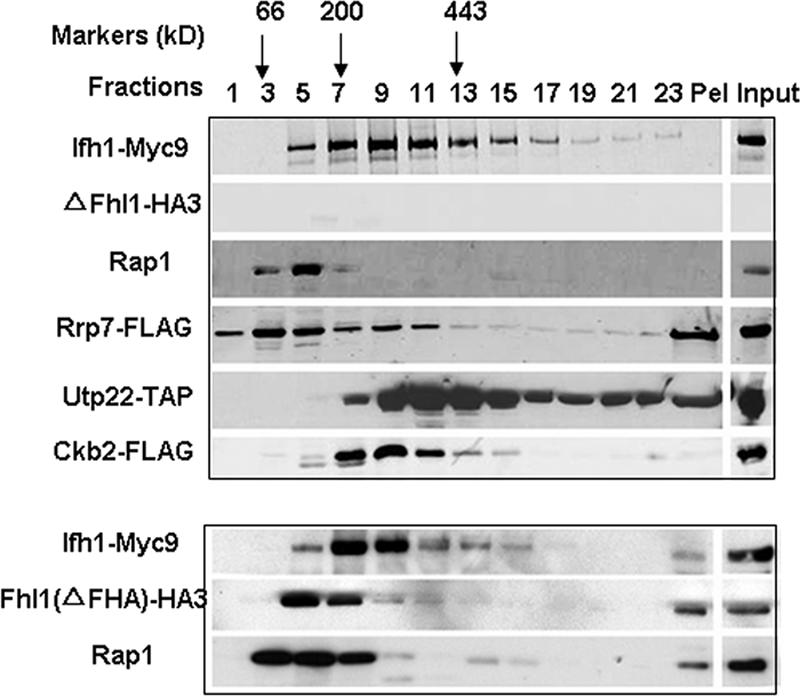
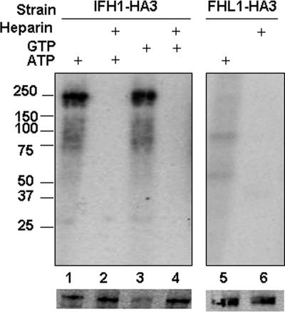
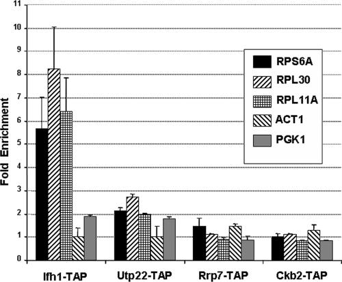
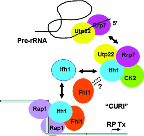
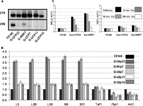
It has become clear that in Saccharomyces cerevisiae the transcription of ribosomal protein genes, which makes up a major proportion of the total transcription by RNA polymerase II, is controlled by the interaction of three transcription factors, Rap1, Fhl1, and Ifh1. Of these, only Rap1 binds directly to DNA and only Ifh1 is absent when transcription is repressed. We have examined further the nature of this interaction and find that Ifh1 is actually associated with at least two complexes. In addition to its association with Rap1 and Fhl1, Ifh1 forms a complex (CURI) with casein kinase 2 (CK2), Utp22, and Rrp7. Fhl1 is loosely associated with the CURI complex; its absence partially destabilizes the complex. The CK2 within the complex phosphorylates Ifh1 in vitro but no other members of the complex. Two major components of this complex, Utp22 and Rrp7, are essential participants in the processing of pre-rRNA. Depletion of either protein, but not of other proteins in the early processing steps, brings about a substantial increase in ribosomal protein mRNA. We propose a model in which the CURI complex is a key mediator between the two parallel pathways necessary for ribosome synthesis: the transcription and processing of pre-rRNA and the transcription of ribosomal protein genes.


Figures

FIG. 1.
FIG. 1.
Rap1 interacts with both Fhl1 and Ifh1. (A) Co-IP was carried out using rabbit polyclonal anti-Rap1 (αRap1) antibody with extracts prepared from strain DR36 (FHL1-HA3 IFH1-Myc9 double tagged). The immunoprecipitated protein complex was resuspended in SDS loading buffer, boiled, and analyzed by SDS-polyacrylamide gel electrophoresis followed by Western blotting using anti-HA, anti-Myc, or anti-Rap1 antibodies (lane 3). Whole-cell extract was loaded in a separate lane as a loading control (lane 1; Input). Immunoprecipitation with a rabbit polyclonal antibody raised against the yeast protein Nhp2 (αNhp2) was used as a negative control (lane 2). In the right-hand panel, Co-IP was carried out using anti-Rap1 antibody on extracts prepared from DR36 in the presence of 200 μg/ml ethidium bromide (lane 6). In this case, preimmune serum was used as a negative control (lane 5). (B) Co-IP was carried out using rabbit polyclonal anti-Rap1 antibody on extracts prepared from strains harboring HA3-tagged full-length Fhl1 (WT FHL1), with the FH domain deleted (ΔFH), with the FHA domain deleted (ΔFHA), or with the S325R mutant version of Fhl1 (lanes 6, 7, 8, and 9, strains DR47, DR48, DR49, and DR65, respectively). The endogenous copy of FHL1 in these strains is deleted, and Ifh1 is tagged C terminally with Myc9. Rabbit polyclonal antibody against Nhp2 was used as a negative control (lane 5).
FIG. 2.
FIG. 2.
Ifh1 is found as a high-MW complex. (A) Extract prepared from strain DR36 (FHL1-HA3 IFH1-Myc9) was loaded onto a 10 to 30% glycerol gradient and centrifuged for 5 h at 49 krpm using an SW50.1 rotor. Two hundred-microliter fractions were collected. Aliquots (15 μl) from the indicated fractions as well as the whole-cell extract (Input) were analyzed by SDS-polyacrylamide gel electrophoresis followed by Western blotting using anti-HA, anti-Myc, or anti-Rap1 antibodies. The positions of the MW markers bovine serum albumin (66 kDa), β-amylase (200 kDa), and apoferritin (443 kDa), centrifuged in a parallel gradient, are indicated above. Pel, pellet at the bottom of the glycerol gradient. (B) Co-IP was performed on a pool of fractions (Frax) 10 and 12 using anti-Myc (α-Myc) antibody or mouse IgG (α-IgG). This was followed by Western blot analysis of the immunoprecipitated protein complex and probing with anti-HA and anti-Myc antibodies. (C) Co-IP using rabbit polyclonal anti-Rap1 (α-Rap1) antibody or rabbit IgG was performed on a pool of fractions 3 and 5 or 9 and 11 of the glycerol gradient shown in Fig. 2A. Western blotting was performed on the immunoprecipitated protein complex and probed with anti-HA, anti-Myc, or anti-Rap1 antibodies.
FIG. 3.
FIG. 3.
Ifh1 is in a complex with rRNA processing factors. (A) Western blot analysis performed on whole-cell extracts prepared from the indicated wild-type or TAP-tagged strains, each under its own promoter (see Materials and Methods). HRP-conjugated chicken anti-protein A was used to probe for the TAP-tagged proteins. (B) Slot blot analysis performed on serially diluted amounts of whole-cell extracts prepared from the indicated strains. WT, wild type. (C) The first step of TAP purification (i.e., the eluate derived from an IgG-Sepharose column after cleavage with the TEV protease) was performed using untagged or Ifh1-TAP strains (DR13 and DR23, respectively) as described in Materials and Methods. This was then applied on a glycerol gradient as described in the legend to Fig. 2A, and fractions were analyzed by SDS-polyacrylamide gel electrophoresis and silver staining. Ifh1 and its associated proteins were visualized largely in fractions 9 and 11 as shown. Proteins identified by mass spectrometry are indicated.
FIG. 4.
FIG. 4.
Portions of Ckb2, Rrp7, and Utp22 cosediment with Ifh1. (A) Glycerol gradient analysis was performed on extracts prepared from cultures of strains DR113, DR114, and DR115. Aliquots (15 μl) from the indicated fractions were analyzed by SDS-polyacrylamide gel electrophoresis followed by Western blotting using anti-HA, anti-Myc, anti-Rap1, or anti-FLAG antibodies for strains DR114 and DR115. For strain DR113, Western blotting was performed using HRP-conjugated chicken IgG to detect Utp22-TAP. A summary of the glycerol gradient pattern from the three strains is shown. Pel, pellet at the bottom of the glycerol gradient. (B) Whole-cell extract was prepared from strains JD001 (Ifh1-Myc9 Utp22-FLAG) (lanes 1, 3, and 4) and W303 (untagged) (lanes 2, 5, and 6) and subjected to immunoprecipitation with anti-myc (α Myc) or anti-FLAG (α-FLAG) antibody, and the bound fraction was subjected to Western (upper two panels) and Northern (lower panel) analyses. α Utp22:FLAG, anti-Utp22-FLAG. Lanes 1 and 2, input (6.5%); lanes 3 and 5, IP with anti-Myc; lanes 4 and 6, IP with anti-FLAG.
FIG. 5.
FIG. 5.
The CURI complex interacts with Fhl1. (A) Co-IP using IgG-Sepharose was performed on a pool of fractions 9 and 11 of the glycerol gradients from extracts prepared from strains DR113 (UTP22-TAP) and DR47 (as a negative control). Western blotting was performed on the immunoprecipitated proteins using anti-Myc, anti-HA, and HRP-conjugated chicken IgG. (B and C) Co-IP using anti-FLAG rabbit polyclonal antibody was performed on a pool of indicated fractions of the glycerol gradients from extracts prepared from strains DR114 (RRP7-FLAG) and DR115 (CKB2-FLAG). This was followed by Western blotting of the immunoprecipitated protein complexes using the indicated antibodies.
FIG. 6.
FIG. 6.
Fhl1 but not Rap1 interacts with components of the CURI complex in whole-cell extracts. (A) Co-IP was performed using anti-HA (α-HA) antibody or anti-mouse IgG (α-IgG) on strains DR114 (Rrp7-FLAG) and DR115 (Ckb2-FLAG), respectively. This was followed by Western blot analysis and probing with anti-HA and anti-FLAG antibodies to detect the indicated proteins. (B and C) Co-IP followed by Western blot analysis was performed using IgG-Sepharose on strains DR113 (Utp22-TAP) (B) and DR115 (C), respectively. In panel B, immunoprecipitation with IgG-Sepharose performed on strain DR47 was used as a negative control. In panel C, immunoprecipitation with mouse IgG was performed as a negative control. (D) Co-IP was performed using anti-rabbit Rap1 (α-Rap1) antibody or rabbit IgG on whole-cell extracts prepared from DR114 (Rrp7-FLAG) (lanes 1 to 3) or DR127 (Utp22-FLAG) (lanes 4 to 6) strains. The indicated input and immunoprecipitated proteins were detected by Western blot analysis.
FIG. 7.
FIG. 7.
Fhl1 influences the stability of the CURI complex. Glycerol gradient analysis was performed on extracts prepared from DR116, DR117, and DR118 strains. In each, FHL1 is deleted and Utp22, Rrp7, and Ckb2 are tagged as indicated. Aliquots (15 μl) from the indicated fractions as well as the whole-cell extract (Input) were analyzed by SDS-polyacrylamide gel electrophoresis followed by Western blotting using anti-HA, anti-Myc, anti-Rap1, or anti-FLAG antibodies for strains DR117 and DR118. For strain DR116, Western blotting was performed using HRP-conjugated chicken IgG to detect Utp22-TAP. A summary of the glycerol gradient patterns from the three strains is shown. In the lower panel, a glycerol gradient was performed on extracts from strain DR49 (ΔFHA), followed by Western blotting using anti-HA, anti-Myc, or anti-Rap1 antibodies. Pel, pellet at the bottom of the glycerol gradient.
FIG. 8.
FIG. 8.
Phosphorylation of Ifh1 by CK2. Fractions of the glycerol gradient containing the CURI complex from strain YZ146 (Ifh1-HA3) were incubated with γ-32P-labeled ATP (lanes 1 and 2) or GTP (lanes 3 and 4), in the absence (lanes 1 and 3) or presence (lanes 2 and 4) of heparin, followed by immunoprecipitation with anti-HA and analysis on two parallel SDS gels. Lanes 5 and 6 are identical to lanes 1 and 2, except that the strain used was DR23 (Fhl1-HA3). One SDS gel (upper panel) was subjected to autoradiography, and the other (lower panel) was Western blotted and probed with anti-HA. MW markers are indicated.
FIG. 9.
FIG. 9.
Utp22, Rrp7, and Ckb2 are not present on the promoters of RP genes. ChIP was performed on strains harboring TAP-tagged IFH1 (DR14) and strains from Open Biosystems, Inc., carrying TAP-tagged versions of CKB2, RRP7, and UTP22 (see Materials and Methods.) Following immunoprecipitation, real-time PCR was performed on the samples using primers for the promoters of the indicated genes. Primers specific for the promoters of PGK1 and ACT1 were used as negative controls.
FIG. 10.
FIG. 10.
Model of CURI coupling rRNA and RP production. Ifh1 participates in (at least) two interactions. To the left is an RP promoter to which two molecules of Rap1 bind, simultaneously bending the DNA and clearing it of nucleosomes. Fhl1 and Ifh1 bind (not necessarily directly to the DNA) to promote transcription (Tx). When there is active rRNA transcription, Utp22 and Rrp7 are busy processing pre-rRNA. Ifh1 is free to interact with Fhl1 (and Rap1) to facilitate transcription of RP genes. However, when rRNA transcription slows, Utp22 and Rrp7 become available to bind CK2 and Ifh1 in the CURI complex, preventing Ifh1 from associating with RP genes and slowing their transcription. The role of CK2 is thus far unspecified. The arrangement within the CURI complex remains arbitrary pending further experiments. As described more thoroughly in the Discussion section, the association of Fhl1 with CURI and its role in facilitating the exchange of Ifh1 between CURI and the RP genes remain unclear.
FIG. 11.
FIG. 11.
Depletion of Utp22 or Rrp7 leads to overexpression of RP mRNAs. Cells of the indicated genotype (where G-UTP22 means that transcription of UTP22 mRNA was under GAL control, etc.) were grown in YP-Gal, collected by filtration, and then grown in YP-raffinose for 16 h, by which time growth had slowed significantly, limited for Utp7, -11, -14, or -22 or Rrp7. Cultures were harvested, and RNA was prepared and subjected to Northern analysis using 1 μg/lane for the rRNA probes and 7.5 μg/lane for the mRNA probes. (A) 20S and 27S pre-rRNAs. (B) The PhosphorImager data from the mRNAs of the indicated proteins were compared to the level of U3 RNA and then to the wild-type strain (YZ146). (C) Cells of the indicated genotype were grown in YP-raffinose as in panel B. At time zero, a sample was taken and galactose was added to the remainder of the cultures. Samples were taken at the indicated times, and RNA was prepared and subjected to Northern analysis as in panel B. The PhosphorImager data were normalized to the ACT1 signal and then to the wild type.

Similar articles

Cited by

References

    1. Barz, T., K. Ackermann, G. Dubois, R. Eils, and W. Pyerin. 2003. Genome-wide expression screens indicate a global role for protein kinase CK2 in chromatin remodeling. J. Cell Sci. 116:1563-1577. - PubMed
    1. Baudin-Baillieu, A., D. Tollervey, C. Cullin, and F. Lacroute. 1997. Functional analysis of Rrp7p, an essential yeast protein involved in pre-rRNA processing and ribosome assembly. Mol. Cell. Biol. 17:5023-5032. - PMC - PubMed
    1. Bernstein, K. A., J. E. G. Gallagher, B. M. Mitchell, S. Granneman, and S. J. Baserga. 2004. The small-subunit processome is a ribosome assembly intermediate. Eukaryot. Cell 3:1619-1626. - PMC - PubMed
    1. DeRisi, J. L., V. R. Iyer, and P. O. Brown. 1997. Exploring the metabolic and genetic control of gene expression on a genomic scale. Science 278:680-686. - PubMed
    1. Dragon, F., J. E. Gallagher, P. A. Compagnone-Post, B. M. Mitchell, K. A. Porwancher, K. A. Wehner, S. Wormsley, R. E. Settlage, J. Shabanowitz, Y. Osheim, A. L. Beyer, D. F. Hunt, and S. J. Baserga. 2002. A large nucleolar U3 ribonucleoprotein required for 18S ribosomal RNA biogenesis. Nature 417:967-970. - PubMed

Publication types

MeSH terms