

doi: 10.1073/pnas.0702225104.
Epub 2007 Apr 18.
Patterning of frontal cortex subdivisions by Fgf17
Affiliations
- PMID: 17442747
- PMCID: PMC1863435
- DOI: 10.1073/pnas.0702225104
Item in Clipboard
Patterning of frontal cortex subdivisions by Fgf17
Proc Natl Acad Sci U S A.
.
Abstract
The frontal cortex (FC) is the seat of higher cognition. The genetic mechanisms that control formation of the functionally distinct subdivisions of the FC are unknown. Using a set of gene expression markers that distinguish subdivisions of the newborn mouse FC, we show that loss of Fgf17 selectively reduces the size of the dorsal FC whereas ventral/orbital FC appears normal. These changes are complemented by a rostral shift of sensory cortical areas. Thus, Fgf17 functions similar to Fgf8 in patterning the overall neocortical map but has a more selective role in regulating the properties of the dorsal but not ventral FC.
Conflict of interest statement
The authors declare no conflict of interest.
Figures
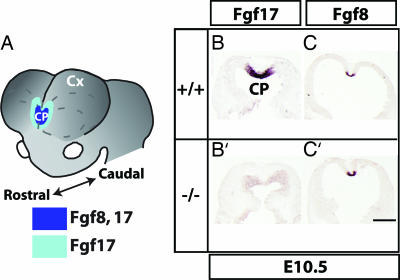
Fgf8 and Fgf17 expression overlap in the forebrain rostral patterning center, and Fgf8 expression is maintained in the Fgf17−/− mutant. (A) Fgf17 and Fgf8 RNA expression in the rostral forebrain patterning center. CP, commissural plate; Cx, cortex. (B, B′, C, and C′) Fgf17 and Fgf8 in situ hybridization (ISH) on horizontal sections from embryonic day 10.5 Fgf17+/+ (B and C) and Fgf17−/− (B′ and C′) forebrain. Rostral is at the top. (Scale bar: 0.5 mm.)
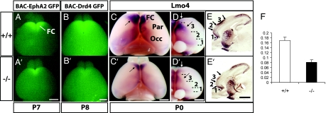
Reduced FC size in Fgf17−/− mice. Arrows signify shifted boundaries, and arrowheads signify maintained boundaries. (A and A′) Dorsal views of P7 Fgf17+/+ and Fgf17−/− brains positive for the BAC-EphA2 GFP transgene. The GFP+ domain that marks the FC was reduced in Fgf17−/− mutants. (B and B′) Dorsal views of P8 Fgf17+/+ and Fgf17−/− brains positive for the BAC-Drd4 GFP transgene. (C and C′) Dorsal views of Lmo4 whole-mount ISH on P0 Fgf17+/+ and Fgf17−/− brains. Par, parietal cortex; Occ, occipital cortex. (D and D′) Frontal views of the same brains in C and C′ reveal gene expression boundaries that distinguish three early FC subdivisions that we have labeled 1–3. (E and E′) Sagittal sections processed for Lmo4 ISH on P0 Fgf17+/+ and Fgf17−/− brains reveal sharp gene expression boundaries within the FC. White arrows in D and D′ indicate the approximate plane of section in E and E′. (F) Ratio of Lmo4+ dorsal FC area to total cortex area in Fgf17+/+ (n = 4) and Fgf17−/− (n = 4) P0 hemispheres (Student's t test; t = 5.21 and P < 0.01). (Scale bars: 1 mm.)
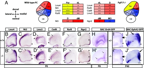
Selective changes in dorsal FC molecular properties revealed by a panel of gene expression markers. Arrows signify shifted boundaries, and arrowheads signify maintained boundaries. (A) Schema of wild-type and Fgf17−/− mutant FC subdivisions based on a panel of gene expression markers at P0. Dorsal and ventral FC subdivisions are shaded in red and blue, respectively. The parietal cortex is shaded in yellow. The table focuses on key wild-type subdivision distinctions with levels of expression for each gene: +++, strong expression; ++, moderate expression; +, weak expression; −, no detectable expression. See Table 1 for corresponding anatomical areas and SI Tables 2 and 3 for a more detailed analysis. (B–G and B′–G′) ISH for Lmo4, Nt3, Lmo3, Cad6, Rzr-β, and Ngn2 on Fgf17+/+ and Fgf17−/− littermate P0 coronal sections. Note the shift in dorsal expression borders (arrows) but maintenance of ventral borders (arrowheads). (H, H′, I, and I′) Anti-GFP immunohistochemistry on coronal sections from P8 (H and H′) and P40 (I and I′) mice containing the BAC-Drd4 GFP transgene. Note that the expression is much broader in the FC at P8 but is restricted in the mPFC at P40. (J and J′) Anti-GFP immunohistochemistry on coronal sections from P40 mice containing the BAC-EphA2 GFP transgene. D, dorsal FC; dlO, dorsolateral orbital cortex; dM, dorsomedial FC; LO, lateral orbital cortex; MO, medial orbital cortex; Par, parietal cortex; VO, ventral orbital cortex. (Scale bars: 0.5 mm.)
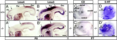
Rostral shift of the neocortical map in Fgf17−/− mutants. Rostral is to the left. Arrows signify shifted boundaries, and arrowheads signify maintained boundaries. (A, A′, B, and B′) Lmo4 and Lmo3 ISH on sagittal sections from Fgf17+/+ and Fgf17−/− P0 brains mark complementary cortical domains: frontal/occipital (FC/Occ) and parietal (Par) cortex, respectively. (Scale bar: 0.5 mm.) (C, C′, D, and D′) Cytochrome oxidase (CO) and anti-serotonin (5-HT) immunohistochemistry on tangential sections of flattened P7 cortices reveal a rostrodorsal shift of primary sensory areas. S1, somatosensory; V1, visual; A1, auditory. (Scale bar: 1 mm.)
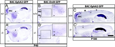
Persistence of changes in FC molecular regionalization in mature Fgf17−/− mice. Arrows signify shifted boundaries, and arrowheads signify maintained boundaries. (A, A′, B, and B′) Sagittal sections of brains from P40 mice carrying either the BAC-EphA2 GFP or BAC-Drd4 GFP transgene processed for anti-GFP immunohistochemistry. Fiber staining in the dorsal striatum in BAC-EphA2 GFP+ mice corresponds to projections from the FC (asterisks in A and A′). Boxed areas, consisting of dorsomedial (Md) and ventromedial (Mv) subdivisions of the PFC, are shown in a magnified view (Right Insets in B and B′). (Scale bars: 0.5 mm.) (C and C′) Sagittal sections of cortex from adult (P180) BAC-EphA2+, Fgf17+/+, and Fgf17−/− mice processed for anti-GFP immunohistochemistry. The broken arrow approximates the sensory (S)–motor (M) boundary. (Scale bar: 1 mm.)
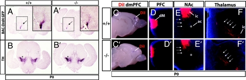
FC connectivity in Fgf17−/− mice. (A and A′) Reduction in FC projections to the ventral midbrain revealed by anti-GFP immunohistochemistry on P0 coronal sections from Fgf17+/+ and Fgf17−/− mice containing the BAC-Drd4 GFP transgene. Note the reduced staining of fibers emanating from the cerebral peduncle (arrows). Insets show magnified views of the boxed areas. (B and B′) Anti-tyrosine hydroxylase (TH) immunohistochemistry on sections adjacent to those shown in A and A′. Staining of the substantia nigra pars compacta and ventral tegmental area (asterisks) was similar between genotypes. (C and C′) DiI crystal placements in the dorsomedial FC of P3 Fgf17+/+ and Fgf17−/− brains viewed from the medial side. (D and D′) Restricted DiI-labeled field in the dorsomedial (dM) PFC in coronal section rostral to the crystal placement. Medial is to the left. (E and E′) Projections to the nucleus accumbens (NAc) (arrows). Anterograde-labeled fibers in the internal capsule (ic) restricted to the ventromedial striatum are also present. The anterior commisure provides a landmark (asterisk). Medial is to the left. (F and F′) Corticothalamic fibers (arrows) emanate from the internal capsule (ic) and are present in similar locations in both genotypes. A dotted line designates the thalamic midline.
Similar articles
-
Frontal cortex subdivision patterning is coordinately regulated by Fgf8, Fgf17, and Emx2.J Comp Neurol. 2008 Jul 10;509(2):144-55. doi: 10.1002/cne.21709. J Comp Neurol. 2008. PMID: 18459137 Free PMC article.
-
Organizing activity of Fgf8 on the anterior telencephalon.Dev Growth Differ. 2017 Dec;59(9):701-712. doi: 10.1111/dgd.12411. Epub 2017 Nov 10. Dev Growth Differ. 2017. PMID: 29124740
-
FGF15 promotes neurogenesis and opposes FGF8 function during neocortical development.Neural Dev. 2008 Jul 14;3:17. doi: 10.1186/1749-8104-3-17. Neural Dev. 2008. PMID: 18625063 Free PMC article.
-
Area patterning of the mammalian cortex.Neuron. 2007 Oct 25;56(2):252-69. doi: 10.1016/j.neuron.2007.10.010. Neuron. 2007. PMID: 17964244 Review.
-
Genetic regulation of prefrontal cortex development and function.Novartis Found Symp. 2007;288:165-73; discussion 173-7, 276-81. Novartis Found Symp. 2007. PMID: 18494258 Review.
Cited by
-
Mutations in FGF17, IL17RD, DUSP6, SPRY4, and FLRT3 are identified in individuals with congenital hypogonadotropic hypogonadism.Am J Hum Genet. 2013 May 2;92(5):725-43. doi: 10.1016/j.ajhg.2013.04.008. Am J Hum Genet. 2013. PMID: 23643382 Free PMC article.
-
Instability restricts signaling of multiple fibroblast growth factors.Cell Mol Life Sci. 2015 Jun;72(12):2445-59. doi: 10.1007/s00018-015-1856-8. Epub 2015 Feb 18. Cell Mol Life Sci. 2015. PMID: 25854632 Free PMC article.
-
OTX2 Transcription Factor Controls Regional Patterning within the Medial Ganglionic Eminence and Regional Identity of the Septum.Cell Rep. 2015 Jul 21;12(3):482-94. doi: 10.1016/j.celrep.2015.06.043. Epub 2015 Jul 9. Cell Rep. 2015. PMID: 26166575 Free PMC article.
-
Three phases of DiGeorge/22q11 deletion syndrome pathogenesis during brain development: patterning, proliferation, and mitochondrial functions of 22q11 genes.Int J Dev Neurosci. 2011 May;29(3):283-94. doi: 10.1016/j.ijdevneu.2010.08.005. Epub 2010 Sep 15. Int J Dev Neurosci. 2011. PMID: 20833244 Free PMC article. Review.
-
Transcriptional and epigenetic mechanisms of early cortical development: An examination of how Pax6 coordinates cortical development.J Comp Neurol. 2016 Feb 15;524(3):609-29. doi: 10.1002/cne.23866. Epub 2015 Aug 25. J Comp Neurol. 2016. PMID: 26304102 Free PMC article. Review.
References
-
- Fuster JM. Neuron. 2001;30:319–333. - PubMed
-
- Uylings HB, Groenewegen HJ, Kolb B. Behav Brain Res. 2003;146:3–17. - PubMed
-
- Heidbreder CA, Groenewegen HJ. Neurosci Biobehav Rev. 2003;27:555–579. - PubMed
-
- Rakic P. Science. 1988;241:170–176. - PubMed
-
- O'Leary DD, Nakagawa Y. Curr Opin Neurobiol. 2002;12:14–25. - PubMed
Publication types
MeSH terms
Substances
Grants and funding
LinkOut - more resources
Full Text Sources
Molecular Biology Databases
Miscellaneous
