. 2007 Jun;81(12):6459-70.
doi: 10.1128/JVI.00380-07. Epub 2007 Apr 11.

US3 of herpes simplex virus type 1 encodes a promiscuous protein kinase that phosphorylates and alters localization of lamin A/C in infected cells

Affiliations

US3 of herpes simplex virus type 1 encodes a promiscuous protein kinase that phosphorylates and alters localization of lamin A/C in infected cells

Fan Mou et al. J Virol. 2007 Jun.

Abstract

The herpes simplex virus type 1 (HSV-1) US3 gene encodes a serine/threonine kinase that, when inactivated, causes capsids to aggregate aberrantly between the inner and outer nuclear membranes (INM and ONM, respectively) within evaginations/extensions of the perinuclear space. In both Hep2 cells and an engineered cell line derived from Hep2 cells expressing lamin A/C fused to enhanced green fluorescent protein (eGFP-lamin A/C), lamin A/C localized mostly in a reticular pattern with small regions of the INM devoid of eGFP-lamin A/C when they were either mock infected or infected with wild-type HSV-1(F). Cells infected with HSV-1(F) also contained some larger diffuse regions lacking lamin A/C. Proteins UL31 and UL34, markers of potential envelopment sites at the INM and perinuclear virions, localized within the regions devoid of lamin A/C and also in regions containing lamin A/C. Similar to previous observations with Vero cells (S. L. Bjerke and R. J. Roller, Virology 347:261-276, 2006), the proteins UL34 and UL31 localized exclusively in very discrete regions of the nuclear lamina lacking lamin A/C in the absence of US3 kinase activity. To determine how US3 alters lamin A/C distribution, US3 was purified and shown to phosphorylate lamin A/C at multiple sites in vitro, despite the presence of only one putative US3 kinase consensus site in the lamin A/C sequence. US3 kinase activity was also sufficient to invoke partial solubilization of lamin A/C from permeabilized Hep2 cell nuclei in an ATP-dependent manner. Two-dimensional electrophoretic analyses of lamin A/C revealed that lamin A/C is phosphorylated in HSV-infected cells, and the full spectrum of phosphorylation requires US3 kinase activity. These data suggest that US3 kinase activity regulates HSV-1 capsid nuclear egress at least in part by phosphorylation of lamin A/C.


Figures

FIG. 1.
FIG. 1.
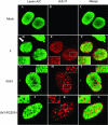
Confocal analyses of a Hep2-derived cell line stably expressing eGFP-lamin A/C. Cells were either mock infected (Mock) or infected with 5.0 PFU/cell of wild-type HSV-1(F) (designated F) or mutant US3(K220A), lacking pUS3 kinase activity. Cells were fixed and permeabilized at 16 h postinfection and stained with polyclonal anti-pUL34 chicken polyclonal antibody, followed by Texas Red-conjugated secondary antibody, and examined with confocal microscopy. (A) Analysis of optical sections taken through the middle of cells. (B) Optical sections taken at the bottom of cells. Regions indicated in white rectangles in panels D, E, F, J, K, and L are digitally magnified in the panel immediately below them (panels G, H, I, L, M and N, respectively). Coincident signals in the merged images (rightmost column) are indicated by a yellow color. An arrow indicates one region of the infected cell containing less lamin A/C than surrounding areas.
FIG. 2.
FIG. 2.
Digital confocal images of Hep2 cells immunostained with antibodies to lamin A/C and pUL31. Hep2 cells were mock infected (Mock) or infected with a US3 deletion mutant (dUs3), a mutant lacking US3 kinase activity [US3 (K220A)], or wild-type virus HSV-1(F) (designated F). Sixteen hours after infection, the cells were fixed, permeabilized, and reacted with preadsorbed rabbit polyclonal antiserum against pUL31 or chicken immunoglobulin Y directed against lamin A/C. Bound antibodies were revealed with Texas Red-conjugated anti-rabbit antibody or FITC-conjugated anti-chicken antibodies. Optical sections closest to the glass coverslips are shown. An arrow indicates regions of the nuclear rim that are mostly devoid of lamin A/C immunostaining. White boxes indicate areas of interest that are magnified in an inset in the same panel.
FIG. 3.
FIG. 3.
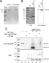
Analysis of in vitro kinase activities of purified GST-pUS3 and the mutant GST-pUS3(K220A). (A) Denaturing polyacrylamide gel containing purified GST-pUS3 and GST-pUS3(K220A) and stained with Coomassie brilliant blue. (B) In vitro kinase reaction. The fusion proteins indicated at the top of the left panel were mixed with 100 ng GST-US3. After 30 min at 30°C in the presence of [γ-32P]ATP, the reaction mixtures were resolved on a denaturing polyacrylamide gel and visualized by Coomassie (CBB) staining (left panel) and autoradiography (right panel). The position of the approximately 90,000 apparent Mr GST-US3 fusion protein is shown in the right panel. GST-UL34 (amino acids 205 to 275) is a negative control inasmuch as it does not contain the phosphorylation consensus site in pUL34. (C) In vitro kinase activities of GST-pUS3 and GST-pUS3(K220A). Purified GST-pUS3 (0.1 μg) or 0.5 μg GST-pUS3(K220A) was incubated with [γ-32P]ATP in the presence or absence of partially purified GST-pUL31 as a potential substrate, followed by subsequent incubation in the presence or absence of λ-Ppase. The reaction components were denatured, electrophoretically separated, and stained with Coomassie brilliant blue (CBB, bottom panel) or dried and autoradiographed (upper panel). For comparative purposes, the molecular weight standards from the bottom panel were copied and aligned with the top panel, using Adobe Photoshop. Sizes of the standards are indicated to the left of the figure in thousands [K]).
FIG. 4.
FIG. 4.
Phosphorylation of lamin A by US3 kinase in vitro. Full-length lamin A fused to GST was partially purified and reacted with either purified GST-pUS3 or GST-pUS3(K220A) in the presence of [γ-32P]ATP, followed by incubation in the presence or absence of λ-Ppase. The reaction components were separated on a polyacrylamide gel and stained with Coomassie brilliant blue (CBB, lower panel) and autoradiographed (upper panel).
FIG. 5.
FIG. 5.
(A) Schematic diagram of lamin A that primary structure and five subdomains fused to GST. The amino acids (AA) in each peptide subdomain are indicated, assuming that the start methionine codon is 1. (Reprinted from reference .) (B) Full-length (FL) lamin A or lamin A fragments fused to GST and reacted with GST-pUS3 kinase. The five GST fusion proteins bearing fragments of lamin A as detailed for panel A were purified, mixed with US3-GST and [γ-32P]ATP, and electrophoretically separated and stained with Coomassie brilliant blue (lower panel) and autoradiographed (upper panel). H, head; R1, rod1; R2, rod2; T1, tail 1; T2, tail2; lane C, GST-pUL34 (amino acids 205 to 275).
FIG. 6.
FIG. 6.
Immunoblots of total and solubilized lamin A/C from permeabilized Hep2 cell nuclei reacted with wild-type and mutant US3-encoded kinases. Purified and permeabilized Hep2 cell nuclei were incubated for 30 min in the presence or absence of the indicated fusion proteins and ATP. As a loading control, a sample of the total material (L) was collected immediately after the reactions. S1 was collected after centrifugation at low speed. A second supernatant fraction (S2) was collected after high-speed centrifugation. Proteins in the various fractions were denatured in SDS, electrophoretically separated, transferred to nitrocellulose, and probed with lamin A/C-specific antibody. Bound immunoglobulin was detected by a horseradish peroxidase-conjugated secondary antibody and chemiluminescence. The intensity of the chemiluminescence signals of the lamin A and C bands were quantified using a Syngene Chemi-Genius imaging system and associated GeneTools software, and the amount of chemiluminescence was pooled. The reported percentages represent the intensity of chemiluminescence in that lane relative to that detected in the first lane (sample L of GST-pUs3 plus ATP).
FIG. 7.
FIG. 7.
Two-dimensional gel electrophoresis and immunoblots of lamin A/C isoforms from mock-infected and HSV-infected cells. Hep2 cells were mock infected or infected with wild-type HSV-1(F) (panels labeled F) or mutant Us3(K220A). Proteins were extracted in buffer containing 9 M urea to solubilize lamins and separated by isoelectric focusing in the first dimension using nonlinear pH gradients from pH 3.0 to 10.0 (A and B) or linear pH gradients 5.0 to 8.0 (C) and by size on denaturing polyacrylamide gels in the second dimension. The proteins were then transferred to nitrocellulose and probed with polyclonal chicken antibody directed against lamin A/C or a rabbit antibody against lamin B1 and lamin B2. Bound immunoglobulin was detected by appropriate horseradish peroxidase-conjugated secondary antibodies. Chemiluminescence generated by the addition of appropriate substrates was recorded on X-ray film. In some experiments (λ-Ppase), proteins were reacted with λ-Ppase before electrophoretic separation. For comparative purposes, the three blots in panels A and B were aligned by position of lamin B-specific spots and by distance from the cationic and anionic poles. The positions of the terminal poles on the pH strip of the four immunoblots shown in panel C are also aligned. For orientation, the positions of the lamin A-, B-, and C-specific spots are indicated to the left of the top immunoblot shown in panels A and C.

Similar articles

Cited by

References

    1. Benetti, L., and B. Roizman. 2004. Herpes simplex virus protein kinase US3 activates and functionally overlaps protein kinase A to block apoptosis. Proc. Natl. Acad. Sci. USA 101:9411-9416. - PMC - PubMed
    1. Bjerke, S. L., J. M. Cowan, J. K. Kerr, A. E. Reynolds, J. D. Baines, and R. J. Roller. 2003. Effects of charged cluster mutations on the function of herpes simplex virus type 1 UL34 protein. J. Virol. 77:7601-7610. - PMC - PubMed
    1. Bjerke, S. L., and R. J. Roller. 2006. Roles for herpes simplex virus type 1 U(L)34 and U(S)3 proteins in disrupting the nuclear lamina during herpes simplex virus type 1 egress. Virology 347:261-276. - PMC - PubMed
    1. Chang, Y. E., C. Van Sant, P. W. Krug, A. E. Sears, and B. Roizman. 1997. The null mutant of the UL31 gene of herpes simplex virus 1: construction and phenotype of infected cells. J. Virol. 71:8307-8315. - PMC - PubMed
    1. Collas, P. 1999. Sequential PKC- and Cdc2-mediated phosphorylation events elicit zebrafish nuclear envelope disassembly. J. Cell Sci. 112:977-987. - PubMed

Publication types

LinkOut - more resources