. 2007 Apr 9;177(1):73-86.
doi: 10.1083/jcb.200612094.

A role for cell sex in stem cell-mediated skeletal muscle regeneration: female cells have higher muscle regeneration efficiency

Affiliations

A role for cell sex in stem cell-mediated skeletal muscle regeneration: female cells have higher muscle regeneration efficiency

Bridget M Deasy et al. J Cell Biol. .

Abstract

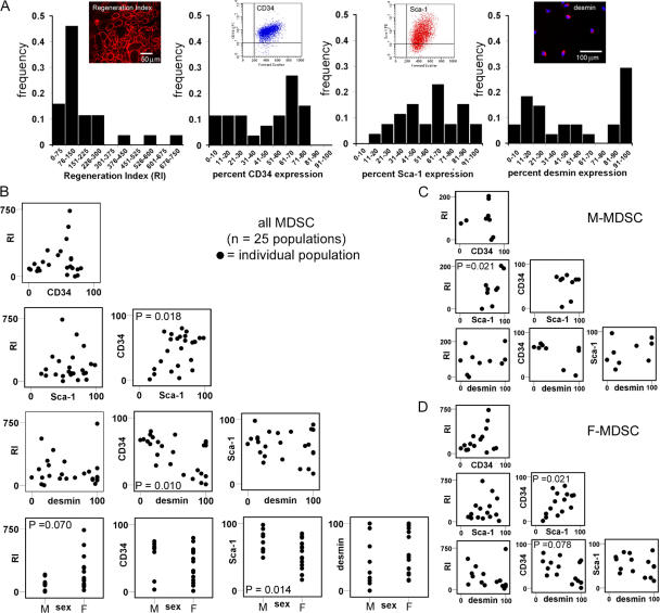
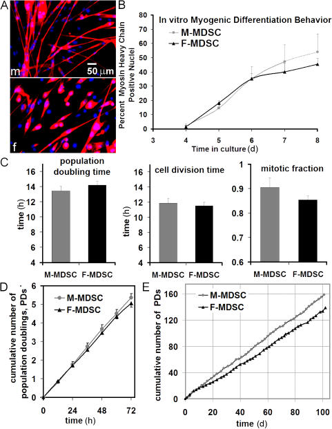
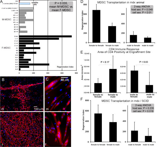
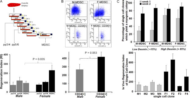
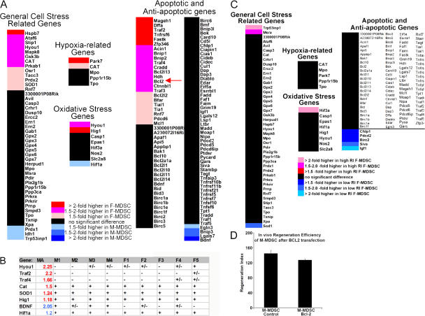
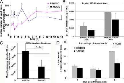
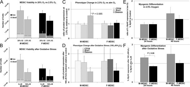
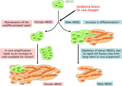
We have shown that muscle-derived stem cells (MDSCs) transplanted into dystrophic (mdx) mice efficiently regenerate skeletal muscle. However, MDSC populations exhibit heterogeneity in marker profiles and variability in regeneration abilities. We show here that cell sex is a variable that considerably influences MDSCs' regeneration abilities. We found that the female MDSCs (F-MDSCs) regenerated skeletal muscle more efficiently. Despite using additional isolation techniques and cell cloning, we could not obtain a male subfraction with a regeneration capacity similar to that of their female counterparts. Rather than being directly hormonal or caused by host immune response, this difference in MDSCs' regeneration potential may arise from innate sex-related differences in the cells' stress responses. In comparison with F-MDSCs, male MDSCs have increased differentiation after exposure to oxidative stress induced by hydrogen peroxide, which may lead to in vivo donor cell depletion, and a proliferative advantage for F-MDSCs that eventually increases muscle regeneration. These findings should persuade researchers to report cell sex, which is a largely unexplored variable, and consider the implications of relying on cells of one sex.


Figures

Figure 1.
Figure 1.
Variability among muscle-derived cell populations. (A) We examined 25 populations of MDSCs for five variables: in vivo muscle regeneration efficiency (RI), CD34 expression, Sca-1 expression, desmin expression, and cell sex. Histograms show the distribution of each parameter for the 25 different populations. In vivo regeneration was quantified as the number of dystrophin-positive myofibers (red) present in the skeletal muscle of dystrophin-deficient mdx mice after the transplantation of MDSCs. Flow plots are shown for CD34 and Sca-1 expression, and immunofluorescence is shown for desmin (desmin, red; nuclei, blue). (B) We performed correlation analysis to identify significant relationships between variables. The scatter plots are shown. Each population is represented as a dot on the scatter plots. P-values are shown for the correlation coefficient when a linear relationship was found to exist between the two given variables. Only cell sex correlated with in vivo regeneration efficiency (R = 0.367; P = 0.070; M, male; F, female); our subsequent t test comparison of the regeneration efficiency of M-MDSCs with that of F-MDSCs revealed a significant difference (P = 0.035). We also detected a significant correlation between Sca-1 expression and cell sex (R = −0.490; P = 0.014), with significantly more M- than F-MDSCs expressing Sca-1 (P = 0.001; t test). (C and D) Examination of the correlation matrices for M- (C) and F-MDSCs (D) again revealed the heterogeneity of the populations. In particular, the higher level of Sca-1 expression by M-MDSCs correlated positively with a higher RI (P = 0.021), which suggests that the significantly higher levels of Sca-1 expression by M-MDSCs were not directly related to their low in vivo RI (see plot of RI vs. Sca-1 for M-MDSCs in D).
Figure 2.
Figure 2.
M- and F-MDSCs demonstrate similar in vitro stem cell characteristics. (A) M- and F-MDSCs underwent myogenic differentiation, as shown by MyHC expression (red; m, M-MDSC; f, F-MDSC). (B) We did not detect a significant difference in the extent or rate of myogenic differentiation as measured by the percentage of nuclei that colocalized with MyHC in low serum–containing medium (P > 0.05). (C) PDT, cell division time, and mitotic fraction were similar for M- and F-MDSCs. (D) Short-term proliferation kinetics were similar for M- and F-MDSCs. (E) Long-term proliferation kinetics showed that both M- and F-MDSCs can be expanded for >150 PDs in vitro. However, we observed a trend toward faster proliferation by M-MDSCs cultured for longer periods of time. Error bars represent SD.
Figure 3.
Figure 3.
F-MDSCs induce more efficient skeletal muscle regeneration than M-MDSCs. (A) Quantitation of engraftment in terms of the RI (RI = number of dystrophin-positive fibers per 105 donor cells). The overall mean RI is significantly higher for F-MDSCs (black bars; RI = 230 ± 52; n = 15 F-MDSC populations; two to six muscles per population) than for M-MDSCs (gray bars; RI = 109 ± 18; mean ± SEM; n = 10 M-MDSC populations; two to six muscles per population; P = 0.035; t test). Sham-injected muscles (PBS; red bar) and RI values from previous studies of myoblasts (Qu-Petersen et al., 2002) and male myogenic progenitor cells (Jankowski et al., 2002) are also shown. (B) Representative engraftments of transplanted cells to mouse mdx skeletal muscle show dystrophin-positive fibers (red) within dystrophic muscle (nuclei stained with Hoechst). M-MDSCs produced fewer dystrophin-positive fibers than did F-MDSCs. (C) Dystrophin expression in vitro confirms the ability of both M- and F-MDSCs to express the dystrophin gene after cell fusion. (D)We performed sex-matched and sex-crossed transplantations. Two-way parametric ANOVA indicated significant differences in the RI associated with the sex of the donor cells (P < 0.001) and the sex of the host (P = 0.048). Mean RI and SEM (error bars) are shown (female to female, RI = 686 ± 120; female to male, RI = 347 ± 69; male to female, RI = 160 ± 75; male to male, RI = 105 ± 25; n = 6–12 per group). (E) Immune response at the site of transplantation in immune-competent mdx mice was quantified as the cross-sectional area (micrometers squared) of CD4 expression in tissue sections. There was significantly more CD4 positivity after male to female cross transplantation than after sex-matched male to male transplantation (mean ± SEM; t test; n = 4). (F) Transplantation of MDSCs into mdx/SCID mice. Two-way nonparametric ANOVA revealed no effect on the RI as a result of the sex of the host (P = 0.235), but there was a significant effect as a result of the sex of the cells (P = 0.018). RIs are reported as mean ± SEM (female to female, RI = 546 ± 166; female to male, RI = 381 ± 145; male to female, RI = 216 ± 39; male to male, RI = 115 ± 11; n = 4 per group).
Figure 4.
Figure 4.
Examination of alternate M-MDSC sources. (A) The preplate technique to isolate subfractions of muscle-derived cells is based on their adhesion characteristics from muscle biopsy. Male myoblast subfractions also demonstrated low regeneration efficiency. There was no significant difference between the RIs of male pp3/4 (74 ± 32; n = 4) and pp5/6 (122 ± 38; n = 3) when compared with the RI of M-MDSCs (109 ± 18; n = 10 for all M-MDSC populations shown in Fig. 2). There also was no significant difference between the RIs of male myoblasts and female myoblasts (female pp3/4 RI = 79 ± 14, n = 4; female pp5/6 RI = 107 ± 10, n = 3; P > 0.05). The bars for the M- and F-MDSCs represent the mean RIs for all male and female populations shown in Fig. 2 (95 ± 21 [n = 10] and 230 ± 52 [n = 15], respectively). (B) M- and F-MDSCs were sorted by FACS to obtain a subpopulation expressing the stem cell marker CD34. The presorted population had similar expression levels for desmin/Sca-1 and CD34. Cells were purified to contain >96% CD34+ cells only. Transplantation of purified M-MDSCs expressing CD34 resulted in a significantly lower RI (266 ± 61) than did transplantation of F-MDSCs expressing CD34 (417 ± 56; P = 0.052; t test). All RIs are reported as means ± SEM (error bars), and p-values are for t test comparisons. (C, top) 2 wk after single-cell cloning, there was no significant difference in the percentage of single-cell clones derived from M- or F-MDSC populations that yielded myogenic colonies as defined by myotube formation (73–90%; P = 0.776). We detected no effect as a result of the cell sex (P = 0.756). Parent populations were either low in desmin expression (<15% positive cells within the population) or high in desmin expression (>85% positive). (C, bottom) We did not observe any male clone that exhibited a higher RI as compared with the parent population. We transplanted four M-MDSC clonal populations (M1, M2, M3, and M4) and four F-MDSC clonal populations (F1, F2, F3, and F4; mean ± SEM is shown; n = 3 muscle transplantations per clone). In contrast, we found that three of the four F-MDSCs display a high-regenerating potential as observed with their parent population.
Figure 5.
Figure 5.
Differences in hypoxic and oxidative stress genes. (A) Microarray gene ontology analysis of M- and F-MDSCs. An increase in the expression of cell stress–related genes, including oxidative stress and antiapoptotic genes, was identified for F-MDSCs in comparison with M-MDSCs (colored bars represent significant differences; P < 0.05). Of 45 general cellular stress–related genes, 29% of the genes were significantly increased in F-MDSCs, 7% were significantly higher in M-MDSCs, and 64% showed no significant difference. Of 99 apoptosis-related genes, 23% of the genes were significantly increased in F-MDSCs, 4% were higher in M-MDSCs, and 72% showed no significant difference. (B) Gene levels of several key genes were examined using RT-PCR analysis. The results of several rounds of RT-PCR showing the gene symbol (Gene), the increase as seen by microarray analysis (MA; blue indicates that it is higher in M-MDSCs, and red indicates that it is higher in F-MDSCs), and the results of nine screened populations (four male populations, M1–M4, and five female populations, F1–F5; + indicates detection at 25 or 28 cycles, +/− denotes genes that were only observed at 30 cycles, and – represents genes that were not observed at 30 cycles). (C) We also examined the microarray data for differences in F-MDSCs that yielded a high level of muscle regeneration (high RI) as compared with F-MDSCs that yielded a low level of regeneration (low RI). High RIs are shown as red bars, and low RIs are shown blue bars. P < 0.05. (D) We found that the antiapoptotic factor Bcl2 was twofold higher in F-MDSCs than in M-MDSCs (A, arrow). We hypothesized that low Bcl2 expression may be related to reduced survival after cell transplantation, and we transfected M-MDSCs to overexpress Bcl2 (M-MDSC–Bcl2). However, we did not observe a significant difference in the RI of M-MDSC–Bcl2 (RI = 128 ± 17) as compared with the M-MDSC control (RI = 145 ± 48) at 2 wk after transplantation into dystrophic muscle (P = 0.415).
Figure 6.
Figure 6.
Examination of the transplantation site at early time points. (A) M- and F-MDSCs were transduced with the retroviral lacZ gene and transplanted into the gastrocnemius muscles of sex-matched mice, and the muscles were harvested at several time points between 16 h and 14 d. We quantified the number of lacZ nuclei that were detected at the transplantation site. At 16, 24, 48, and 72 h, there were significantly more nuclei detected in M-MDSC transplantations as compared with F-MDSC transplantations (*, P < 0.05 at all time points). (B) We digested the skeletal muscle after 24 and 48 h after transplantation, and we quantified the amount of lacZ gene by RT-PCR to support the histological staining results. Similar to the histological analysis, we detected more β-gal transcripts in muscles transplanted with M-MDSCs as compared with F-MDSC transplantations. (C) We stained M- and F-MDSCs using monochlorobimane (MCB) and found significantly more intracellular glutathione in M-MDSCs as compared with F-MDSCs (P < 0.01). (D) We quantified the percentage of lacZ + donor cells that are located within regenerating muscle fibers at 1, 2, and 5 d after MDSC transplantation. At 5 d after transplantation, we observed a trend toward significantly more donor cell fusion or differentiation with M-MDSCs as compared with F-MDSC transplantations to mdx muscle (P = 0.068). Error bars represent SD.
Figure 7.
Figure 7.
Behavioral response to low oxygen and oxidative stress. (A) After exposure to physiologic oxygen for 24 h (2.5% O2), we observed a trend toward reduced cell numbers in both M-MDSCs and F-MDSCs (P = 0.18 and P = 0.079, respectively). There was no significant difference in the amount of cell viability or cell death in M-MDSCs as compared with F-MDSCs (ratio of the percentage of cells in low oxygen to atmospheric oxygen; P = 0.652). (B) After exposure to oxidative stress (100 μM H2O2), we observed a significant decrease in cell numbers of both M-MDSCs and F-MDSCs (P = 0.039 and P = 0.001, respectively). There was no difference in the percent change in cell viability for M-MDSCs vs. F-MDSCs after oxidative stress (ratio of the percentage of cells in 100 μM to atmospheric oxygen; P = 0.325). Bars are means ± SEM (error bars). (C) We examined the expression of CD34, Sca-1, and desmin in the populations after exposure to low oxygen. We did not observe a change in CD34 or Sca-1 expression in M- or F-MDSCs that were exposed to 2.5% O2 as compared with cells cultured in atmospheric O2. We observed a trend in increased desmin expression in M-MDSCs that had initially low levels of desmin (P = 0.065). There was no similar change in desmin expression in a comparable population of F-MDSCs. (D) After treatment with hydrogen peroxide, we observed trends toward an increase in CD34 and Sca-1 in all populations; however, there was no significant difference in the phenotype of M-MDSCs as compared with F-MDSCs (P > 0.05). Bars are means ± SD. (E) We induced myogenic differentiation and subsequently exposed cells to 2.5% oxygen for 24 and 48 h. We quantified the percentage of nuclei that colocalized with cells expressing the myogenic marker fast MyHC. We observed a trend toward more myogenic differentiation with M-MDSCs as compared with F-MDSCs. (F) We observed an increase in myogenic differentiation with M-MDSCs that were treated with 100 μM H2O2 as compared with their untreated controls at both 24 and 48 h. In comparison with their female counterparts, this change was significant at 24 h (P = 0.06). Bars are means ± SEM.
Figure 8.
Figure 8.
Schematic of proposed sex-related differences in response to cell stress and role in skeletal muscle regeneration. Sex-related differences affect the response of MDSCs to low oxygen or oxidative stress. F-MDSCs may respond by maintaining a low level of proliferation. In a myogenic environment with low oxygen or oxidative stress, F-MDSCs do not appear to readily differentiate. Conversely, M-MDSCs demonstrate increased myogenic differentiation in the presence of low oxygen or oxidative stress. In vivo, we hypothesize that the increased differentiation will result in the rapid depletion of donor M-MDSCs as cells fuse to form terminally differentiated multinucleated myotubes/fibers. F-MDSCs may be less proliferative at early time points after implantation and demonstrate an increase in cell numbers starting at 3 d after implantation. Ultimately, the tendency to maintain the undifferentiated phenotype or resist differentiation would be a mechanism that allows for in vivo expansion of F-MDSCs that are subsequently available at later time points for muscle regeneration.

Similar articles

Cited by

References

    1. Aristotle. 350 BC. Historia Animalium: Books VII–X. 1991. edition. D.M. Balme, editor. Harvard University Press, Cambridge, MA. 435–437.
    1. Avery, B., C.B. Jorgensen, V. Madison, and T. Greve. 1992. Morphological development and sex of bovine in vitro-fertilized embryos. Mol. Reprod. Dev. 32:265–270. - PubMed
    1. Aviv, A. 2002. Telomeres, sex, reactive oxygen species, and human cardiovascular aging. J. Mol. Med. 80:689–695. - PubMed
    1. Aviv, A., J. Shay, K. Christensen, and W. Wright. 2005. The longevity gender gap: are telomeres the explanation? Sci. Aging Knowledge Environ. 2005:pe16. - PubMed
    1. Beauchamp, J.R., J.E. Morgan, C.N. Pagel, and T.A. Partridge. 1994. Quantitative studies of efficacy of myoblast transplantation. Muscle Nerve. 1:S261.

Publication types