Indian Hedgehog produced by postnatal chondrocytes is essential for maintaining a growth plate and trabecular bone
- PMID: 17409191
- PMCID: PMC1851055
- DOI: 10.1073/pnas.0608449104
Indian Hedgehog produced by postnatal chondrocytes is essential for maintaining a growth plate and trabecular bone
Abstract
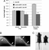
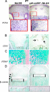
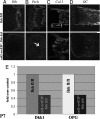
Indian hedgehog (Ihh) is essential for chondrocyte and osteoblast proliferation/differentiation during prenatal endochondral bone formation. The early lethality of various Ihh-ablated mutant mice, however, prevented further analysis of its role in postnatal bone growth and development. In this study, we describe the generation and characterization of a mouse model in which the Ihh gene was successfully ablated from postnatal chondrocytes in a temporal/spatial-specific manner; postnatal deletion of Ihh resulted in loss of columnar structure, premature vascular invasion, and formation of ectopic hypertrophic chondrocytes in the growth plate. Furthermore, destruction of the articular surface in long bones and premature fusion of growth plates of various endochondral bones was evident, resulting in dwarfism in mutant mice. More importantly, these mutant mice exhibited continuous loss of trabecular bone over time, which was accompanied by reduced Wnt signaling in the osteoblastic cells. These results demonstrate, for the first time, that postnatal chondrocyte-derived Ihh is essential for maintaining the growth plate and articular surface and is required for sustaining trabecular bone and skeletal growth.
Conflict of interest statement
The authors declare no conflict of interest.
Figures


Similar articles
-
Recent Insights into Long Bone Development: Central Role of Hedgehog Signaling Pathway in Regulating Growth Plate.Int J Mol Sci. 2019 Nov 20;20(23):5840. doi: 10.3390/ijms20235840. Int J Mol Sci. 2019. PMID: 31757091 Free PMC article. Review.
-
Partial rescue of postnatal growth plate abnormalities in Ihh mutants by expression of a constitutively active PTH/PTHrP receptor.Bone. 2010 Feb;46(2):472-8. doi: 10.1016/j.bone.2009.09.009. Epub 2009 Sep 15. Bone. 2010. PMID: 19761883 Free PMC article.
-
Conditional Deletion of Indian Hedgehog in Limb Mesenchyme Results in Complete Loss of Growth Plate Formation but Allows Mature Osteoblast Differentiation.J Bone Miner Res. 2015 Dec;30(12):2262-72. doi: 10.1002/jbmr.2582. Epub 2015 Jul 29. J Bone Miner Res. 2015. PMID: 26094741
-
Core binding factor beta (Cbfβ) controls the balance of chondrocyte proliferation and differentiation by upregulating Indian hedgehog (Ihh) expression and inhibiting parathyroid hormone-related protein receptor (PPR) expression in postnatal cartilage and bone formation.J Bone Miner Res. 2014 Jul;29(7):1564-1574. doi: 10.1002/jbmr.2275. J Bone Miner Res. 2014. PMID: 24821091 Free PMC article.
-
Indian Hedgehog, a critical modulator in osteoarthritis, could be a potential therapeutic target for attenuating cartilage degeneration disease.Connect Tissue Res. 2014 Aug;55(4):257-61. doi: 10.3109/03008207.2014.925885. Epub 2014 Jun 13. Connect Tissue Res. 2014. PMID: 24844414 Review.
Cited by
-
Exogenous Indian hedgehog antagonist damages intervertebral discs homeostasis in adult mice.J Orthop Translat. 2022 Oct 6;36:164-176. doi: 10.1016/j.jot.2022.09.009. eCollection 2022 Sep. J Orthop Translat. 2022. PMID: 36263384 Free PMC article.
-
Skeletal stem cells: origins, definitions, and functions in bone development and disease.Life Med. 2022 Dec 8;1(3):276-293. doi: 10.1093/lifemedi/lnac048. eCollection 2022 Dec. Life Med. 2022. PMID: 36811112 Free PMC article. Review.
-
Recent Insights into Long Bone Development: Central Role of Hedgehog Signaling Pathway in Regulating Growth Plate.Int J Mol Sci. 2019 Nov 20;20(23):5840. doi: 10.3390/ijms20235840. Int J Mol Sci. 2019. PMID: 31757091 Free PMC article. Review.
-
Chondrocyte β-catenin signaling regulates postnatal bone remodeling through modulation of osteoclast formation in a murine model.Arthritis Rheumatol. 2014 Jan;66(1):107-20. doi: 10.1002/art.38195. Arthritis Rheumatol. 2014. PMID: 24431282 Free PMC article.
-
Inactivation of Ihh in Sp7-Expressing Cells Inhibits Osteoblast Proliferation, Differentiation, and Bone Formation, Resulting in a Dwarfism Phenotype with Severe Skeletal Dysplasia in Mice.Calcif Tissue Int. 2022 Nov;111(5):519-534. doi: 10.1007/s00223-022-00999-5. Epub 2022 Jun 22. Calcif Tissue Int. 2022. PMID: 35731246
References
-
- Horton W. Growth Genet Horm. 1990;6:1–3.
-
- Gilbert S. Developmental Biology. Sunderland, MA: Sinauer; 1997.
-
- Hall B. Am Sci. 1988;76:174–181.
-
- Erlebacher A, Filvaroff E, Gitelman S, Derynck R. Cell. 1995;80:371–378. - PubMed
-
- de Crombrugghe B, Lefebvre V, Nakashima K. Curr Opin Cell Biol. 2001;13:721–727. - PubMed
Publication types
MeSH terms
Substances
Grants and funding
LinkOut - more resources
Full Text Sources
Other Literature Sources
Molecular Biology Databases

