. 2007 Apr 9;177(1):127-37.
doi: 10.1083/jcb.200610018. Epub 2007 Apr 2.

Phosphorylation and regulation of a G protein-coupled receptor by protein kinase CK2

Affiliations

Phosphorylation and regulation of a G protein-coupled receptor by protein kinase CK2

Ignacio Torrecilla et al. J Cell Biol. .

Abstract

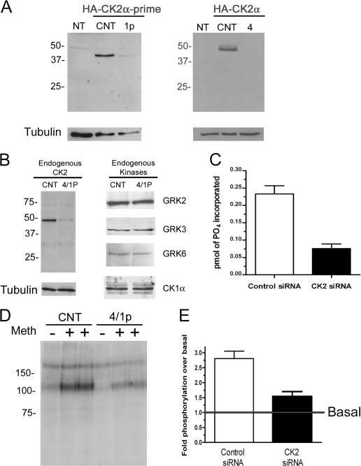
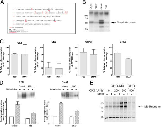
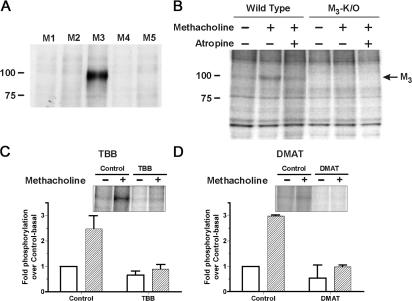
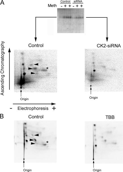
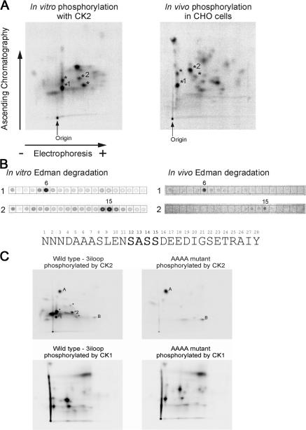
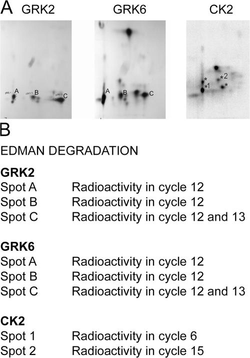
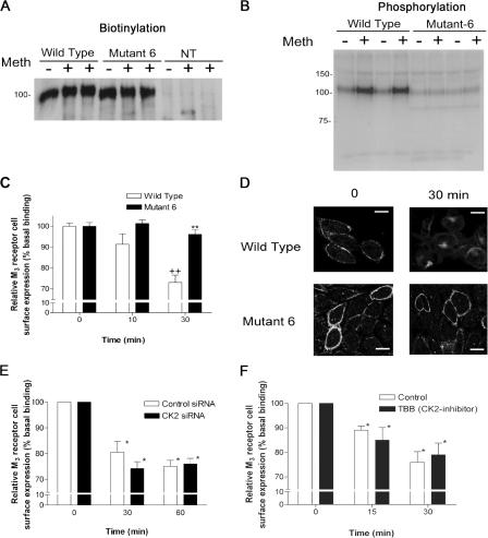
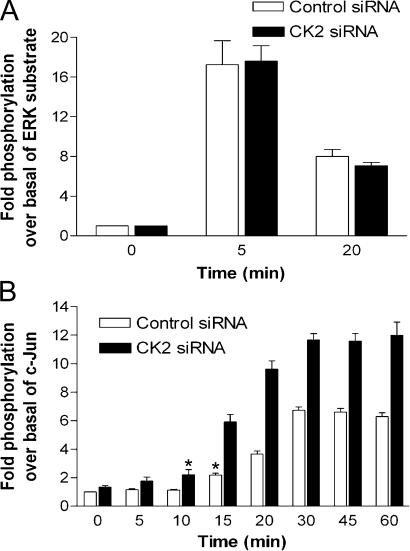
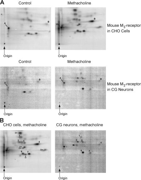
We demonstrate a role for protein kinase casein kinase 2 (CK2) in the phosphorylation and regulation of the M3-muscarinic receptor in transfected cells and cerebellar granule neurons. On agonist occupation, specific subsets of receptor phosphoacceptor sites (which include the SASSDEED motif in the third intracellular loop) are phosphorylated by CK2. Receptor phosphorylation mediated by CK2 specifically regulates receptor coupling to the Jun-kinase pathway. Importantly, other phosphorylation-dependent receptor processes are regulated by kinases distinct from CK2. We conclude that G protein-coupled receptors (GPCRs) can be phosphorylated in an agonist-dependent fashion by protein kinases from a diverse range of kinase families, not just the GPCR kinases, and that receptor phosphorylation by a defined kinase determines a specific signalling outcome. Furthermore, we demonstrate that the M3-muscarinic receptor can be differentially phosphorylated in different cell types, indicating that phosphorylation is a flexible regulatory process where the sites that are phosphorylated, and hence the signalling outcome, are dependent on the cell type in which the receptor is expressed.


Figures

Figure 1.
Figure 1.
CK2 siRNAs decrease agonist-driven M3-muscarinic receptor phosphorylation. (A) Western blot analysis (using anti-HA antibody) of transfected HA-CK2α′ (left) and HA-CK2α (right) expression in CHO cells cotransfected with the indicated siRNA duplexes. α-Tubulin was used as a loading control. NT, nontransfected. (B) CHO cells were transfected with the indicated siRNAs and used to determine expression levels of endogenous CK2α subunit, GRK2, GRK3, GRK6, and CK1α by immunoblotting with the indicated antibodies. (C) CK2 kinase activity in lysates from the siRNA transfected CHO cells was determined by the phosphorylation of a substrate peptide. Data correspond to mean ± SD from three independent experiments. (D) Phosphorylation of the M3 receptor in CHO-M3 cells transfected with siRNA duplexes CK2α-4 and CK2α′-1p or scrambled control siRNAs (CNT) and stimulated with 100 μM methacholine for 5 min. (E) Quantitative analysis of phosphorylation from D. Phosphate content of M3 receptor was normalized to the basal level of phosphorylation. Data correspond to mean ± SD from three independent experiments.
Figure 2.
Figure 2.
Pharmacological inhibition of CK2 decreases agonist-mediated M3-muscarinic receptor phosphorylation. (A) The third intracellular loop of the M3-muscarinic showing the consensus sites for CK1α, CK2, and the GRKs as predicted using GPS (http://973-proteinweb.ustc.edu.cn/gps/gps_web/; Xue et al., 2005). (B) In vitro phosphorylation of a GST-H3iloop fusion protein by CK1, CK2, GRK2, and GRK6. The position of the fusion protein as determined by Coomassie staining is shown. (C) In vitro phosphorylation of a GST-H3iloop fusion protein by CK1, CK2, GRK2, and GRK6 in the presence of 1 μM TBB and DMAT (CK2 inhibitors). Results are expressed as a percentage of controls (phosphorylation without the inhibitor). (D) M3-muscarinic receptor phosphorylation in CHO-M3 cells in the absence (control) or presence of TBB and DMAT (20 μM, 1.5 h). Cells were stimulated with 100 μM methacholine for 5 min. The inset shows representative autoradiographs. Quantification of three independent experiments was normalized to the basal phosphorylation of control cells. Bars represent mean ± SD of at least three replicates. (E) Phosphorylation of the M3-muscarinic receptor contained in membrane preparations from CHO-M3 cells (or as a control CHO cells) in the presence and absence of CK2 and 0.1 mM methacholine. After a 10-min phosphorylation reaction, membranes were solubilized and the M3-muscarinic receptor was immunoprecipitated. The results shown are representative of three independent experiments.
Figure 3.
Figure 3.
Pharmacological inhibition of CK2 decreases phosphorylation of the M3-muscarinic receptor in mouse CG neurons. (A) Immunoprecipitation of biotinylated mouse M1–M5 muscarinic receptors using an in-house anti-mouse M3 antibody. Equal amounts of receptors in each lane were ensured by parallel [3H]-NMS binding assays. (B) Phosphorylation of the M3-muscarinic receptor in CG neurons from wild-type and M3-knockout (K/O) mice. Neurons were metabolically labeled with [32P]-orthophosphate and stimulated with 100 μM methacholine for 5 min in the presence or absence of 20 μM atropine as indicated. (C) Phosphorylation assays in CG neurons treated with 10 μM TBB for 30 min or 20 μM DMAT for 15 min and stimulated with 100 μM methacholine for 5 min. The inset shows representative autoradiographs. Phosphorylation of the M3-receptor was quantified and normalized to the basal phosphorylation of the receptor in control cells without inhibitor. Data represent mean ± SD of at least three replicates.
Figure 4.
Figure 4.
CK2 inhibition decreases agonist-induced phosphorylation of only a subset of phosphoacceptor sites in the M3-muscarinic receptor. (A) Chymotryptic phosphopeptide maps of the M3-muscarinic receptor immunoprecipitated from CHO-M3 cells that had been transfected with scrambled control or CK2 siRNAs (CK2α-4/CK2α′-1p) and stimulated with 100 μM methacholine for 5 min. Indicated are the origins of sample application, direction of electrophoresis, and chromatography. Spots marked by the arrowheads and asterisks are those that increase in phosphorylation in control-stimulated cells but not in CK2 siRNA–treated cells. The arrows further represent spots that are in common with the in vitro CK2 phosphopeptide map shown in Fig. 5 A. (B) Same as A, but 20 μM TBB (1.5 h) was used to inhibit CK2 activity. Data is representative of at least three experiments.
Figure 5.
Figure 5.
CK2 phosphorylates the SASSDEED motif in the third intracellular loop of the M3-muscarinic receptor. (A) Chymotryptic phosphopeptide map of GST-H3iloop phosphorylated by CK2 in vitro (left) and of the M3-muscarinic receptor phosphorylated in vivo by endogenous kinases in CHO-M3 cells (right). Indicated are the origins of sample application, direction of electrophoresis, and chromatography. Asterisks point out phosphopeptides that migrate to similar positions in both maps. Maps depicted are representative of at least three experiments. (B) Peptides 1 and 2 were subjected to Edman degradation, and the product of each cycle was spotted onto filter paper and exposed to determine which cycle contained the phosphorylated residue. The sequence shown is a predicted receptor chymotryptic peptide where the serine in position 15 is at a consensus CK2 phosphorylation site. (C) Comparison of the chymotryptic phosphopeptide map of the wild-type third intracellular loop fusion protein (left) and a fusion protein where the SASSDEED motif was been mutated to AAAADEED (right; AAAA mutant). These fusion proteins were phosphorylated with either CK2 (top) or CK1 (bottom). A representative experiment of at least three determinations is shown. The data shown were performed in parallel.
Figure 6.
Figure 6.
Comparison of the phosphorylation of GST-H3iloop with GRK2, GRK6, and CK2. The GST fusion protein containing the third intracellular loop of the human M3-muscarinic receptor (GST-H3iloop) was phosphorylated with GRK2, GRK6, or CK2, and chymotryptic phosphopeptide maps were generated. (A) Chymotryptic phosphopeptide maps resulting from GRK2, GRK6, or CK2 phosphorylation. (B) The phosphopeptides labeled A, B, and C from the GRK phosphorylations and the peptides labeled 1 and 2 from the CK2 phosphorylation were subjected to Edman degradation, and the cycle in which radioactivity was detected is given. Note that the CK2 data is reproduced from Fig. 5 to enable easy comparison against the GRK phosphopeptide maps.
Figure 7.
Figure 7.
Internalization of the M3-muscarinic receptor is dependent on non–CK2-mediated receptor phosphorylation. (A) Immunoprecipitation of biotinylated wild-type and mutant-6 M3-muscarinic receptors from CHO cells. Equal quantities of receptor (as determined by [3H]-NMS assay) were used in the immunoprecipitation. NT, nontransfected cells. (B) Phosphorylation of wild-type and mutant-6 receptors in response to 100 μM methacholine for 5 min. (C) Internalization of the M3-muscarinic receptor was determined by stimulation with 100 μM methacholine for the times indicated followed by the quantification of cell surface receptor expression using [3H]-NMS binding at 4°C. The histograms represent the percentage binding and correspond to mean ± SD of three experiments. Statistical comparison was performed using a one-way ANOVA with Bonferroni posttest. ++, P < 0.001 between nonstimulated and methacholine treated cells; **, P < 0.001 between wild-type and mutant-6 cells. (D) Representative experiment of the redistribution of M3 receptors in response to methacholine treatment. After stimulation, cells were fixed and stained with the anti-M3 antibody followed by FITC fluorescence analysis. Bars, 10 μm. (E) Internalization of the M3-muscarinic receptor in CHO-M3 cells transfected with scrambled control siRNA or CK2 siRNA (CK2α-4/CK2α′-1p) as measured by [3H]-NMS binding at 4°C. (F) Internalization of the M3-muscarinic receptor in CHO-M3 cells treated with 20 μM TBB for 1.5 h. Statistical comparison was performed using a one-way ANOVA with Bonferroni posttest. *, P > 0.05 (no significant difference between treated and nontreated cells).
Figure 8.
Figure 8.
CK2 regulates M3-muscarinic receptor activation of Jun-kinase but not ERK1/2. CHO-M3 cells treated with control scrambled or CK2 siRNAs (CK2α-4/CK2α-1p) were stimulated with 100 μM methacholine for the indicated times, and cell extracts were obtained and used in an ERK1/2 kinase assay where the ERK substrate was an EGFr peptide (A) or a Jun-kinase assay where GST–c-Jun fusion protein was a substrate for Jun-kinase (B). Phosphate incorporated into substrates GST–c-Jun or EGFr per milligram of protein in cell extracts was normalized to the basal level of phosphorylation (time 0). Data correspond to mean ± SD from at least three experiments. *, P < 0.05 (significant difference between stimulated levels and control).
Figure 9.
Figure 9.
The M3-muscarinic receptor is differentially phosphorylated in different cell types. (A) CHO cells expressing the mouse M3-muscarinic receptor (top) or CG neurons (bottom) were 32P-labeled and treated with or without 100 μM methacholine for 5 min. The receptors were then immunoprecipitated, and a tryptic phosphopeptide map was generated. These maps are representative of three CHO and two CG neurons replicates with very similar results. (B) Comparison of receptor tryptic phosphopeptides (phosphorylation signatures) from methacholine-stimulated CHO (left) and CG cells (right). These maps are the same as shown in A except that the numbered phosphopeptides indicate those that migrate to similar positions in both maps, whereas the open arrowheads represent phosphopeptides that are specific to CHO cells and the closed arrowhead a phosphopeptide specific to receptors derived from CG neurons.

Similar articles

Cited by

References

    1. Blaukat, A., A. Pizard, A. Breit, C. Wernstedt, F. Alhenc-Gelas, W. Muller-Esterl, and I. Dikic. 2001. Determination of bradykinin B2 receptor in vivo phosphorylation sites and their role in receptor function. J. Biol. Chem. 276:40431–40440. - PubMed
    1. Budd, D.C., A. Rae, and A.B. Tobin. 1999. Activation of the mitogen-activated protein kinase pathway by a Gq/11-coupled muscarinic receptor is independent of receptor internalization. J. Biol. Chem. 274:12355–12360. - PubMed
    1. Budd, D.C., J.E. McDonald, and A.B. Tobin. 2000. Phosphorylation and regulation of a Gq/11-coupled receptor by casein kinase 1alpha. J. Biol. Chem. 275:19667–19675. - PubMed
    1. Budd, D.C., G.B. Willars, J.E. McDonald, and A.B. Tobin. 2001. Phosphorylation of the Gq/11-coupled m3-muscarinic receptor is involved in receptor activation of the ERK-1/2 mitogen-activated protein kinase pathway. J. Biol. Chem. 276:4581–4587. - PubMed
    1. Budd, D.C., J. McDonald, N. Emsley, K. Cain, and A.B. Tobin. 2003. The C-terminal tail of the M3-muscarinic receptor possesses anti-apoptotic properties. J. Biol. Chem. 278:19565–19573. - PubMed

Publication types

MeSH terms

Substances