. 2007 Apr 9;177(1):39-49.
doi: 10.1083/jcb.200608066. Epub 2007 Apr 2.

The role of karyopherins in the regulated sumoylation of septins

Affiliations

The role of karyopherins in the regulated sumoylation of septins

Taras Makhnevych et al. J Cell Biol. .

Abstract

In the yeast Saccharomyces cerevisiae, several components of the septin ring are sumoylated during anaphase and then abruptly desumoylated at cytokinesis. We show that septin sumoylation is controlled by the interactions of two enzymes of the sumoylation pathway, Siz1p and Ulp1p, with the nuclear transport machinery. The E3 ligase Siz1p is imported into the nucleus by the karyopherin Kap95p during interphase. In M phase, Siz1p is exported from the nucleus by the karyopherin Kap142p/Msn5p and subsequently targeted to the septin ring, where it participates in septin sumoylation. We also show that the accumulation of sumoylated septins during mitosis is dependent on the interactions of the SUMO isopeptidase Ulp1p with Kap121p and Kap95p-Kap60p and the nuclear pore complex (NPC). In addition to sequestering Ulp1 at the NPC, Kap121p is required for targeting Ulp1p to the septin ring during mitosis. We present a model in which Ulp1p is maintained at the NPC during interphase and transiently interacts with the septin ring during mitosis.


Figures
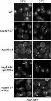
Figure 1.
Figure 1.
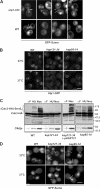
Nuclear import of Siz1-GFP is dependent on Kap95p. The endogenous SIZ1 gene was tagged with GFP in the indicated haploid strains, and the localization of Siz1-GFP was visualized by fluorescence confocal microscopy in cells grown at 23°C or after a 3-h incubation at 37°C. Siz1-GFP localization is also shown in a kap95-14 mutant arrested with nocodazole (Noc) and incubated at the indicated temperatures. Arrows point to the location of the labeled septin rings in representative cells. The arrowheads point to NE-bound Siz1-GFP in the kap95-14 mutant. Bar, 5 μm.
Figure 2.
Figure 2.
Msn5p mediates nuclear export of Siz1-GFP and septin sumoylation. (A) Nuclear export of Siz1p during mitosis requires Msn5p. WT and msn5Δ cells producing Siz1-GFP were arrested in mitosis using nocodazole, and the localization of the GFP fusion was detected by confocal microscopy. Arrows point to the location of the septin ring. (B and C) Septin sumoylation is inhibited in an msn5Δ mutant. (B) The sumoylation state of endogenously tagged Cdc3-HA was evaluated in whole-cell lysates derived from WT and an msn5-null strain (msn5Δ) arrested with nocodazole. Proteins from the indicated strains were analyzed by SDS-PAGE and Western blotting using antibodies directed against HA. The region of the blot containing sumoylated Cdc3-HA is indicated ([Cdc3-HA]-Smt3n). Identical blots were probed with anti-Clb2p antibodies to confirm cell cycle arrest and compare protein levels. The positions of molecular mass markers in this and subsequent figures are show in kilodaltons. (C) The localization pattern of GFP-Sumo was visualized in nocodazole-arrested WT and the msn5-null (msn5Δ) strains using fluorescence confocal microscopy. Bars, 5 μm.
Figure 3.
Figure 3.
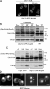
Kap121p is required for septin sumoylation during mitosis. (A) Septin sumoylation is altered in cells expressing the ulp1-333 mutation. Asynchronous cultures of YRW121 (WT) and YRW122 (ulp1-333) cells were grown at 23°C and examined by confocal microscopy. These cells also express GFP-SMT3-ΔC, which encodes mature GFP-Sumo (Wykoff and O'Shea, 2005), bypassing the requirement for Ulp1p-dependent processing. M phase (left) and postcytokinesis mother/daughter pairs (right three columns) are shown. Arrows point to labeled septin rings. (B) Ulp1p localization at the nuclear rim is diminished at the nonpermissive temperature in kap121-34, but not kap95-14, containing mutants. The endogenous ULP1 gene was tagged with GFP in the indicated haploid strains. Strains were grown at 23°C or shifted to 37°C for 3 h, and the localization of Ulp1-GFP was examined by confocal microscopy. (C) Cdc3p sumoylation is inhibited at the nonpermissive temperature in the presence of a kap121-34, but not a kap95-14, mutation. Levels of sumoylated Cdc3-HA in WT, kap121-34, and kap95-14 mutants were detected as in Fig. 2 using anti-HA antibodies. Cells were arrested with α-factor, and G1 phase arrest was confirmed by visual inspection of cell morphology. Cultures were then shifted to 37°C and either maintained in α-factor (αF) or switched to media containing hydroxyurea (HU) or nocodazole (Noc). The region of the blot containing sumoylated Cdc3-HA is indicated ([Cdc3-HA]-Smt3n). Blots were also probed with anti-Clb2p antibodies to confirm cell cycle M phase arrest and compare protein levels. Note that Cdc3-HA sumoylation could be rescued in the kap121-34–containing strain by introducing a plasmid-born copy of KAP121 (pKAP121). (D) GFP-Sumo concentration at the septin ring is inhibited at the nonpermissive temperature in kap121-34, but not kap95-14, mutant cells. WT, kap121-34, and kap95-14 mutants producing GFP-Sumo were arrested in M phase with nocodazole and incubated at 23 and 37°C. The GFP fusion was visualized by confocal microscopy. Arrows point to the positions of septin rings. Bars, 5 μm.
Figure 4.
Figure 4.
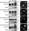
Binding of Ulp1p to kaps is required for septin sumoylation and regulating Ulp1p association with the septin ring. Plasmids containing coding sequences for Ulp1-GFP and various Ulp1p deletion derivatives, including Ulp1Δ1-150-GFP, Ulp1Δ150-340-GFP, and Ulp1Δ340-403-GFP, were introduced into a ulp1Δ deletion mutant expressing CDC3-HA. (A, C, E, and G) Ulp1p deletion mutants lacking kap binding domains alter septin sumoylation. Cells were arrested with α-factor and then either maintained in α-factor (αF) or washed to remove α-factor and incubated in media containing hydroxyurea (HU) or nocodazole (Noc) at 30°C. Levels of Cdc3-HA sumoylation in the indicated strains were detected using anti-HA antibodies as in Fig. 2. The region of the blot containing sumoylated Cdc3-HA is indicated ([Cdc3-HA]-Smt3n). Blots were also probed with anti-Clb2p antibodies to confirm M phase arrest and compare protein levels. (B, D, F, and H) Localization of the various Ulp1-GFP derivatives was visualized in logarithmically growing cells using confocal microscopy. Arrows indicate the localization of Ulp1Δ150-340 to the bud neck. Bar, 5 μm.
Figure 5.
Figure 5.
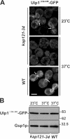
Kap121p is required for the association of Ulp1Δ150-340 with the septin ring. (A) WT and kap121-34 cells were transformed with pULP1Δ150-340-GFP. Logarithmically growing cultures were arrested in M phase with nocodazole. The distribution of Ulp1Δ150-340-GFP was examined by confocal microscopy at 23°C or 2 h after cultures were shifted to 37°C. Arrows indicate the localization of Ulp1Δ150-340 to the bud neck. Bar, 5 μm. (B) Cellular levels of Ulp1Δ150-340-GFP were examined by SDS-PAGE and Western blotting of whole-cell lysates using α-GFP antibodies. Blots were also probed with anti-Gsp1p antibodies to compare protein levels.
Figure 6.
Figure 6.
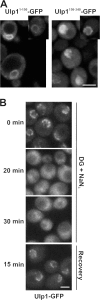
The association of Ulp1p with nuclear pores is energy dependent. (A) The kap binding domains of Ulp1p are targeted to distinct locations. WT cells expressing plasmid-born ULP11-150-GFP or ULP1150-340-GFP were grown to early log phase in selective media, and the distribution of each GFP fusion was examined using confocal microscopy. Individual cells in the culture exhibit different levels of the GFP fusions representative of high and low producers. Insets show cells containing low to moderate levels of the fusion protein. (B) Cells synthesizing Ulp1-GFP (ULP1-GFP integrated at the ULP1 locus) were synchronized in M phase using nocodazole. Cells were washed and placed in media lacking glucose and containing 100 mM 2-deoxyglucose (DG), 10 mM sodium azide (NaN3), and nocodazole. The localization of the GFP fusion was visualized by confocal microscopy after 0, 20, and 30 min of incubation at 30°C. After the 30-min incubation, cells were washed and released into YPD media containing nocodazole (recovery), and the localization of Ulp1-GFP was visualized. Bars, 5 μm.
Figure 7.
Figure 7.
Anchoring of Ulp1p to the nuclear pores inhibits septin desumoylation. WT Ulp1p was replaced by a plasmid encoding the Ulp1C-GFP-Nup60 fusion protein in a strain expressing CDC3-HA (YRW110). (A) YRW110 cells were arrested in M phase using nocodazole and then treated with 100 mM 2-deoxyglucose (DG) and 10 mM sodium azide (NaN3) for the indicated times, as in Fig. 6 B. Cells were examined by confocal microscopy. (B) In YRW110 and an isogenic WT strain, the sumoylation state of Cdc3-HA was evaluated in nocodazole-arrested cells or after treatment with the metabolic poisons for the indicated times. (C) Cdc3-HA sumoylation in Ulp1C-GFP-Nup60– or Ulp1-GFP–containing cells was compared at various points in the cell cycle. Cells were arrested with α-factor, and G1 phase arrest was confirmed by visual inspection of cell morphology. Cultures were then maintained in α-factor (αF) or switched to media containing hydroxyurea (HU) or nocodazole (Noc). Samples were analyzed by Western blotting of whole-cell lysates using anti-HA antibodies. Anti-Clb2p blots were used to confirm cell cycle arrest and compare protein levels. (D) Ulp1C-GFP-Nup60 is restricted to the NE in YRW110 (A), allowing us to examine GFP-Sumo labeling of septins in this strain background (YMR123). Shown are images of postcytokinesis mother/daughter pairs. Arrows point to the positions of labeled septin rings. Bars, 5 μm.
Figure 8.
Figure 8.
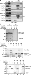
Ulp1p is associated with Kap121p and Nup53p. (A) Kap121p, Kap95p, Kap60p, and Nup53p copurify with Ulp1p. A yeast strain expressing TAP-tagged ULP1 (ULP1-TAP integrated at the ULP1 locus) and KAP95-GFP (pKAP95-GFP) was synchronized in either G1 (with α-factor) or M phase (with nocodazole). Cell extracts were prepared, and the Ulp1-TAP fusion was affinity purified using IgG-Sepharose. Beads were washed with lysis buffer containing 50 mM MgCl2 (W), and Ulp1-TAP–associated proteins were eluted with a step gradient of 200, 500, and 1,000 mM MgCl2. Proteins were analyzed by Western blotting using antibodies directed against Kap121p, Kap60p, Nup53p, and GFP (to detect Kap95p). Lanes L and UB contain a sample of the fraction loaded on the IgG-Sepharose and the unbound fraction, respectively. (B–D) Kap121p, Ulp1p, and Nup53p form a trimeric complex in vitro. GST-Ulp1 (B), GST-Ulp11-150 (C), or GST (D) was bound to glutathione–Sepharose beads. Bound proteins were then incubated with combinations of purified recombinant Kap121p or Nup53p. These proteins do not bind GST alone (D). In addition, purified recombinant Nup53p was incubated with bead-bound complexes of Kap121p–Ulp1p (B, lane 3) or Kap121p–Ulp11-150 (C, lane 5). In each case, the beads were washed extensively and bound proteins were subsequently eluted from the beads using SDS-PAGE sample buffer. Proteins present in bead-bound (B) and unbound (UB) samples were separated by SDS-PAGE and visualized by Coomassie blue staining.
Figure 9.
Figure 9.
Desumoylation of Cdc3-HA after mitosis is delayed in a nup53 mutant. WT cells or a strain containing the nup53-Δ405-430 mutant and expressing CDC3-HA were arrested in M phase with nocodazole (Noc). Cells were then released from arrest and allowed to exit mitosis. At the indicated times, Cdc3-HA sumoylation and Clb2p degradation were evaluated by Western blotting of whole-cells lysates using anti-HA and anti-Clb2p antibodies.

Similar articles

Cited by

References

    1. Boustany, L.M., and M.S. Cyert. 2002. Calcineurin-dependent regulation of Crz1p nuclear export requires Msn5p and a conserved calcineurin docking site. Genes Dev. 16:608–619. - PMC - PubMed
    1. Delorme, E. 1989. Transformation of Saccharomyces cerevisiae by electroporation. Appl. Environ. Microbiol. 55:2242–2246. - PMC - PubMed
    1. Dilworth, D.J., A. Suprapto, J.C. Padovan, B.T. Chait, R.W. Wozniak, M.P. Rout, and J.D. Aitchison. 2001. Nup2p dynamically associates with the distal regions of the yeast nuclear pore complex. J. Cell Biol. 153:1465–1478. - PMC - PubMed
    1. Ghaemmaghami, S., W.K. Huh, K. Bower, R.W. Howson, A. Belle, N. Dephoure, E.K. O'Shea, and J.S. Weissman. 2003. Global analysis of protein expression in yeast. Nature. 425:737–741. - PubMed
    1. Hang, J., and M. Dasso. 2002. Association of the human SUMO-1 protease SENP2 with the nuclear pore. J. Biol. Chem. 277:19961–19966. - PubMed

Publication types

MeSH terms