Role of PD-1 and its ligand, B7-H1, in early fate decisions of CD8 T cells
- PMID: 17392506
- PMCID: PMC1896112
- DOI: 10.1182/blood-2006-12-062422
Role of PD-1 and its ligand, B7-H1, in early fate decisions of CD8 T cells
Abstract
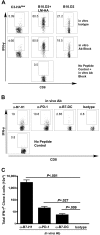
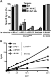
Expression of the PD-1 receptor on T cells has been shown to provide an important inhibitory signal that down-modulates peripheral effector responses in normal tissues and tumors. Furthermore, PD-1 up-regulation on chronically activated T cells can maintain them in a partially reversible inactive state. The function of PD-1 in the very early stages of T-cell response to antigen in vivo has not been fully explored. In this study, we evaluate the role of PD-1 and its 2 B7 family ligands, B7-H1 (PD-L1) and B7-DC (PD-L2), in early fate decisions of CD8 T cells. We show that CD8 T cells specific for influenza hemagglutinin (HA) expressed as a self-antigen become functionally tolerized and express high levels of surface PD-1 by the time of their first cell division. Blockade of PD-1 or B7-H1, but not B7-DC, at the time of self-antigen encounter mitigates tolerance induction and results in CD8 T-cell differentiation into functional cytolytic T lymphocytes (CTLs). These findings demonstrate that, in addition to modulating effector functions in the periphery, B7-H1:PD-1 interactions regulate early T-cell-fate decisions.
Figures


Similar articles
-
Target-dependent B7-H1 regulation contributes to clearance of central nervous system infection and dampens morbidity.J Immunol. 2009 May 1;182(9):5430-8. doi: 10.4049/jimmunol.0803557. J Immunol. 2009. PMID: 19380790 Free PMC article.
-
The programmed death-1 ligand 1:B7-1 pathway restrains diabetogenic effector T cells in vivo.J Immunol. 2011 Aug 1;187(3):1097-105. doi: 10.4049/jimmunol.1003496. Epub 2011 Jun 22. J Immunol. 2011. PMID: 21697456 Free PMC article.
-
Cutting Edge: Programmed death (PD) ligand-1/PD-1 interaction is required for CD8+ T cell tolerance to tissue antigens.J Immunol. 2006 Dec 15;177(12):8291-5. doi: 10.4049/jimmunol.177.12.8291. J Immunol. 2006. PMID: 17142723
-
PD-1 and its ligands in tolerance and immunity.Annu Rev Immunol. 2008;26:677-704. doi: 10.1146/annurev.immunol.26.021607.090331. Annu Rev Immunol. 2008. PMID: 18173375 Free PMC article. Review.
-
The right place at the right time: novel B7 family members regulate effector T cell responses.Curr Opin Immunol. 2002 Jun;14(3):384-90. doi: 10.1016/s0952-7915(02)00342-4. Curr Opin Immunol. 2002. PMID: 11973139 Review.
Cited by
-
Immune microenvironment in papillary thyroid carcinoma: roles of immune cells and checkpoints in disease progression and therapeutic implications.Front Immunol. 2024 Sep 3;15:1438235. doi: 10.3389/fimmu.2024.1438235. eCollection 2024. Front Immunol. 2024. PMID: 39290709 Free PMC article. Review.
-
From foes to friends: rethinking the role of lymph nodes in prostate cancer.Nat Rev Urol. 2024 Nov;21(11):687-700. doi: 10.1038/s41585-024-00912-9. Epub 2024 Aug 2. Nat Rev Urol. 2024. PMID: 39095580 Review.
-
Advancements in nanomedicine delivery systems: unraveling immune regulation strategies for tumor immunotherapy.Nanomedicine (Lond). 2024;19(21-22):1821-1840. doi: 10.1080/17435889.2024.2374230. Epub 2024 Jul 16. Nanomedicine (Lond). 2024. PMID: 39011582 Review.
-
Efficacy of neoadjuvant chemotherapy combined with surgery in patients with nonsmall cell lung cancer: A meta-analysis.Clin Respir J. 2024 May;18(5):e13756. doi: 10.1111/crj.13756. Clin Respir J. 2024. PMID: 38725310 Free PMC article.
-
Unraveling the Role of Epithelial Cells in the Development of Chronic Rhinosinusitis.Int J Mol Sci. 2023 Sep 18;24(18):14229. doi: 10.3390/ijms241814229. Int J Mol Sci. 2023. PMID: 37762530 Free PMC article. Review.
References
-
- Greenwald RJ, Freeman GJ, Sharpe AH. The B7 family revisited. Annu Rev Immunol. 2005;23:515–548. - PubMed
-
- Zhu B, Guleria I, Khosroshahi A, et al. Differential role of programmed death-ligand 1 [corrected] and programmed death-ligand 2 [corrected] in regulating the susceptibility and chronic progression of experimental autoimmune encephalomyelitis [published erratum appears in J Immunol. 2006;176:5683]. J Immunol. 2006;176:3480–3489. - PubMed
-
- Curiel TJ, Wei S, Dong H, et al. Blockade of B7-H1 improves myeloid dendritic cell-mediated antitumor immunity. Nat Med. 2003;9:562–567. - PubMed
-
- Hirano F, Kaneko K, Tamura H, et al. Blockade of B7-H1 and PD-1 by monoclonal antibodies potentiates cancer therapeutic immunity. Cancer Res. 2005;65:1089–1096. - PubMed
Publication types
MeSH terms
Substances
LinkOut - more resources
Full Text Sources
Other Literature Sources
Research Materials

