. 2007 Jun;81(11):5788-806.
doi: 10.1128/JVI.00140-07. Epub 2007 Mar 28.

Kaposi's sarcoma-associated herpesvirus/human herpesvirus 8 ORF50/Rta lytic switch protein functions as a tetramer

Affiliations

Kaposi's sarcoma-associated herpesvirus/human herpesvirus 8 ORF50/Rta lytic switch protein functions as a tetramer

Wei Bu et al. J Virol. 2007 Jun.

Abstract

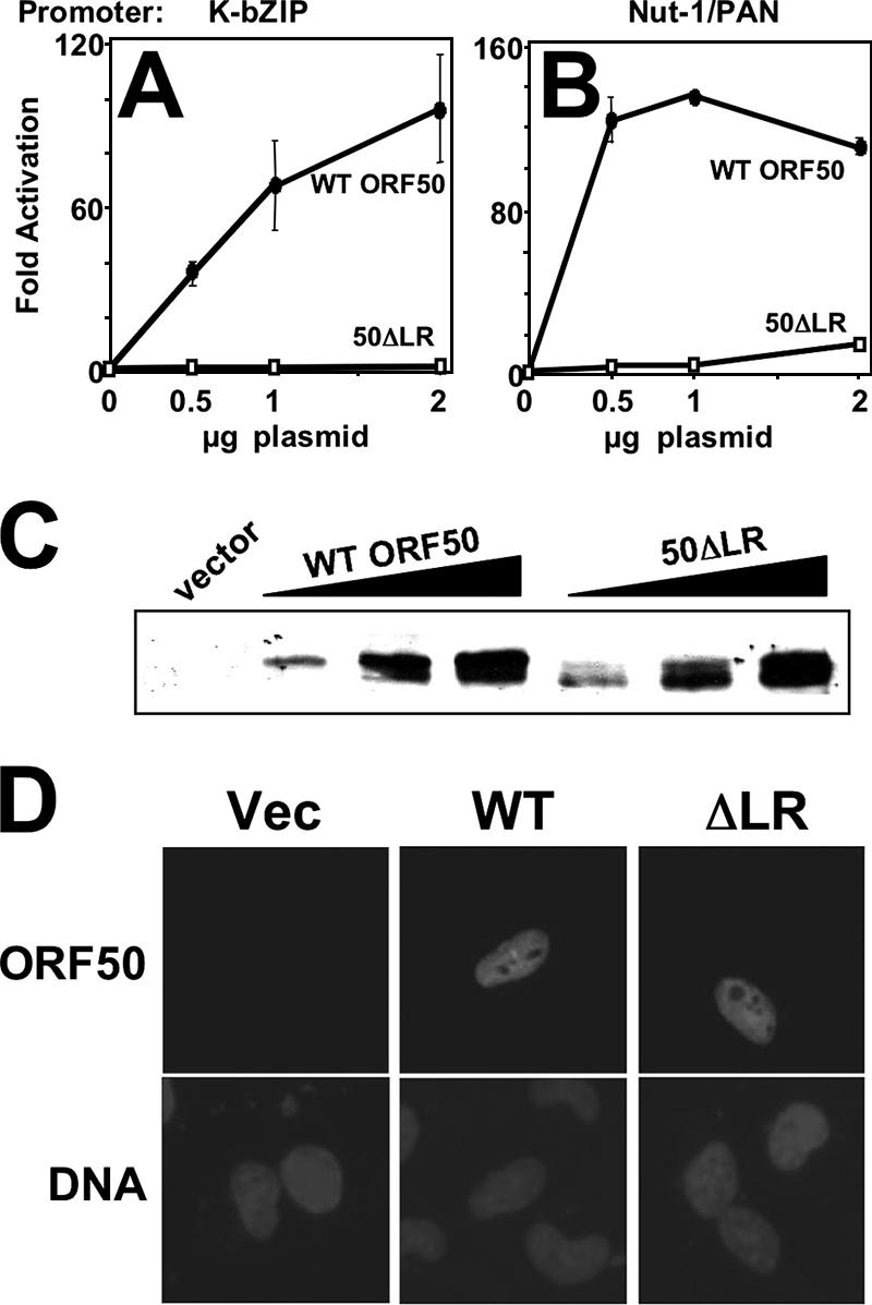
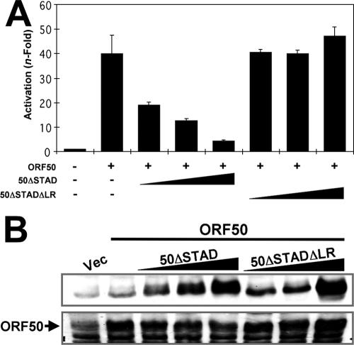
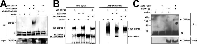
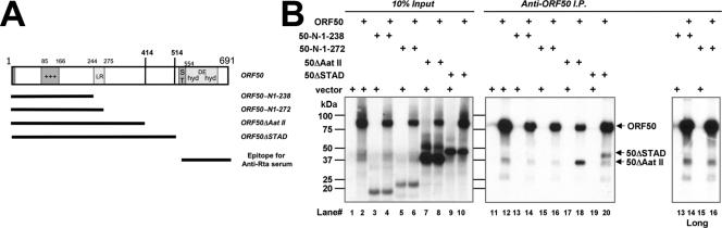
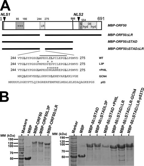
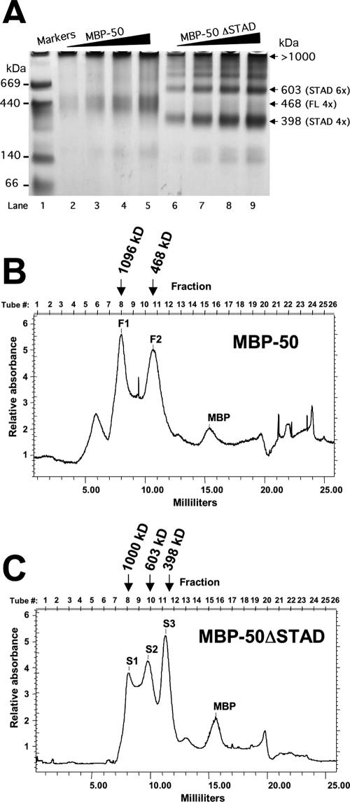
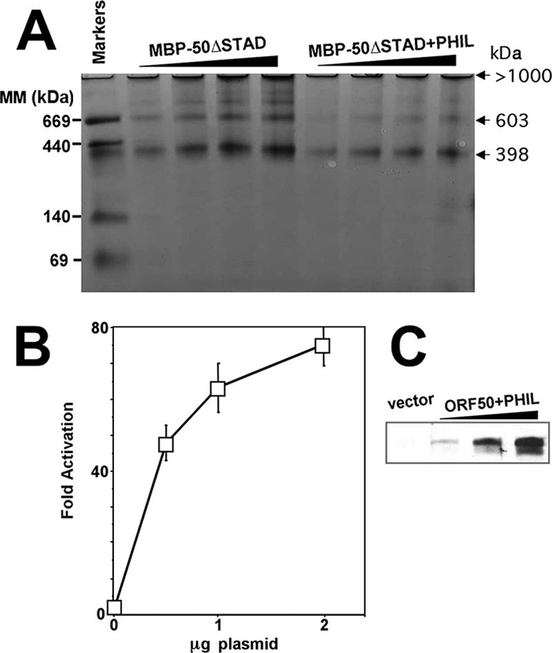
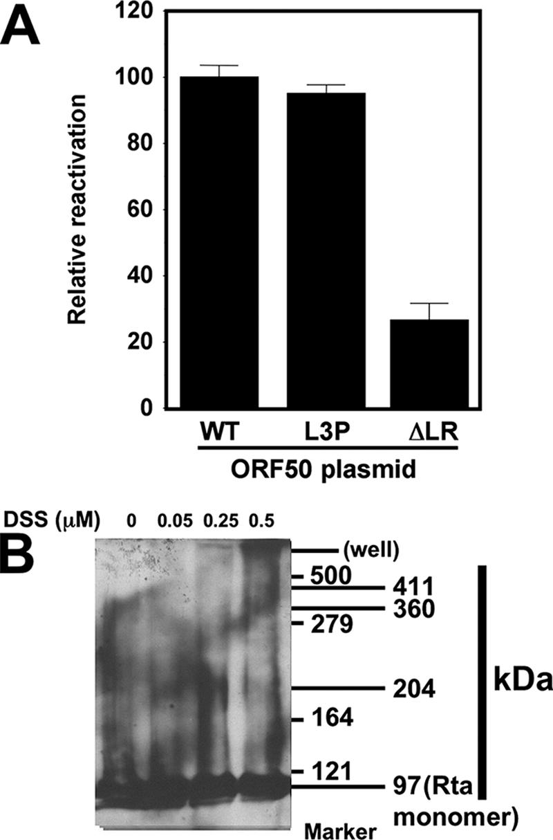
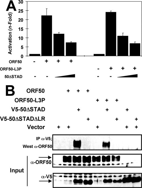
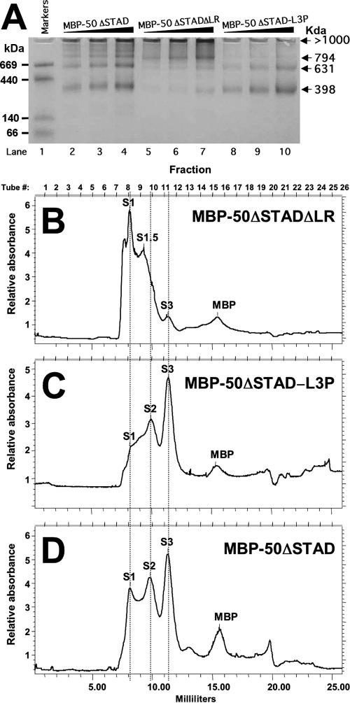
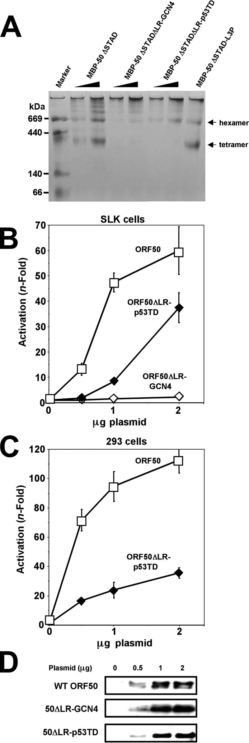
The Kaposi's sarcoma-associated herpesvirus open reading frame 50 (ORF50) protein (called Rta), is necessary and sufficient for reactivation of the virus from latency. We previously demonstrated that a truncated mutant of ORF50 lacking its C-terminal transcriptional activation domain, called ORF50DeltaSTAD, formed mixed multimers with wild-type (WT) ORF50 and functioned as a dominant negative inhibitor of reactivation. For this report, we investigated the requirements for multimerization of ORF50/Rta in transactivation and viral reactivation. We analyzed multimerization of WT, mutant, and chimeric ORF50 proteins, using Blue Native polyacrylamide gel electrophoresis and size exclusion chromatography. WT and mutant ORF50 proteins form tetramers and higher-order multimers, but not monomers, in solution. The proline-rich, N-terminal leucine heptapeptide repeat (LR) of ORF50 (amino acids [aa] 244 to 275) is necessary but not sufficient for oligomer formation and functions in concert with the central portion of ORF50/Rta (aa 245 to 414). The dominant negative mutant ORF50DeltaSTAD requires the LR to form mixed multimers with WT ORF50 and inhibit its function. In the context of the WT ORF50/Rta protein, mutagenesis of the LR, or replacement of the LR by heterologous multimerization domains from the GCN4 or p53 proteins, demonstrates that tetramers of Rta are sufficient for transactivation and viral reactivation. Mutants of Rta that are unable to form tetramers but retain the ability to form higher-order multimers are reduced in function or are nonfunctional. We concluded that the proline content, but not the leucine content, of the LR is critical for determining the oligomeric state of Rta.


Figures

FIG. 1.
FIG. 1.
Primary amino acid sequence of KSHV ORF50/Rta protein. (A) Structure/function map of ORF50/Rta. A schematic of the primary amino acid sequence of the FL ORF50/Rta protein is shown. The numbers on the diagram refer to amino acid positions; the numbers to the right in parentheses indicate references. The bars with arrowheads indicate the positions of single-amino acid mutations that eliminate ubiquitin E3 ligase activity and binding to C/EBPα and DNA. The bars underneath ORF50 represent domains with functions identified in the list at the right. See the text for the relevant references. Abbreviations: NLS, nuclear localization sequence; AatII, position of truncation of cDNA by restriction digestion by the AatII enzyme; ST, serine/threonine-rich sequence; hyd, hydrophobic; DE, acidic amino acid-rich; HDAC1, histone deacetylase 1. Symbols: *, location of single-amino-acid mutation that eliminates binding to C/EBPα; +++, basic amino acid-rich sequence. (B) Alignment of KSHV ORF50/Rta LR with other gammaherpesvirus ORF50 proteins. Numbers refer to the animo acid position. Arrows point to leucines in the heptapeptide repeat. White lettering on a black background indicates amino acid identity. The bar under the viral sequences indicates the core of highest identity among all the indicated proteins. “heptad” indicates the lettering of canonical amino acid positions in heptad repeats. Abbreviations: M. mulatta, Macaca mulatta (either RRV/H26-95 or RRV/17577 isolates); M. fuscata, Macaca fuscata (GenBank accession number NC_007016) (S. G. Hansen, N. Avery, M. K. Axthelm, and S. W. Wong, unpublished data); HVS, herpesvirus saimiri; MHV-68, murine gammaherpesvirus 68; EHV-2, equine herpesvirus 2; AHV-1, alcelaphine herpesvirus 1; EBV, Epstein-Barr virus; GCN4, S. cerevisiae GCN4 leucine zipper.
FIG. 2.
FIG. 2.
The LR is required for KSHV Rta to activate transcription. Human endothelial SLK cells were cotransfected with the indicated amounts of vector expressing WT ORF50 or ORF50ΔLR and a luciferase reporter vector for the K-bZIP (A) or Nut-1/PAN (B) promoter. Total DNA was normalized by the addition of empty expression vectors. The cells were harvested and lysed at 40 h posttransfection. Luciferase activity was normalized to β-galactosidase activity, and n-fold activation is given relative to that of the reporter vector transfected alone (0 μg plasmid). Vertical bars indicate standard deviations. Data represent results of triplicate transfection experiments repeated at least twice. See Materials and Methods for details. (C) Equivalent amounts of WT ORF50 and ORF50ΔLR were expressed in transfected SLK cells. The extracts used for the experiments shown in panel A were analyzed by SDS-PAGE/Western blotting, using anti-Rta serum. (D) WT ORF50 and ORF50ΔLR are both expressed in SLK cell nuclei. SLK cells were transfected as for panel A and analyzed by immunofluorescence 40 h posttransfection, using anti-Rta primary serum, fluorescein isothiocyanate-conjugated secondary serum, and DAPI (4′,6′-diamidino-2-phenylindole) to stain nuclear DNA. Vec, vector.
FIG. 3.
FIG. 3.
Deletion of the LR does not eliminate interaction of ORF50/Rta with RBP-Jk or promoter DNA. (A) GST-RBP-Jk or the GST moiety alone was purified and tested for binding to RRLs programmed with plasmids expressing the indicated proteins. The proteins were labeled cotranslationally by addition of L-[35S]-methionine to the RRLs. Bound proteins were visualized by autoradiography after being displayed by SDS-PAGE. Five percent of the input protein was also analyzed as a size reference. (B) WT ORF50 (50WT) or ORF50ΔLR (50ΔLR) was expressed and purified as a fusion to the MBP and tested in increasing amounts for binding to 32P-labeled oligos of the Rta binding sites from the indicated promoters. 0, lanes containing labeled DNA mixed with buffer in the absence of protein.
FIG. 4.
FIG. 4.
The LR is required for ORF50ΔSTAD to function as a dominant negative transcriptional inhibitor of WT ORF50/Rta. (A) SLK cells were cotransfected with the indicated plasmids. The cells were harvested, and extracts were analyzed as for Fig. 2. (B) Top panel, equivalent amounts of V5-ORF50ΔSTAD and V5-ORF50ΔSTADΔLR were expressed in transfected SLK cells. Bottom panel, WT ORF50 expression levels did not change in the presence of ORF50ΔSTAD or ORF50ΔSTADΔLR. Extracts from the transfection experiments for panel A were analyzed by SDS-PAGE/Western blotting, using anti-V5 primary serum (top panel) or anti-Rta primary serum (bottom panel). The bands seen in the vector-only lane (Vec) represent background bands in the cell extracts.
FIG. 5.
FIG. 5.
The LR is required for ORF50ΔSTAD to interact with WT ORF50/Rta. (A) 293 cells were cotransfected with the indicated plasmids, and total cellular extracts were prepared 48 h posttransfection. Equal amounts of protein were immunoprecipitated, using anti-V5 serum, and the proteins were analyzed by SDS-PAGE/Western blotting, using anti-Rta primary serum. Ten percent of the total cell extract for each immunoprecipitation was analyzed for the input on the Western blot (Input). (B) RRLs were programmed with the indicated plasmids in the presence of [35S]methionine to label synthesized proteins. Equal amounts of RRL were immunoprecipitated, using anti-Rta serum, and the proteins were analyzed by SDS-PAGE/autoradiography. Ten percent of the RRL for each immunoprecipitation was analyzed for the input lanes (10% Input). (C) 293 cells were cotransfected with the indicated plasmids, and immunoprecipitation was performed as for panel A, using anti-Gal4 primary antibody. Western blotting was performed using anti-V5 primary antibody. I.P., immunoprecipitation; Ig, immunoglobulin band; +, transfected plasmid.
FIG. 6.
FIG. 6.
The LR is necessary but not sufficient for homomultimerization of ORF50/Rta. (A) Schematic of ORF50-truncation proteins. The numbers refer to amino acids, and the bars underneath ORF50 represent sequences included in the ORF50-truncated mutants identified in the list at the right. The bottom bar indicates the region of ORF50/Rta used as antigen for generation of the Rta-specific antiserum (53) used for the experiments for panel B. ST, serine/threonine-rich sequence; hyd, hydrophobic; DE, acidic amino acid-rich; +++, basic amino acid-rich sequence. (B) Immunoprecipitation of RRLs. RRLs were programmed with the indicated plasmids and analyzed as for Fig. 5B. The panel to the far right shows a longer exposure for lanes 13 to 16. I.P., immunoprecipitation; +, transfected plasmid.
FIG. 7.
FIG. 7.
Design and expression of MBP fusions of ORF50. (A) Schematic of ORF50/Rta constructs. The numbers refer to amino acids, and the bars underneath ORF50 represent sequences included in the ORF50 mutants identified in the list at the right. The four amino acid sequences at the bottom were inserted at the LR (aa 244 to 275) in the WT, L3P mutant, or chimeric (GCN4 or p53) proteins. ST, serine/threonine-rich sequence; hyd, hydrophobic; DE, acidic amino acid-rich; +++, basic amino acid-rich sequence; *, amino acid position of the mutations in chimeric proteins. (B) MBP fusions of ORF50 stained with Gel Code Blue stain (Pierce). The indicated proteins were expressed and purified from E. coli, as described in Materials and Methods. Equal amounts of protein were displayed by SDS-PAGE, and the gels were fixed and stained with Gel Code Blue (Pierce).
FIG. 8.
FIG. 8.
Multimerization of MBP-ORF50 and MBP-50ΔSTAD. (A) BN-PAGE. One to three micrograms of MBP-50 or MBP-50ΔSTAD was analyzed by BN-PAGE on 4% to 12% Bis-Tris gradient gels, as described in Materials and Methods. When the dye front reached the bottom, the gel was destained and analyzed by capturing a digital image in visible light. The migration position of the molecular mass markers is shown at the left. The numbered arrows on the right show migration positions of the MBP fusion protein complexes; apparent molecular masses were determined by gel exclusion chromatography as for panels B and C. (B and C) Gel filtration chromatography. MBP-50 (B) or MBP-50ΔSTAD (C) was separated by gel filtration chromatography, using a Superdex200 HR 10/30 column, as described in Materials and Methods. Fractions (0.5 ml) were collected, and protein content was measured by optical density at 280 nm. The peaks are labeled F1 and F2 for full-length ORF50 and S1, S2, and S3 for ORF50ΔSTAD. The molecular mass of each peak is shown above each elution profile; the molecular mass was determined by comparison to the elution profile of thymoglobulin, ferritin, and catalase (Table 1). MBP fusion proteins, or MBP alone, was identified in the indicated peaks by Western blotting, using anti-MBP antibody (data not shown).
FIG. 9.
FIG. 9.
The MBP-50-L3P mutant primarily forms tetramers in solution. MBP-50-L3P (A) and MBP-ORF50 (B) were analyzed by gel filtration chromatography as described in the legend to Fig. 8. ORF50 and ORF50-L3P were analyzed for transactivation of the K-bZIP promoter (C) and expression (D) as described for Fig. 2.
FIG. 10.
FIG. 10.
ORF50+PHIL functions with WT ORF50 transcriptional activity. (A) MBP-50ΔSTAD and MBP-50ΔSTAD+PHIL were analyzed by BN-PAGE as described for Fig. 8A. (B) ORF50+PHIL transactivates transcription with WT ORF50 activity. The indicated plasmids were cotransfected into SLK cells with the K-bZIP/RAP reporter plasmid, extracts were generated, and transactivation was analyzed as described in the legend to Fig. 2A. (C) Equivalent amounts of WT ORF50 and ORF50+PHIL were expressed in transfected SLK cells. Extracts from the experiments for panel A were analyzed by SDS-PAGE/Western blotting, using anti-Rta serum.
FIG. 11.
FIG. 11.
Tetramers of ORF50/Rta are sufficient to reactivate KSHV from latency. (A) BCBL-1 cells were electroporated in duplicate with plasmids expressing the indicated proteins and analyzed for reactivation by indirect immunofluorescence 72 h later. The cells were scored by fluorescent microscopic visualization as the percentage of Rta single-positive cells that also expressed K8.1 (Rta/K8.1 double-positive cells; no cells expressed K8.1 in the absence of Rta). To eliminate spontaneously reactivating cells, the identical calculation was performed on cells electroporated with an empty control vector, pcDNA3.1; that percentage was subtracted from the values for all other transfections. Those corrected percentages were then normalized to that of WT ORF50-transfected cells, which was adjusted to 100%. (B) Total cellular extracts from uninduced or TPA-induced (24 h) BCBL-1 cells were treated with the cross-linker DSS and then analyzed by SDS-PAGE/Western blotting, using Rta-specific antiserum as the primary antibody. The first column of numbers on the right shows the migration positions of the protein molecular mass markers. The second column shows the migration positions of Rta-containing complexes.
FIG. 12.
FIG. 12.
ORF50ΔSTAD inhibits ORF50-L3P transactivation using a mechanism similar to that of WT ORF50. (A) ORF50ΔSTAD inhibits ORF50-L3P transactivation in a dominant negative fashion. SLK cells were cotransfected with the indicated plasmids. The cells were harvested, and extracts were analyzed as described in the legend to Fig. 2. (B) ORF50ΔSTAD binds directly to ORF50-L3P in transfected cells. 293 cells were cotransfected with the indicated plasmids, and total cellular extracts were prepared 48 h posttransfection. Equal amounts of protein were immunoprecipitated, using anti-V5 serum, and the proteins were analyzed by SDS-PAGE/Western blotting, using anti-Rta primary serum. Ten percent of the total cell extract for each immunoprecipitation was analyzed for the input Western blots (Input). Antibodies used for the input Western blots are indicated to the left of the bottom panels. IP, immunoprecipitation.
FIG. 13.
FIG. 13.
The LR is required for ORF50 to form tetramers. The indicated proteins were analyzed by BN-PAGE (A) or gel filtration chromatography (B, C, and D) as described in the legend to Fig. 8.
FIG. 14.
FIG. 14.
Tetramerization is required for full ORF50 transcriptional activity. (A) Multimerization of ORF50 chimeric proteins. The indicated proteins were analyzed by BN-PAGE as described in the legend to Fig. 8A. The arrows to the right indicate migration of tetramers and hexamers of MBP-50ΔSTAD and MBP50ΔSTADΔLR, which were included as molecular mass controls. (B and C) Transactivation by ORF50 decreases as the extent of multimerization increases. SLK (B) and 293 (C) cells were transfected with the indicated amounts of plasmids together with the K-bZIP/RAP reporter vector. Extracts were prepared and analyzed as described in the legend to Fig. 2. (D) Equivalent amounts of WT ORF50, 50ΔLR-GCN4, and 50ΔLR-p53TD were expressed in SLK cells. Extracts from the transfection experiment for panel B were analyzed by SDS-PAGE/Western blotting, using anti-Rta primary serum.

Similar articles

Cited by

References

    1. Ambroziak, J., D. Blackbourn, B. Herndier, R. Glogan, J. Gullet, A. McDonald, E. Lennette, and J. Levy. 1995. Herpesvirus-like sequences in HIV-infected and uninfected Kaposi's sarcoma patients. Science 268:582-583. - PubMed
    1. Baker, S. J., S. Markowitz, E. R. Fearon, J. K. Willson, and B. Vogelstein. 1990. Suppression of human colorectal carcinoma cell growth by wild-type p53. Science 249:912-915. - PubMed
    1. Barlow, D. J., and J. M. Thornton. 1988. Helix geometry in proteins. J. Mol. Biol. 201:601-619. - PubMed
    1. Berra, E., A. Ginouves, and J. Pouyssegur. 2006. The hypoxia-inducible-factor hydroxylases bring fresh air into hypoxia signalling. EMBO Rep. 7:41-45. - PMC - PubMed
    1. Boneschi, V., L. Brambilla, E. Berti, S. Ferrucci, M. Corbellino, C. Parravicini, and S. Fossati. 2001. Human herpesvirus 8 DNA in the skin and blood of patients with Mediterranean Kaposi's sarcoma: clinical correlations. Dermatology 203:19-23. - PubMed

Publication types

MeSH terms