. 2007 Mar 28;2(3):e328.
doi: 10.1371/journal.pone.0000328.

Ultrastructural characterization of the giant volcano-like virus factory of Acanthamoeba polyphaga Mimivirus

Affiliations

Ultrastructural characterization of the giant volcano-like virus factory of Acanthamoeba polyphaga Mimivirus

Marie Suzan-Monti et al. PLoS One. .

Abstract

Acanthamoeba polyphaga Mimivirus is a giant double-stranded DNA virus defining a new genus, the Mimiviridae, among the Nucleo-Cytoplasmic Large DNA Viruses (NCLDV). We used utrastructural studies to shed light on the different steps of the Mimivirus replication cycle: entry via phagocytosis, release of viral DNA into the cell cytoplasm through fusion of viral and vacuolar membranes, and finally viral morphogenesis in an extraordinary giant cytoplasmic virus factory (VF). Fluorescent staining of the AT-rich Mimivirus DNA showed that it enters the host nucleus prior to the generation of a cytoplasmic independent replication centre that forms the core of the VF. Assembly and filling of viral capsids were observed within the replication centre, before release into the cell cytoplasm where progeny virions accumulated. 3D reconstruction from fluorescent and differential contrast interference images revealed the VF emerging from the cell surface as a volcano-like structure. Its size dramatically grew during the 24 h infectious lytic cycle. Our results showed that Mimivirus replication is an extremely efficient process that results from a rapid takeover of cellular machinery, and takes place in a unique and autonomous giant assembly centre, leading to the release of a large number of complex virions through amoebal lysis.


Conflict of interest statement

Competing Interests: The authors have declared that no competing interests exist.

Figures

Figure 1
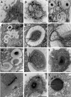
Figure 1. Ultrastructural aspects of the early steps of Mimivirus replication cycle.
Transmission electron microscopy pictures were taken at 0 h p.i. (A–C) or at 4 h p.i. (D–L). (A) Mimivirus particle being phagocytosed by an amoeba; bar = 2 µm. (B) Several single viral particles within intra-cytoplasmic vacuoles; bar = 2 µm. (C) Higher magnification of the boxed area in B showed the open vertex of an empty particle (arrow); bar = 1 µm. (D) Close contact of the membranes of two vacuoles (arrow), one with several Mimivirus particles and the other with a single viral particle; bars = 1 µm. (E) Extrusion of the internal Mimivirus membrane toward the vacuole membrane; bar = 200 nm. (F) Higher magnification of the contact zone between viral and vacuole membranes (arrow); bar = 100 nm. (G) Full closed, empty with open vertex (arrow) and opening Mimivirus particles; bar = 500 nm. (H) Higher magnification of the opening Mimivirus particle in G. The fused viral and vacuole membranes were clearly visible (arrow); bar = 100 nm. (I) Condensed electron dense material inside the cell nucleus (arrow); bar = 2 µm. (J) Higher magnification of the condensed electron dense material between the nuclear membrane and the nucleolus (arrow); bar = 500 nm. (K) An electron dense structure (arrow), distinct from the cell nucleus was observed; bar = 5 µm. (L) Higher magnification of this heterogeneous structure, surrounded with mitochondria; bar = 200 nm.
Figure 2
Figure 2. Ultrastructural aspects of the late steps of Mimivirus replication cycle - capsid assembly.
(A) At 8 h p.i. the virus factory (VF) appeared composed of a dense replication centre surrounded by new viral particles. Nu : cell nucleus; bar = 5 µm. (B) At 12 h p.i. the cell cytoplasm was filled with newly synthesised viruses. The cell nucleus (Nu) was expelled to the periphery; bar = 3 µm. (C–G) Pictures were taken at 8, 12 or 16 h p.i. (C) Different stages of viral particles morphogenesis from the replication centre : beginning of hexagonal capsid assembly (white and black arrowheads); complete empty capsid (thick closed black arrow); filling of empty capsids with condensed electron dense material (thick open black and white arrows); release of full closed viral particles surrounded by fibrils at the periphery of the virus factory; bar = 500 nm. (D–G) Different aspects of viral capsid assembly : beginning of capsid assembly (D bar = 500 nm; G bar = 100 nm); almost complete capsids detaching from the replication center (E bar = 100 nm; F bar = 200 nm); complete capsid being filled with electron dense material (F) or complete viral particle without fibril (G). Membranes were observed beneath the capsid layer (F, G long black arrow). The vertex (small black arrows) was on the external side, opposite to the attachment and filling side (C–G).
Figure 3
Figure 3. Ultrastructural aspects of the late steps of Mimivirus replication cycle – encapsidation of viral DNA.
Pictures were taken at 8, 12 or 16 h p.i. (A) Different stages of capsid assembly and DNA encapsidation : complete empty capsid (arrowhead); progressive stages of viral DNA insertion (black arrows) through a portal opposite to the vertex (white arrows); bar = 500 nm. (B) Viral DNA insertion into a capsid (arrowhead) and two different detaching steps of full complete viruses from the replication centre (arrows); bar = 500 nm. (C) Higher magnification of the complete viruses seen in B; bar = 200 nm; (D) Insertion of condensed viral DNA into a viral capsid; bar = 200 nm.
Figure 4
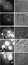
Figure 4. Mimivirus infectious cycle.
Mimivirus infected A. polyphaga were stained with DAPI at different times points p.i. and representative pictures are shown. A, B : non infected amoebae; C, D: 0 h p.i. Mimivirus particles inside the cytoplasm and near the cell nucleus could be seen; E, F: 4 h p.i. The heterogeneous structure of the VF appeared near the cell nucleus. No viral particles were detectable in the cytoplasm; G, H : 8 h p.i. The intensively stained VF appeared as an homogeneous structure and neosynthesized viral particles accumulated around the VF; I, J : 18 h p.i. The VF was still intensely stained with quite a different structure, whereas the cell cytoplasm was completely filled with new viral particles. Fluorescence (left column) and DIC (right column) images of the same slide field were taken with a 63×/1.4 oil lens. Fluorescence pictures were taken with an exposure time of 1 sec (A) and 64 msec with gain 2 (C, E, G, I). Inset pictures corresponded to the same as E, G and I taken at a different exposure time 64 msec (E, G) and 16 msec respectively (I).
Figure 5
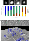
Figure 5. Evolution and 3D reconstruction of Mimivirus factory.
A: Characterisation and quantification of the different types of replication centre. Fluorescence pictures were taken at 4 h (I, exposure time 32 msec), 8 h (II and III, exposure time 64 and 128 msec respectively) and 16 h (IV, exposure time 32 msec) p.i. Bar = 10 µm. Histogram : a total of 717 DAPI-stained cells were analysed to quantify the proportion of each replication centre time at the indicated time points. B: DIC pictures of an APM infected A. polyphaga at 8 h p.i. taken with a 63×/1.4 oil lens. Different sections according to the depth of focus are shown downward (upper part, from a to d). Bar = 10 µm. The lower part represented a 3D reconstruction combining DIC and DAPI staining of the VF present in different infected cells of a microscope field. The dotted line box framed the cell analysed in the upper part. Blue : DAPI-staining.
Figure 6
Figure 6. Quantification of the kinetics of Mimivirus factory formation.
(A) Fluorescence picture of a representative field from A. polyphaga at 5 h p.i., stained with DAPI (40× magnification/0.7 lens). The nuclei are marked around their edge with a blue line to allow quantification of their areas compared to those of the Mimivirus factories. (B) Quantification curve showing the normalized parameter (y-axis) as a function of time p.i. (x-axis). Red line: nuclei intensity; green line: nuclei surface fraction; blue line: Mimivirus factory surface fraction. Evolution of the mean fluorescence intensity over time p.i. in cell nuclei (C) or in virus factories (D), at 100 and 4 msec exposure time respectively, using R software; *** = p<0.01 (Wilcoxon test).
Figure 7
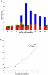
Figure 7. Increase in total cellular DNA content in A. polyphaga during the Mimivirus infection cycle.
A. Estimation of the total amount of cellular DNA by fluorescence intensity quantification of DAPI staining in Mimivirus infected (bars) or uninfected amoebae (hatched bars). Total intensity = staining area (pixels) × mean intensity (intensity/pixel). The total intensity at different time points was divided by the intensity at time = 0 h p.i. for normalization. The bar height represents the variation of DNA content compared to the t0 timepoint for different cellular compartments. Red bar: nucleus; green bar: cytoplasm; blue bar: virus factory. B. The increase of total cellular DNA was extrapolated from 0 to 8 h p.i. and fitted with the exponential equation: y = A0.e(Rx). Data showed a high predictability with the exponential growth model (R2 = 0.94).
Figure 8
Figure 8. Schematic representation of APM replication cycle.
Mimivirus entry through a phagocytic vacuole (1). Fusion of phagocytic vacuoles (2) and delivery of Mimivirus genetic material into the cell cytoplasm (3). Mimivirus DNA entry into the host nucleus (3), where the first round of DNA replication might begin (4). At 3 h p.i. Mimivirus DNA came out the host nucleus to form the VF replication centre (5). At 5 h p.i. the VF size showed a 50% increase and viral proteins began to be detected. Proviral capsid assembly and viral capsids budding from the VF central core could be observed (6). Empty or DNA filled capsids accumulated nearby the central core, resulting in a growing VF with viral particles free in the cytoplasm (7). Complete viral capsids surrounded by fibrils might be released through cell lysis (8).

Similar articles

Cited by

References

    1. La Scola B, Audic S, Robert C, Jungang L, de Lamballerie X, et al. A giant virus in amoebae. Science. 2003;299:2033. - PubMed
    1. Suzan-Monti M, La Scola B, Raoult D. Genomic and evolutionary aspects of Mimivirus. Virus Res. 2006;117:145–155. - PubMed
    1. Xiao C, Chipman PR, Battisti AJ, Bowman VD, Renesto P, et al. Cryo-electron microscopy of the giant Mimivirus. J Mol Biol. 2005;353:493–496. - PubMed
    1. Raoult D, Audic S, Robert C, Abergel C, Renesto P, et al. The 1.2-megabase genome sequence of Mimivirus. Science. 2004;306:1344–1350. - PubMed
    1. Iyer LM, Aravind L, Koonin EV. Common origin of four diverse families of large eukaryotic DNA viruses. J Virol. 2001;75:11720–11734. - PMC - PubMed

Publication types