Monoclonal antibodies capable of discriminating the human inhibitory Fcgamma-receptor IIB (CD32B) from the activating Fcgamma-receptor IIA (CD32A): biochemical, biological and functional characterization
- PMID: 17386079
- PMCID: PMC2265948
- DOI: 10.1111/j.1365-2567.2007.02588.x
Monoclonal antibodies capable of discriminating the human inhibitory Fcgamma-receptor IIB (CD32B) from the activating Fcgamma-receptor IIA (CD32A): biochemical, biological and functional characterization
Abstract
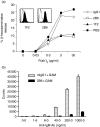
Human CD32B (FcgammaRIIB), the low-affinity inhibitory Fcgamma receptor (FcgammaR), is highly homologous in its extracellular domain to CD32A (FcgammaRIIA), an activating FcgammaR. Available monoclonal antibodies (mAb) against the extracellular region of CD32B recognize both receptors. Through immunization of mice transgenic for human CD32A, we generated a set of antibodies specific for the extracellular region of CD32B with no cross-reactivity with CD32A, as determined by enzyme-linked immunosorbent assay and surface plasmon resonance with recombinant CD32A and CD32B, and by fluorescence-activated cell sorting analysis of CD32 transfectants. A high-affinity mAb, 2B6, was used to explore the expression of CD32B by human peripheral blood leucocytes. While all B lymphocytes expressed CD32B, only a fraction of monocytes and almost no polymorphonuclear cells stained with 2B6. Likewise, natural killer cells, which express CD32C, a third CD32 variant, did not react with 2B6. Immune complexes co-engage the inhibitory receptor with activating Fcgamma receptors, a mechanism that limits cell responses. 2B6 competed for immune complex binding to CD32B as a monomeric Fab, suggesting that it directly recognizes the Fc-binding region of the receptor. Furthermore, when co-ligated with an activating receptor, 2B6 triggered CD32B-mediated inhibitory signalling, resulting in diminished release of inflammatory mediators by FcepsilonRI in an in vitro allergy model or decreased proliferation of human B cells induced by B-cell receptor stimulation. These antibodies form the basis for the development of investigational tools and therapeutics with multiple potential applications, ranging from adjuvants in FcgammaR-mediated responses to the treatment of allergy and autoimmunity.
Figures


Similar articles
-
Fc optimization of therapeutic antibodies enhances their ability to kill tumor cells in vitro and controls tumor expansion in vivo via low-affinity activating Fcgamma receptors.Cancer Res. 2007 Sep 15;67(18):8882-90. doi: 10.1158/0008-5472.CAN-07-0696. Cancer Res. 2007. PMID: 17875730
-
Therapeutic control of B cell activation via recruitment of Fcgamma receptor IIb (CD32B) inhibitory function with a novel bispecific antibody scaffold.Arthritis Rheum. 2010 Jul;62(7):1933-43. doi: 10.1002/art.27477. Arthritis Rheum. 2010. PMID: 20506263
-
A shift in the balance of inhibitory and activating Fcgamma receptors on monocytes toward the inhibitory Fcgamma receptor IIb is associated with prevention of monocyte activation in rheumatoid arthritis.Arthritis Rheum. 2004 Dec;50(12):3878-87. doi: 10.1002/art.20672. Arthritis Rheum. 2004. PMID: 15593228
-
FcgammaR: The key to optimize therapeutic antibodies?Crit Rev Oncol Hematol. 2007 Apr;62(1):26-33. doi: 10.1016/j.critrevonc.2006.12.003. Epub 2007 Jan 19. Crit Rev Oncol Hematol. 2007. PMID: 17240158 Review.
-
A role of FcgammaRIIB in the development of collagen-induced arthritis.Biomed Pharmacother. 2004 Jun;58(5):292-8. doi: 10.1016/j.biopha.2004.04.005. Biomed Pharmacother. 2004. PMID: 15194165 Review.
Cited by
-
Neutropenia in Cynomolgus Monkeys With Anti-Drug Antibodies Associated With Administration of Afucosylated Humanized Monoclonal Antibodies.Toxicol Pathol. 2022 Dec;50(8):910-919. doi: 10.1177/01926233221131510. Epub 2022 Nov 3. Toxicol Pathol. 2022. PMID: 36329562 Free PMC article.
-
SARS-CoV-2 mediated lung inflammatory responses in host: targeting the cytokine storm for therapeutic interventions.Mol Cell Biochem. 2021 Feb;476(2):675-687. doi: 10.1007/s11010-020-03935-z. Epub 2020 Oct 16. Mol Cell Biochem. 2021. PMID: 33064288 Free PMC article. Review.
-
Evaluating Anti-CD32b F(ab) Conformation Using Molecular Dynamics and Small-Angle X-Ray Scattering.Biophys J. 2018 Jul 17;115(2):289-299. doi: 10.1016/j.bpj.2018.03.040. Biophys J. 2018. PMID: 30021105 Free PMC article.
-
Abatacept does not induce direct gene expression changes in antigen-presenting cells.J Clin Immunol. 2009 Jul;29(4):479-89. doi: 10.1007/s10875-009-9282-z. Epub 2009 Mar 4. J Clin Immunol. 2009. PMID: 19259798
-
Regulation of hematopoietic cell function by inhibitory immunoglobulin G receptors and their inositol lipid phosphatase effectors.Immunol Rev. 2008 Aug;224:44-57. doi: 10.1111/j.1600-065X.2008.00663.x. Immunol Rev. 2008. PMID: 18759919 Free PMC article. Review.
References
-
- Ravetch JV, Bolland S. IgG Fc receptors. Annu Rev Immunol. 2001;19:275–90. - PubMed
-
- Daeron M, Latour S, Malbec O, et al. The same tyrosine-based inhibition motif, in the intracytoplasmic domain of Fc gamma RIIB, regulates negatively BCR-, TCR-, and FcR-dependent cell activation. Immunity. 1995;3:635–46. - PubMed
-
- Hunter S, Indik ZK, Kim MK, Cauley MD, Park JG, Schreiber AD. Inhibition of Fcgamma receptor-mediated phagocytosis by a nonphagocytic Fcgamma receptor. Blood. 1998;91:1762–8. - PubMed
-
- Daeron M. Fc receptor biology. Annu Rev Immunol. 1997;15:203–34. - PubMed
MeSH terms
Substances
LinkOut - more resources
Full Text Sources
Other Literature Sources
Miscellaneous

