Proteolytic degradation of SCOP in the hippocampus contributes to activation of MAP kinase and memory
- PMID: 17382888
- PMCID: PMC1899088
- DOI: 10.1016/j.cell.2006.12.047
Proteolytic degradation of SCOP in the hippocampus contributes to activation of MAP kinase and memory
Abstract
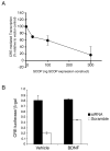
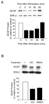
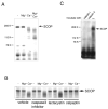
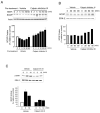
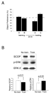
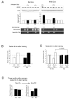
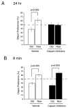
Because activation of ERK1/2 MAP kinase (MAPK) is critical for hippocampus-dependent memory, there is considerable interest in mechanisms for regulation of MAPK during memory formation. Here we report that MAPK and CREB-mediated transcription are negatively regulated by SCOP (suprachiasmatic nucleus [SCN] circadian oscillatory protein) and that SCOP is proteolyzed by calpain when hippocampal neurons are stimulated by brain-derived neurotrophic factor (BDNF), KCl depolarization, or NMDA. Moreover, training for novel object memory decreases SCOP in the hippocampus. To determine if hippocampus-dependent memory is influenced by SCOP in vivo, we generated a transgenic mouse strain for the inducible overexpression of SCOP in the forebrain. Overexpression of SCOP completely blocked memory for novel objects. We conclude that degradation of SCOP by calpain contributes to activation of MAPK during memory formation.
Figures


Comment in
-
SCOPing out proteases in long-term memory.Cell. 2007 Mar 23;128(6):1029-30. doi: 10.1016/j.cell.2007.02.034. Cell. 2007. PMID: 17382874
Similar articles
-
SCOPing out proteases in long-term memory.Cell. 2007 Mar 23;128(6):1029-30. doi: 10.1016/j.cell.2007.02.034. Cell. 2007. PMID: 17382874
-
Developmentally regulated NMDA receptor-dependent dephosphorylation of cAMP response element-binding protein (CREB) in hippocampal neurons.J Neurosci. 2000 May 15;20(10):3529-36. doi: 10.1523/JNEUROSCI.20-10-03529.2000. J Neurosci. 2000. PMID: 10804193 Free PMC article.
-
A calpain-2 selective inhibitor enhances learning & memory by prolonging ERK activation.Neuropharmacology. 2016 Jun;105:471-477. doi: 10.1016/j.neuropharm.2016.02.022. Epub 2016 Feb 18. Neuropharmacology. 2016. PMID: 26907807 Free PMC article.
-
Suprachiasmatic nucleus circadian oscillatory protein, a novel binding partner of K-Ras in the membrane rafts, negatively regulates MAPK pathway.J Biol Chem. 2003 Apr 25;278(17):14920-5. doi: 10.1074/jbc.M213214200. Epub 2003 Feb 19. J Biol Chem. 2003. PMID: 12594205
-
SCOP/PHLPP and its functional role in the brain.Mol Biosyst. 2010 Jan;6(1):38-43. doi: 10.1039/b911410f. Epub 2009 Sep 30. Mol Biosyst. 2010. PMID: 20024065 Free PMC article. Review.
Cited by
-
SCOP/PHLPP1β in the basolateral amygdala regulates circadian expression of mouse anxiety-like behavior.Sci Rep. 2016 Sep 19;6:33500. doi: 10.1038/srep33500. Sci Rep. 2016. PMID: 27640726 Free PMC article.
-
The N-terminal region of p27 inhibits HIF-1α protein translation in ribosomal protein S6-dependent manner by regulating PHLPP-Ras-ERK-p90RSK axis.Cell Death Dis. 2014 Nov 20;5(11):e1535. doi: 10.1038/cddis.2014.496. Cell Death Dis. 2014. PMID: 25412313 Free PMC article.
-
PHLPPing the Script: Emerging Roles of PHLPP Phosphatases in Cell Signaling.Annu Rev Pharmacol Toxicol. 2021 Jan 6;61:723-743. doi: 10.1146/annurev-pharmtox-031820-122108. Epub 2020 Sep 30. Annu Rev Pharmacol Toxicol. 2021. PMID: 32997603 Free PMC article. Review.
-
Calpain-2-mediated PTEN degradation contributes to BDNF-induced stimulation of dendritic protein synthesis.J Neurosci. 2013 Mar 6;33(10):4317-28. doi: 10.1523/JNEUROSCI.4907-12.2013. J Neurosci. 2013. PMID: 23467348 Free PMC article.
-
Suppression of survival signalling pathways by the phosphatase PHLPP.FEBS J. 2013 Jan;280(2):572-83. doi: 10.1111/j.1742-4658.2012.08537.x. Epub 2012 Mar 16. FEBS J. 2013. PMID: 22340730 Free PMC article.
References
-
- Agell N, Bachs O, Rocamora N, Villalonga P. Modulation of the Ras/Raf/MEK/ERK pathway by Ca(2+), and calmodulin. Cell Signal. 2002;14:649–654. - PubMed
-
- Athos J, Impey S, Pineda VV, Chen X, Storm DR. Hippocampal CRE-mediated gene expression is required for contextual memory formation. Nat Neurosci. 2002;5:1119–1120. - PubMed
-
- Atkins CM, Selcher JC, Petraitis JJ, Trzaskos JM, Sweatt JD. The MAPK cascade is required for mammalian associative learning. Nat Neurosci. 1998;1:602–609. - PubMed
-
- Berninger B, Garcia DE, Inagaki N, Hahnel C, Lindholm D. BDNF and NT-3 induce intracellular Ca2+ elevation in hippocampal neurones. Neuroreport. 1993;4:1303–1306. - PubMed
Publication types
MeSH terms
Substances
Grants and funding
LinkOut - more resources
Full Text Sources
Medical
Molecular Biology Databases
Miscellaneous

