Axonal sprouting into the denervated spinal cord and synaptic and postsynaptic protein expression in the spinal cord after transplantation of bone marrow stromal cell in stroke rats
- PMID: 17362881
- PMCID: PMC1950288
- DOI: 10.1016/j.brainres.2007.02.047
Axonal sprouting into the denervated spinal cord and synaptic and postsynaptic protein expression in the spinal cord after transplantation of bone marrow stromal cell in stroke rats
Abstract
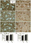
We investigated whether compensatory reinnervation in the corticospinal tract (CST) and the corticorubral tract (CRT) is enhanced by the administration of bone marrow stromal cells (BMSCs) after experimental stroke. Adult male Wistar rats were subjected to permanent right middle cerebral artery occlusion (MCAo). Phosphate-buffered saline (PBS, control, n=7) or 3x10(6) BMSCs in PBS (n=8) were injected into a tail vein at 1 day postischemia. The CST of the left sensorimotor cortices was labeled with DiI 2 days prior to MCAo. Functional recovery was measured. Rats were sacrificed at 28 days after MCAo. The brain and spinal cord were removed and processed for vibratome sections for laser-scanning confocal analysis and paraffin sections for immunohistochemistry. Normal rats (n=4) exhibited a predominantly unilateral pattern of innervation of CST and CRT axons. After stroke, bilateral innervation occurred through axonal sprouting of the uninjured CRT and CST. Administration of BMSCs significantly increased the axonal restructuring on the de-afferented red nucleus and the denervated spinal motoneurons (p<0.05). BMSC treatment also significantly increased synaptic proteins in the denervated motoneurons. These results were highly correlated with improved functional outcome after stroke (r>0.81, p<0.01). We conclude that the transplantation of BMSCs enhances axonal sprouting and rewiring into the denervated spinal cord which may facilitate functional recovery after focal cerebral ischemia.
Figures


Similar articles
-
Contralesional axonal remodeling of the corticospinal system in adult rats after stroke and bone marrow stromal cell treatment.Stroke. 2008 Sep;39(9):2571-7. doi: 10.1161/STROKEAHA.107.511659. Epub 2008 Jul 10. Stroke. 2008. PMID: 18617661 Free PMC article.
-
Effects of treating traumatic brain injury with collagen scaffolds and human bone marrow stromal cells on sprouting of corticospinal tract axons into the denervated side of the spinal cord.J Neurosurg. 2013 Feb;118(2):381-9. doi: 10.3171/2012.11.JNS12753. Epub 2012 Nov 30. J Neurosurg. 2013. PMID: 23198801
-
Bone marrow stromal cells promote skilled motor recovery and enhance contralesional axonal connections after ischemic stroke in adult mice.Stroke. 2011 Mar;42(3):740-4. doi: 10.1161/STROKEAHA.110.607226. Epub 2011 Feb 9. Stroke. 2011. PMID: 21307396 Free PMC article.
-
Molecular, cellular and functional events in axonal sprouting after stroke.Exp Neurol. 2017 Jan;287(Pt 3):384-394. doi: 10.1016/j.expneurol.2016.02.007. Epub 2016 Feb 10. Exp Neurol. 2017. PMID: 26874223 Free PMC article. Review.
-
Axonal remodeling of the corticospinal tract during neurological recovery after stroke.Neural Regen Res. 2021 May;16(5):939-943. doi: 10.4103/1673-5374.297060. Neural Regen Res. 2021. PMID: 33229733 Free PMC article. Review.
Cited by
-
Cell based therapies for ischemic stroke: from basic science to bedside.Prog Neurobiol. 2014 Apr;115:92-115. doi: 10.1016/j.pneurobio.2013.11.007. Epub 2013 Dec 12. Prog Neurobiol. 2014. PMID: 24333397 Free PMC article. Review.
-
Axon sprouting in adult mouse spinal cord after motor cortex stroke.Neurosci Lett. 2009 Jan 30;450(2):191-5. doi: 10.1016/j.neulet.2008.11.017. Epub 2008 Nov 13. Neurosci Lett. 2009. PMID: 19022347 Free PMC article.
-
Magnetic resonance imaging and cell-based neurorestorative therapy after brain injury.Neural Regen Res. 2016 Jan;11(1):7-14. doi: 10.4103/1673-5374.169603. Neural Regen Res. 2016. PMID: 26981068 Free PMC article.
-
Beneficial effects of gfap/vimentin reactive astrocytes for axonal remodeling and motor behavioral recovery in mice after stroke.Glia. 2014 Dec;62(12):2022-33. doi: 10.1002/glia.22723. Epub 2014 Jul 15. Glia. 2014. PMID: 25043249 Free PMC article.
-
MRI of neuronal recovery after low-dose methamphetamine treatment of traumatic brain injury in rats.PLoS One. 2013 Apr 18;8(4):e61241. doi: 10.1371/journal.pone.0061241. Print 2013. PLoS One. 2013. PMID: 23637800 Free PMC article.
References
-
- Bastings EP, Greenberg JP, Good DC. Hand motor recovery after stroke: a transcranial magnetic stimulation mapping study of motor output areas and their relation to functional status. Neurorehabil Neural Repair. 2002;16:275–82. - PubMed
-
- Brosamle C, Schwab ME. Ipsilateral, ventral corticospinal tract of the adult rat: ultrastructure, myelination and synaptic connections. J Neurocytol. 2000;29:499–507. - PubMed
-
- Carmichael ST. Cellular and molecular mechanisms of neural repair after stroke: making waves. Ann Neurol. 2006;59:735–42. - PubMed
-
- Chen H, Chopp M, Zhang ZG, Garcia JH. The effect of hypothermia on transient middle cerebral artery occlusion in the rat. J Cereb Blood Flow Metab. 1992;12:621–8. - PubMed
-
- Chen J, Li Y, Chopp M. Intracerebral transplantation of bone marrow with BDNF after MCAo in rat. Neuropharmacology. 2000;39:711–6. - PubMed
Publication types
MeSH terms
Grants and funding
LinkOut - more resources
Full Text Sources
Other Literature Sources
Medical
Research Materials

