Review
. 2007:76:295-329.
doi: 10.1146/annurev.biochem.76.010307.145803.

Lipid A modification systems in gram-negative bacteria

Affiliations
Review

Lipid A modification systems in gram-negative bacteria

Christian R H Raetz et al. Annu Rev Biochem. 2007.

Abstract

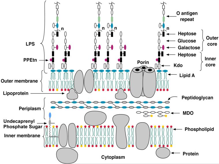
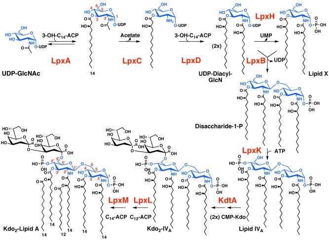
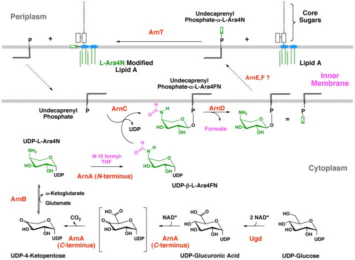
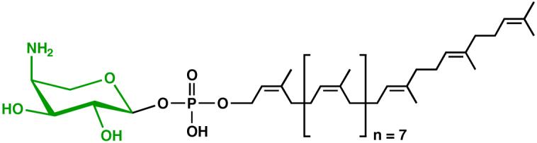
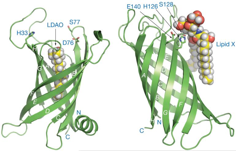
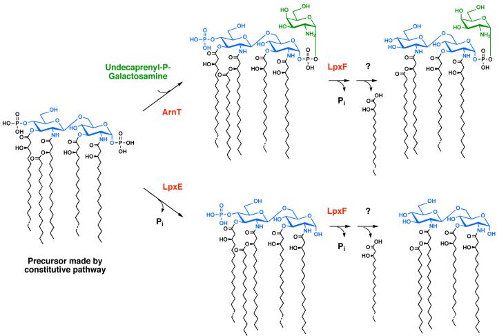
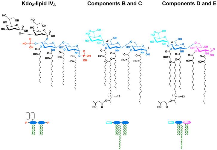
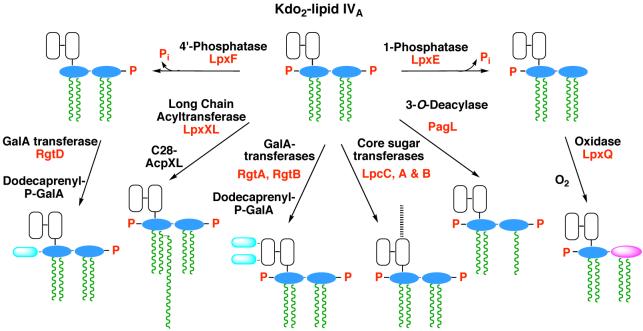
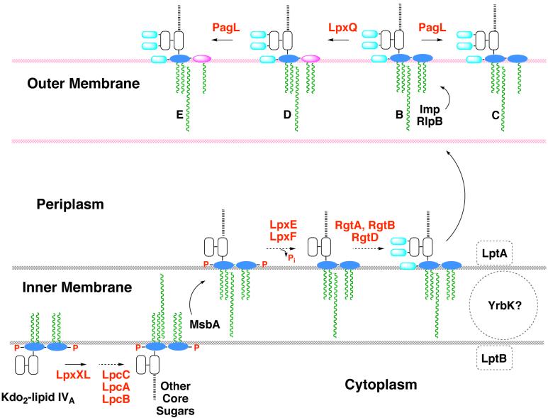
The lipid A moiety of lipopolysaccharide forms the outer monolayer of the outer membrane of most gram-negative bacteria. Escherichia coli lipid A is synthesized on the cytoplasmic surface of the inner membrane by a conserved pathway of nine constitutive enzymes. Following attachment of the core oligosaccharide, nascent core-lipid A is flipped to the outer surface of the inner membrane by the ABC transporter MsbA, where the O-antigen polymer is attached. Diverse covalent modifications of the lipid A moiety may occur during its transit from the outer surface of the inner membrane to the outer membrane. Lipid A modification enzymes are reporters for lipopolysaccharide trafficking within the bacterial envelope. Modification systems are variable and often regulated by environmental conditions. Although not required for growth, the modification enzymes modulate virulence of some gram-negative pathogens. Heterologous expression of lipid A modification enzymes may enable the development of new vaccines.


Figures

Figure 1
Figure 1. Schematic structure of the E. coli K-12 cell envelope
The structure and biosynthesis of LPS (2, 3), peptidoglycan (186), membrane-derived oligosaccharides (239, 240), lipoproteins (241) and phospholipids (242, 243) are reviewed elsewhere. Strains of E. coli K-12 normally do not make O-antigen, unless a mutation in the O-antigen operon is corrected (244). The Kdo2-lipid A region of LPS (the topic of this review) usually represent the minimal substructure required for growth of gram-negative bacteria. Exceptions include some spirochetes and strains of Sphinogmonas, in which the lipid A biosynthesis genes are absent, and Neisseria meningitidis in which lpxA knockouts lacking LPS are viable (142). If the ABC transporter MsbA (the inner membrane flippase for LPS) is over-expressed, E. coli can grow without Kdo (8). These strains still make the tetra-acylated precursor lipid IVA, which is nevertheless required for growth (8). The red phospholipids represent phosphatidylethanolamine and the yellow phosphatidylglycerol. Abbreviations: LPS, lipopolysaccharide; MDO, membrane-derived oligosaccharides; PPEtn, ethanolamine pyrophosphate.
Figure 2
Figure 2. The constitutive pathway for Kdo2-lipid A biosynthesis in E. coli
Each enzyme of the constitutive lipid A pathway is encoded by a single structural gene (2, 69). The glucosamine disaccharide backbone of lipid A is blue. The Kdo disaccharide is black. LpxA, C and D are soluble cytoplasmic proteins, whereas LpxH and B are peripheral membrane proteins (2). The distal enzymes of the pathway, starting with LpxK, are integral inner membrane proteins, the active sites of which face the cytoplasm (2). The red numbers specify the glucosamine ring positions of lipid A and its precursors. The black numbers indicate the predominant fatty acid chain lengths found in E. coli lipid A. The single molecular species shown at the bottom left represents about 90 % of the total lipid A in E. coli, with most of the rest bearing a C12 secondary acyl chain at position 3′ (152). Additional minor acyl chain variants can be detected by high-resolution mass spectrometry (245).
Figure 3
Figure 3. Structure of free LpxA and of LpxA with bound UDP-(3-O-acyl)-GlcNAc
The LpxA homotrimer was solved at 2.6 Å (pdb 1LXA) in the absence of bound ligands (91). Each subunit has its own color. The side view (left) highlights the β-helix domain (91). The LpxA homotrimer was co-crystallized with a ∼25 fold molar excess of UDP-3-O-(R -3-hydroxydecanoyl)-GlcNAc and solved at 1.8 Å (Williams and Raetz, in preparation). The top-down view of this complex (right) reveals the location of the active site and the positioning of the acyl chain, consistent with previous proposals based on site-directed mutagenesis and NMR studies (94, 246).
Figure 4
Figure 4. Biosynthesis and acylation of UDP-GlcNAc3N in L. interrogans
The sugar nucleotide UDP-GlcNAc3N is synthesized in two reactions from UDP-GlcNAc. The intermediate ketone has not yet been characterized, but UDP-GlcNAc3N generated in vitro by GnnA and GnnB can be isolated in mg quantities (89, 95). LpxA from L. interrogans is 41 % identical to E. coli LpxA at the protein level, and its x-ray structure has recently been determined (Williams and Raetz, in preparation). L. interrogans LpxA does not catalyze the acylation of UDP-GlcNAc and is absolutely selective for C12 hydroxyacyl chains (89, 95).
Figure 5
Figure 5. Structures of LpxC inhibitors CHIR-090 and TU-514
A. The slow, tight-binding inhibitor CHIR-090 inhibits diverse LpxC orthologues in the low nM range and displays potent antibiotic activity against many gram-negative bacteria (102). B. The substrate mimetic TU-514 inhibits E. coli LpxC with Ki ∼ 650 nM but has little or no antibiotic activity (100).
Figure 6
Figure 6. NMR structure of LpxC with bound substrate-mimetic inhibitor TU-514
This ribbon diagram is based on the NMR studies of Coggins et al. (107, 108). The recent crystal structure of the same complex is similar, except for slight differences in the orientation of the tetrahydropyran ring (105).
Figure 7
Figure 7. Proteins implicated in O-antigen assembly and in the export of LPS
The proteins in red letters are involved in the export of LPS. The ABC transporter MsbA flips newly-synthesized core-lipid A to the outer surface of the inner membrane (55, 143). O-antigen is assembled separately on undecaprenyl phosphate and is flipped by the putative transporter Wzx (247). O-antigen oligosaccharides are polymerized on the periplasmic surface of the inner membrane by Wzy and Wzz, and transferred to nascent core-lipid A by WaaL (2). In vitro systems for the polymerase and ligase have not been reported. The periplasmic protein LptA (159) is proposed to shuttle LPS to the essential outer membrane protein complex Imp/RlpB, which is required the assembly of LPS into the outer surface of the outer membrane, as judged by accessibility to the ecto-enzymes PagL or PagP (56, 60, 197, 209). With the exception of the lipid-activated ATPase activity of MsbA (154), no in vitro assays have been developed for any of the proposed transporters. The LptA protein may function together with an additional ABC transporter protein termed LptB and the putative trans-membrane protein YrbK (159).
Figure 8
Figure 8. Covalent modifications of Kdo2-lipid A in E. coli K-12 and Salmonella
The known covalent modifications of Kdo2-lipid A (3) are indicated by the substituents with the dashed bonds. The glucosamine disaccharide is blue, the L-Ara4N unit green, and the phosphoethanolamine moieties red. Under some conditions, the positions of the phosphoethanolamine and L-Ara4N substituents are reversed (not shown) (248, 249). Lipid A species with two phosphoethanolamine units or two L-Ara4N moieties may also be present (44). Expression of the enzymes ArnT (47) and EptA(PmrC) is under the control of PmrA/B (50, 52). PagP and PagL are regulated by PhoP/Q (46, 47). LpxO (magenta), LpxR and EptB are not regulated by either PhoP/Q or PmrA/B (44, 53, 54). Asterisks indicate modification enzymes not found in E. coli K-12. Transfer of the Salmonella genes encoding these enzymes to E. coli results in the expected lipid A modifications.
Figure 9
Figure 9. Biosynthesis of the L-Ara4N unit and its attachment to lipid A
Formation of the L-Ara4N moiety begins with the oxidation of UDP-glucose to UDP-glucuronic acid (172), as first proposed by Zhou et al. (165) Next, the C-terminal domain of ArnA catalyzes an NAD+-dependent oxidative decarboxylation to yield an unusual UDP-4-ketopentose, which is converted to UDP-L-Ara4N (green moiety) by the transaminase ArnB (173). The N-terminal domain of ArnA then uses N-10-formyltetrahydrofolate to add a formyl group (magenta) to UDP-L-Ara4N (174). Next, ArnC/PmrF, a distant orthologue of dolichyl phosphate-mannose synthase, selectively transfers the formylated L-Ara4N residue to undecaprenyl phosphate (174). The ArnD-dependent deformylation of this lipid to undecaprenyl phosphate-α-L-Ara4N (which accumulates in polymyxin-resistant mutants) likely occurs on the inner leaflet of the inner membrane and may prevent reversal of the ArnC reaction (174). After transport to the outer surface of the inner membrane by a process that may involve the inner membrane proteins ArnE and ArnF (Yan and Raetz, in preparation), the polytopic membrane protein ArnT transfers the L-Ara4N moiety (shown as a green rectangle) to lipid A. Given the dual function the ArnA holoenzyme (174-176), the possibility of substrate channeling from ArnA to ArnC deserves consideration. However, ArnA and ArnB do not associate with each other in vitro.
Figure 10
Figure 10. Structure of undecaprenyl phosphate-α-L-Ara4N
This lipid accumulates in polymyxin-resistant mutants of E. coli and Salmonella (48).
Figure 11
Figure 11. The outer membrane lipid A palmitoyltransferase PagP (left) and the lipid A 3-O-deacylase PagL (right)
The disordered loop connecting the first and second β-strands of PagP (pdb 1THQ), and the bound lipid X with PagL (pdb 2ERV) were introduced subsequently and energy minimized. The authors thank Chris Neale and Régis Pomès (University of Toronto) and Lucy Rutten and Jan Tommassen (Utrecht University) for providing the coordinates of energy minimized PagP and PagL, respectively. Abbreviation: LDAO, lauroyldimethylamine-N-oxide.
Figure 12
Figure 12. Extracellular covalent modifications of lipid A in F. tularensis
The proposed modification pathway is based on the genetic characterization of the two phosphatases, LpxE and LpxF (58, 59), which are present in many bacteria that synthesize phosphate-deficient lipid A. The attachment of galactosamine to F. tularensis lipid A involves a polyisoprene phosphate donor, analogous to undecaprenyl phosphate-L-Ara4N in E. coli (Wang and Raetz, in preparation). The F. tularensis system is also unusual in that much of its lipid A is “free”, i.e. not covalently attached to LPS (212). The origin and function of free lipid A have not been established.
Figure 13
Figure 13. Kdo2-lipid IVA versus mature lipid A of R. etli and R. leguminosarum
These bacteria make phosphate-deficient lipid A molecules from Kdo2-lipid IVA (226, 250), using the lipid A phosphatases LpxE and LpxF, as described in the text. Lipid A molecules of Rhizobium typically contain a very long secondary acyl chain at position 2′ (222). Additional unique features include the presence of galacturonic acid (cyan) in place of phosphate at position 4′ and oxidation of the proximal glucosamine unit (blue) in a portion of the molecules to aminogluconate (magenta) (219-221). Partial substituents and micro-heterogeneity of acyl chains lengths are indicated by dashed bonds. Components C and E lack the 3-O linked hydroxyacyl chain. The schematic representations of these structures are shown below the actual chemical structures.
Figure 14
Figure 14. Kdo2-lipid IVA processing in R. etli and R. leguminosarum membranes
The processing enzymes unique to the Rhizbium system can be assayed using E. coli Kdo2-lipid IVA as the model substrate (223, 225, 227-229, 232, 233, 250-252). The enzymes that have been characterized to date are labeled in red according to the genes that encode them. Other colors: glucosamine, blue; galacturonic acid, cyan; aminogluconate, magenta; Kdo, white; other core sugars, black dashed line; fatty acids, green.
Figure 15
Figure 15. Topography of lipid A modifications in R. etli and R. leguminosarum
The proposed topography of the processing enzymes is based on the finding that lipid A dephosphorylation by LpxE and LpxF requires a functional msbA gene, when foreign lpxE or lpxF genes are expressed in E. coli (51, 58, 59). The Rgt proteins require a polyisoprene donor as their co-substrate, consistent with a periplasmic localization (232, 233). PagL and LpxQ are known to be outer membrane proteins (47, 231). The x-ray structure of PagL shows that its active site is oriented towards the outside (209). The orientation of LpxQ is unknown (231). PagP is not present in Rhizobium. Enzymes are indicated in red, and putative transport proteins are shown in black. Letters in the outer membrane refer to lipid A components B, C, D and E, shown in Fig. 13. Other colors: glucosamine, blue; galacturonic acid, cyan; aminogluconate, magenta; Kdo, white; other core sugars, black dashed line; fatty acids, green.

Similar articles

Cited by

References

    1. Fahy E, Subramaniam S, Brown HA, Glass CK, Merrill AH, Jr., et al. J. Lipid Res. 2005;46:839–62. - PubMed
    1. Raetz CRH, Whitfield C. Annu. Rev. Biochem. 2002;71:635–700. - PMC - PubMed
    1. Brade H, Opal SM, Vogel SN, Morrison DC, editors. Endotoxin in Health and Disease. Marcel Dekker, Inc.; New York: 1999. p. 950.
    1. Nikaido H. Microbiol. Mol. Biol. Rev. 2003;67:593–656. - PMC - PubMed
    1. Galloway SM, Raetz CRH. J. Biol. Chem. 1990;265:6394–402. - PubMed

Publication types

MeSH terms

LinkOut - more resources