Comparative Study
. 2007 Feb 27;104(9):3585-90.
doi: 10.1073/pnas.0611671104. Epub 2007 Feb 21.

The pRb/E2F cell-cycle pathway mediates cell death in Parkinson's disease

Affiliations
Comparative Study

The pRb/E2F cell-cycle pathway mediates cell death in Parkinson's disease

Günter U Höglinger et al. Proc Natl Acad Sci U S A. .

Abstract

The mechanisms leading to degeneration of dopaminergic neurons (DNs) in the substantia nigra of patients with Parkinson's disease (PD) are not completely understood. Here, we show, in the postmortem human tissue, that these neurons aberrantly express mitosis-associated proteins, including the E2F-1 transcription factor, and appear to duplicate their nuclear DNA. We further demonstrate that the dopaminergic neurotoxin 1-methyl-4-phenyl-1,2,3,6-tetrahydropyridine injected into mice and application of its active metabolite 1-methyl-4-phenylpyridinium to mesencephalic cultures activate the retinoblastoma-E2F pathway in postmitotic DNs. We also find that cell death rather than mitotic division followed the toxin-induced replication of DNA, as determined by BrdU incorporation in DNs. In addition, blocking E2F-1 transcription protected cultured DNs against 1-methyl-4-phenylpyridinium toxicity. Finally, E2F-1-deficient mice were significantly more resistant to 1-methyl-4-phenyl-1,2,3,6-tetrahydropyridine-induced dopaminergic cell death than their wild-type littermates. Altogether, BrdU incorporation in mature neurons and lack of evidence for newborn neurons argue against neuronal turnover in normal conditions or during pathological states in the substantia nigra. Instead, our results demonstrate that mitosis-like signals are activated in mature DNs in patients with PD and mediate neuronal death in experimental models of the disease. Inhibition of mitosis-like signals may therefore provide strategies for neuroprotection in PD.


Conflict of interest statement

The authors declare no conflict of interest.

Figures
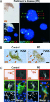
Fig. 1.
Fig. 1.
Aberrant cell-cycle activation in DNs of the substantia nigra in PD. (A) DNA duplication in a DN of a male PD patient. Nuclei of DNs were identified by DAPI staining of DNA (blue), which was embedded in a pocket of neuromelanin (NM). In situ hybridization and confocal microscopy allowed quantification of the number of chromosomes X (CX, red hybridization spots, red numbering) and 18 (C18, green hybridization spots, green numbering). The dopaminergic NM+ neuron in Upper Right contains four copies of chromosome 18 and two copies of chromosome X, indicating that the chromosomes in this cell had been duplicated. The two small nondopaminergic NM-negative nuclei in Lower Right contain two chromosomes 18 and one chromosome X, as expected for a cell not undergoing mitosis. (B) In PD patients, but not in controls, pronounced nuclear PCNA immunoreactivity (brown diaminobenzidine precipitate) was observed (black arrows) in DNs, identified by their cytoplasmic NM content (brown arrowheads). (C) In PD patients, but not in controls, pronounced cytoplasmic E2F-1 immunoreactivity (red) was observed in DNs, identified by their NM content (brown; brown arrowheads). NeuN immunostaining (green) delineated the cell bodies (weak staining) and nuclei (intense staining; green arrowheads). DAPI confirmed the location of the nuclei (blue; blue arrowheads). The fluorescent images are confocal. The merged image superposes E2F-1 (red), NeuN (green), and DAPI (blue) staining. (Scale bars: 10 μm.)
Fig. 2.
Fig. 2.
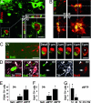
Nuclear DNA synthesis in DNs in the SNc of mice 24 h after MPTP intoxication. (A) Nuclear PCNA immunoreactivity (red) in a DN, identified by TH expression (green) is shown. An overview (ov) and an inset showing a higher magnification of the cell in a confocal reconstruction of the xy, xz, and yz planes are shown. (B) BrdU immunoreactivity (green) in a TH+ neuron (red). Overview (ov) and detail of the boxed area showing the cell in a confocal reconstruction of the xy, xz, and yz planes. The superposition of BrdU and TH appears yellow, demonstrating that both markers colocalize in the same cellular compartment, suggesting a breakdown of the nuclear membrane. Note the disintegrated TH+ cytoplasm and the condensed BrdU+ chromatin, suggesting that the cell is in a state of degeneration. (C and D) Examples of artifactual BrdU-TH colocalization. (C) A BrdU+ nucleus (green) that erroneously appears to belong to a TH+ neuron (red) when seen with conventional microscopy: an overview (ov) and an enlarged detail in the boxed area are shown. However, confocal microscopy of sections 1 μm apart shows that the BrdU+ nucleus is juxtaposed to the TH+ cell and that the nucleus of the TH+ cell (best seen at 5 μm; ∗) is not BrdU+. (D) A BrdU+ nucleus (green) that erroneously appears to belong to a TH+ neuron (white), when observed by confocal microscopy. An overview (ov) and an enlarged detail are shown). However, the lack of counterstaining with the neuronal marker NeuN (red) demonstrates that the BrdU+ nucleus (arrowhead) is not neuronal in nature, in contrast to the neighboring TH+ BrdU cell (arrow). The TH+ immunoreactivity in the vicinity of the BrdU+ nucleus is contained in the neurites of neighboring DNs. (Scale bars: 10 μm.) (E and F) Absolute numbers of PCNA+ TH+ cells (E) and BrdU+ TH+ cells (F) per SNc of mice 24 h after NaCl injection (control) or after intoxication with MPTP in the acute (aMPTP) or subchronic (cMPTP) paradigm. ∗, P < 0.05, ANOVA followed by post hoc test, vs. NaCl. (G) Absolute numbers of BrdU+ TH+ cells per SNc of mice at different time points (d = days) after MPTP intoxication in the acute paradigm (aMPTP). Two-way ANOVA showed a significant effect of treatment (MPTP vs. NaCl, P < 0.001), time (P < 0.01), and treatment × time interaction (P < 0.01). ∗, P < 0.05, post hoc test, vs. NaCl.
Fig. 3.
Fig. 3.
PCNA-expressing dopaminergic cells in the SNc of MPTP mice are not newborn neurons, but preexist. (A) TH+ neurons (green) in the SNc were labeled by striatal injection of the retrograde tracer FluoroGold (FG, blue) 3 days before MPTP intoxication. (B) Overview (ov) showing a cell (arrow) in the SNc of a mouse 24 h after acute MPTP intoxication expressing both TH (green) and PCNA (red). The presence of FluoroGold (blue) demonstrated that the cell existed before the lesion, ruling out the possibility that it was a stem cell-derived newborn dopaminergic cell. (C) An enlarged detail from the boxed area in B shown as a confocal reconstruction in the xy, xz, and yz plane, to confirm the colocalization of TH, PCNA, and FG. [Scale bars: 100 μm (A); 10 μm (B and C).]
Fig. 4.
Fig. 4.
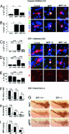
Cell-cycle events do not occur in postmitotic TH+ midbrain neurons in vitro under control conditions (A), but are observed after MPP+ intoxication (B–D). (A) In control cultures, cell cycle-associated proteins, such as cyclin D1 (CyD1, green) were never expressed in TH+ neurons (red). (B) However, after 24 h of intoxication with MPP+, the G1-phase-associated CyD1, the S-phase-associated cyclin E (CyE), the G2-phase-associated cyclin A (CyA), and the M-phase-associated cyclin B (CyB) (green) were expressed in TH+ cells (red). The graph shows the percentage of TH+ cells expressing CyD1, CyE, CyA, and CyB after 24 h of MPP+ intoxication. (C) Immunoreactivity for the S-phase-associated markers p-pRb, E2F-1, PCNA, and BrdU (green) was detected in TH+ DNs (red) after 24 h of MPP+ intoxication. Visualization of chromatin with DAPI (blue) demonstrated the perinuclear expression of E2F-1 (green) in TH+ neurons (red). The graph shows the percentage of TH+ cells immunoreactive for p-pRb, E2F-1, PCNA, and BrdU after 24 h of MPP+ intoxication. (D) A 24-h treatment of cultured neurons with MPP+ led to the appearance of morphologically disintegrated cells (structure, gray) with condensed chromatin (DAPI, white) and dopaminergic phenotype (TH+, red) that were immunopositive for both the activated form of the downstream effector caspase-3 (aC3, blue) and BrdU (green). The graph shows the quantification of the presence (+) or absence (−) of BrdU and aC3 immunoreactivity in TH+ cells after 24 h of MPP+ treatment. [Scale bars: 20 μm (A and B); 10 μm (C and D).]
Fig. 5.
Fig. 5.
E2F-1 signaling is upstream of MPP+-induced caspase-3 activation, and E2F-1 deficiency provides protection against MPTP. (A and B) Caspase activation is not upstream of cell-cycle signaling. Caspase inhibition (CI) with 200 μM zVAD-fmk during a 24-h intoxication of TH+ neurons (red) with MPP+ prevented the appearance of immunoreactivity for the activated form of caspase-3 (aC3, green) (A), but not the incorporation of BrdU (green) (B). DAPI (blue) stains nuclei. The superposition of red and green appears yellow. The superposition of red, green, and blue appears white. The graphs show the percentage of TH+ cells immunoreactive for aC3 (A) and BrdU (B). ∗, P < 0.05; n.s., not significant. (C–E) E2F-1 inhibition protects neurons in vitro from MPP+. Inhibition of E2F-1 expression with antisense (AS), but not sense (S) oligonucleotides during a 24-h intoxication of TH+ DNs (red) with MPP+ attenuated the appearance of immunoreactivity for E2F-1 (green) (C) and aC3 (green) (D) and the MPP+-induced loss of TH+ cell (E). DAPI (blue) stains nuclei. The graphs show the percentage of TH+ cells immunoreactive for E2F-1 (C) and aC3 (D) and the percentage of surviving TH+ cells (E) after transfection with E2F-1 sense (S) or antisense (AS) oligonucleotides and grown for 24 h in the absence (C) or the presence (M) of MPP+. ∗, P < 0.05; ∗∗∗, P < 0.001; n.s., not significant. (F–I) E2F-1 deficiency protects neurons in vivo from MPTP. (F) Dopaminergic (TH+) cell bodies in the SNc, assessed 7 days after MPTP intoxication in both the subchronic (cMPTP) and acute paradigm (aMPTP), were significantly protected in E2F-1−/− mice compared with E2F-1+/+ mice. ∗, P < 0.05; n.s., not significant. (G) Representative photomicrographs of TH+ cell bodies in the SNc in E2F-1−/− mice compared with E2F-1+/+ mice 7 days after MPTP intoxication. (H) The striatal dopamine depletion, assessed 7 days after MPTP intoxication in both paradigms did not differ between E2F-1−/− and E2F-1+/+ mice. (I) The time course of the accumulation and elimination of the toxic MPTP-metabolite MPP+ after an injection of MPTP was identical in E2F-1−/− mice and wild-type littermates (E2F-1+/+). [Scale bars: 10 μm (A-D); 100 μm (E and G).]

Similar articles

Cited by

References

    1. Dauer W, Przedborski S. Neuron. 2003;39:889–909. - PubMed
    1. Jordan-Sciutto KL, Wang G, Murphey-Corb M, Wiley CA. J Neurosci. 2002;22:2185–2195. - PMC - PubMed
    1. Ranganathan S, Bowser R. Am J Pathol. 2003;162:823–835. - PMC - PubMed
    1. Yang Y, Geldmacher DS, Herrup K. J Neurosci. 2001;21:2661–2668. - PMC - PubMed
    1. Yang Y, Mufson EJ, Herrup K. J Neurosci. 2003;23:2557–2563. - PMC - PubMed

Publication types

MeSH terms

Substances