

p63 induces key target genes required for epidermal morphogenesis
- PMID: 17360634
- PMCID: PMC1805532
- DOI: 10.1073/pnas.0611376104
p63 induces key target genes required for epidermal morphogenesis
Abstract
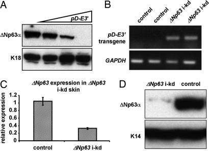
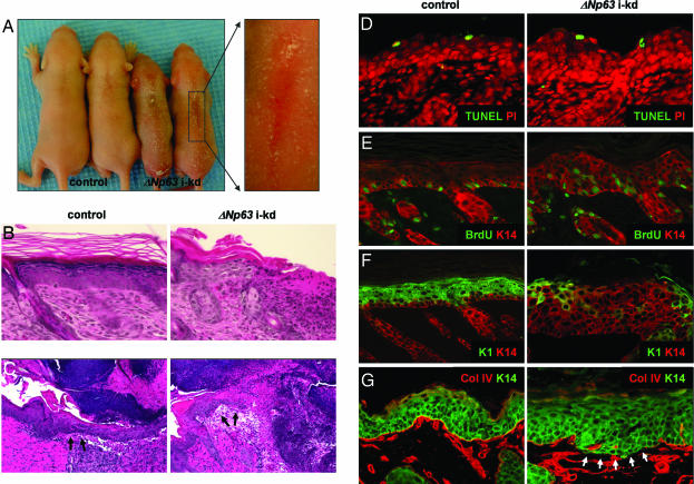
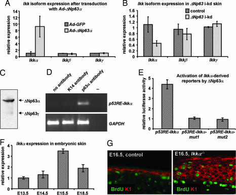
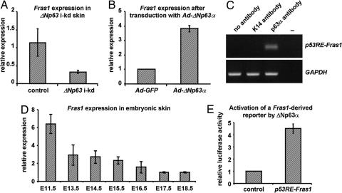
Mice lacking p63, a single gene that encodes a group of transcription factors that either contain (TA) or lack (DeltaN) a transactivation domain, fail to develop stratified epithelia as well as epithelial appendages and limbs. DeltaNp63 isoforms are predominantly expressed during late embryonic and postnatal epidermal development, however, the function of these proteins remains elusive. Using an epidermal-specific inducible knockdown mouse model, we demonstrate that DeltaNp63 proteins are essential for maintaining basement membrane integrity and terminal differentiation of keratinocytes. Furthermore, we have identified two DeltaNp63alpha target genes that mediate these processes. We propose that DeltaNp63alpha initially induces expression of the extracellular matrix component Fras1, which is required for maintaining the integrity of the epidermal-dermal interface at the basement membrane. Subsequently, induction of IkappaB kinase-alpha by DeltaNp63alpha initiates epidermal terminal differentiation resulting in the formation of the spinous layer. Our data provide insights into the role of DeltaNp63alpha in epidermal morphogenesis and homeostasis, and may contribute to our understanding of the pathogenic mechanisms underlying disorders caused by p63 mutations.
Conflict of interest statement
The authors declare no conflict of interest.
Figures
Similar articles
-
Unique domain functions of p63 isotypes that differentially regulate distinct aspects of epidermal homeostasis.Carcinogenesis. 2006 Jan;27(1):53-63. doi: 10.1093/carcin/bgi200. Epub 2005 Aug 4. Carcinogenesis. 2006. PMID: 16081516
-
ΔNp63 knockout mice reveal its indispensable role as a master regulator of epithelial development and differentiation.Development. 2012 Feb;139(4):772-82. doi: 10.1242/dev.071191. Development. 2012. PMID: 22274697 Free PMC article.
-
Abnormal hair follicle development and altered cell fate of follicular keratinocytes in transgenic mice expressing DeltaNp63alpha.Development. 2010 May;137(9):1431-9. doi: 10.1242/dev.045427. Epub 2010 Mar 24. Development. 2010. PMID: 20335364 Free PMC article.
-
Genetic pathways required for epidermal morphogenesis.Eur J Cell Biol. 2004 Dec;83(11-12):625-9. doi: 10.1078/0171-9335-00387. Eur J Cell Biol. 2004. PMID: 15679107 Review.
-
Dynamic life of a skin keratinocyte: an intimate tryst with the master regulator p63.Indian J Exp Biol. 2011 Oct;49(10):721-31. Indian J Exp Biol. 2011. PMID: 22013738 Review.
Cited by
-
IL-13 modulates ∆Np63 levels causing altered expression of barrier- and inflammation-related molecules in human keratinocytes: A possible explanation for chronicity of atopic dermatitis.Immun Inflamm Dis. 2021 Sep;9(3):734-745. doi: 10.1002/iid3.427. Epub 2021 Apr 1. Immun Inflamm Dis. 2021. PMID: 33792188 Free PMC article.
-
Epithelial barrier repair and prevention of allergy.J Clin Invest. 2019 Apr 1;129(4):1463-1474. doi: 10.1172/JCI124608. Epub 2019 Feb 18. J Clin Invest. 2019. PMID: 30776025 Free PMC article. Review.
-
DeltaNp63 regulates stem cell dynamics in the mammalian olfactory epithelium.J Neurosci. 2011 Jun 15;31(24):8748-59. doi: 10.1523/JNEUROSCI.0681-11.2011. J Neurosci. 2011. PMID: 21677159 Free PMC article.
-
Luteolin-7-O-β-d-Glucoside Inhibits Cellular Energy Production Interacting with HEK2 in Keratinocytes.Int J Mol Sci. 2019 May 31;20(11):2689. doi: 10.3390/ijms20112689. Int J Mol Sci. 2019. PMID: 31159225 Free PMC article.
-
DEK promotes HPV-positive and -negative head and neck cancer cell proliferation.Oncogene. 2015 Feb 12;34(7):868-77. doi: 10.1038/onc.2014.15. Epub 2014 Mar 10. Oncogene. 2015. PMID: 24608431 Free PMC article.
References
-
- Dotto GP. Crit Rev Oral Biol Med. 1999;10:442–457. - PubMed
-
- Yang A, Kaghad M, Wang Y, Gillett E, Fleming MD, Dotsch V, Andrews NC, Caput D, McKeon F. Mol Cell. 1998;2:305–316. - PubMed
-
- Mills AA, Zheng B, Wang XJ, Vogel H, Roop DR, Bradley A. Nature. 1999;398:708–713. - PubMed
-
- Yang A, Schweitzer R, Sun D, Kaghad M, Walker N, Bronson RT, Tabin C, Sharpe A, Caput D, Crum C, et al. Nature. 1999;398:714–718. - PubMed
Publication types
MeSH terms
Substances
Grants and funding
LinkOut - more resources
Full Text Sources
Other Literature Sources
Molecular Biology Databases
