

MEK1 and protein phosphatase 4 coordinate Dictyostelium development and chemotaxis
- PMID: 17353263
- PMCID: PMC1899987
- DOI: 10.1128/MCB.02194-06
MEK1 and protein phosphatase 4 coordinate Dictyostelium development and chemotaxis
Abstract
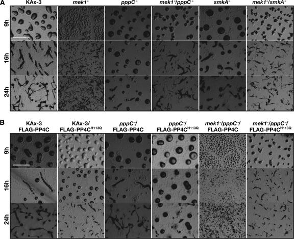
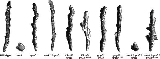
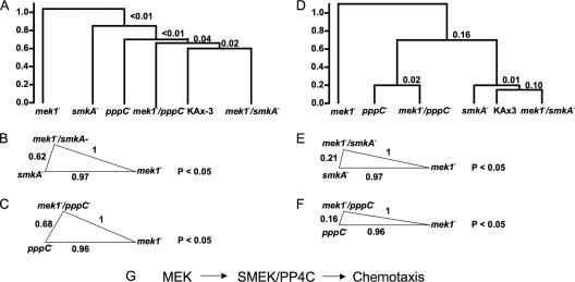
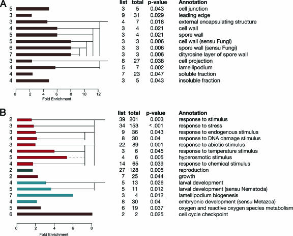
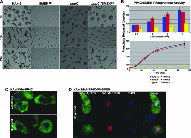
The MEK and extracellular signal-regulated kinase/mitogen-activated protein kinase proteins are established regulators of multicellular development and cell movement. By combining traditional genetic and biochemical assays with a statistical analysis of global gene expression profiles, we discerned a genetic interaction between Dictyostelium discoideum mek1, smkA (named for its role in the suppression of the mek1(-) mutation), and pppC (the protein phosphatase 4 catalytic subunit gene). We found that during development and chemotaxis, both mek1 and smkA regulate pppC function. In other organisms, the protein phosphatase 4 catalytic subunit, PP4C, functions in a complex with the regulatory subunits PP4R2 and PP4R3 to control recovery from DNA damage. Here, we show that catalytically active PP4C is also required for development, chemotaxis, and the expression of numerous genes. The product of smkA (SMEK) functions as the Dictyostelium PP4R3 homolog and positively regulates a subset of PP4C's functions: PP4C-mediated developmental progression, chemotaxis, and the expression of genes specifically involved in cell stress responses and cell movement. We also demonstrate that SMEK does not control the absolute level of PP4C activity and suggest that SMEK regulates PP4C by controlling its localization to the nucleus. These data define a novel genetic pathway in which mek1 functions upstream of pppC-smkA to control multicellular development and chemotaxis.
Figures

Similar articles
-
Depressing time: Waiting, melancholia, and the psychoanalytic practice of care.In: Kirtsoglou E, Simpson B, editors. The Time of Anthropology: Studies of Contemporary Chronopolitics. Abingdon: Routledge; 2020. Chapter 5. In: Kirtsoglou E, Simpson B, editors. The Time of Anthropology: Studies of Contemporary Chronopolitics. Abingdon: Routledge; 2020. Chapter 5. PMID: 36137063 Free Books & Documents. Review.
-
Comparison of Two Modern Survival Prediction Tools, SORG-MLA and METSSS, in Patients With Symptomatic Long-bone Metastases Who Underwent Local Treatment With Surgery Followed by Radiotherapy and With Radiotherapy Alone.Clin Orthop Relat Res. 2024 Dec 1;482(12):2193-2208. doi: 10.1097/CORR.0000000000003185. Epub 2024 Jul 23. Clin Orthop Relat Res. 2024. PMID: 39051924
-
Using Experience Sampling Methodology to Capture Disclosure Opportunities for Autistic Adults.Autism Adulthood. 2023 Dec 1;5(4):389-400. doi: 10.1089/aut.2022.0090. Epub 2023 Dec 12. Autism Adulthood. 2023. PMID: 38116059 Free PMC article.
-
A Blog-Based Study of Autistic Adults' Experiences of Aloneness and Connection and the Interplay with Well-Being: Corpus-Based and Thematic Analyses.Autism Adulthood. 2023 Dec 1;5(4):437-449. doi: 10.1089/aut.2022.0073. Epub 2023 Dec 12. Autism Adulthood. 2023. PMID: 38116056 Free PMC article.
-
Metformin for endometrial hyperplasia.Cochrane Database Syst Rev. 2024 May 2;5(5):CD012214. doi: 10.1002/14651858.CD012214.pub3. Cochrane Database Syst Rev. 2024. PMID: 38695827 Review.
Cited by
-
Global transcriptional responses to cisplatin in Dictyostelium discoideum identify potential drug targets.Proc Natl Acad Sci U S A. 2007 Sep 25;104(39):15406-11. doi: 10.1073/pnas.0705996104. Epub 2007 Sep 18. Proc Natl Acad Sci U S A. 2007. PMID: 17878305 Free PMC article.
-
Protein phosphatase 4 coordinates glial membrane recruitment and phagocytic clearance of degenerating axons in Drosophila.Cell Death Dis. 2017 Feb 23;8(2):e2623. doi: 10.1038/cddis.2017.40. Cell Death Dis. 2017. PMID: 28230857 Free PMC article.
-
The protein phosphatase 4 complex promotes the Notch pathway and wingless transcription.Biol Open. 2017 Aug 15;6(8):1165-1173. doi: 10.1242/bio.025221. Biol Open. 2017. PMID: 28652317 Free PMC article.
-
Moving towards a paradigm: common mechanisms of chemotactic signaling in Dictyostelium and mammalian leukocytes.Cell Mol Life Sci. 2014 Oct;71(19):3711-47. doi: 10.1007/s00018-014-1638-8. Epub 2014 May 21. Cell Mol Life Sci. 2014. PMID: 24846395 Free PMC article. Review.
-
Paxillin and phospholipase D interact to regulate actin-based processes in Dictyostelium discoideum.Eukaryot Cell. 2011 Jul;10(7):977-84. doi: 10.1128/EC.00282-10. Epub 2011 Apr 29. Eukaryot Cell. 2011. PMID: 21531871 Free PMC article.
References
-
- Affolter, M., and C. J. Weijer. 2005. Signaling to cytoskeletal dynamics during chemotaxis. Dev. Cell 9:19-34. - PubMed
-
- Andreeva, A. V., and M. A. Kutuzov. 2001. PPP family of protein Ser/Thr phosphatases: two distinct branches? Mol. Biol. Evol. 18:448-452. - PubMed
-
- Aubry, L., and R. Firtel. 1999. Integration of signaling networks that regulate Dictyostelium differentiation. Annu. Rev. Cell Dev. Biol. 15:469-517. - PubMed
-
- Barford, D. 1996. Molecular mechanisms of the protein serine/threonine phosphatases. Trends Biochem. Sci. 21:407-412. - PubMed
-
- Barford, D., A. K. Das, and M. P. Egloff. 1998. The structure and mechanism of protein phosphatases: insights into catalysis and regulation. Annu. Rev. Biophys. Biomol. Struct. 27:133-164. - PubMed
Publication types
MeSH terms
Substances
Grants and funding
LinkOut - more resources
Full Text Sources
Molecular Biology Databases
Miscellaneous