. 2007 Apr;18(4):1457-63.
doi: 10.1091/mbc.e06-10-0908. Epub 2007 Feb 21.

ERK activity and G1 phase progression: identifying dispensable versus essential activities and primary versus secondary targets

Affiliations

ERK activity and G1 phase progression: identifying dispensable versus essential activities and primary versus secondary targets

Jessie Villanueva et al. Mol Biol Cell. 2007 Apr.

Abstract

The ERK subfamily of MAP kinases is a critical regulator of S phase entry. ERK activity regulates the induction of cyclin D1, and a sustained ERK signal is thought to be required for this effect, at least in fibroblasts. We now show that early G1 phase ERK activity is dispensable for the induction of cyclin D1 and that the critical ERK signaling period is restricted to 3-6 h after mitogenic stimulation of quiescent fibroblasts. Similarly, early G1 phase ERK activity is dispensable for entry into S phase. Moreover, if cyclin D1 is expressed ectopically, ERK activity becomes dispensable throughout the G1 phase. In addition to its effect on cyclin D1, ERK activity is thought to contribute to the down-regulation of p27kip1. We found that this effect is restricted to late G1/S phase. Mechanistic analysis showed that the ERK effect on p27kip1 is mediated by Skp2 and is secondary to its effect on cyclin D1. Our results emphasize the importance of mid-G1 phase ERK activity and resolve primary versus secondary ERK targets within the G1 phase cyclin-dependent kinases.


Figures

Figure 1.
Figure 1.
Early G1 phase ERK activity is dispensable for cyclin D1 gene expression. (A–C) Quiescent MEFs were stimulated with 10% FBS and treated with DMSO or 50 μM U0126 at 0, 2, 4, 6, and 8 h after serum stimulation. All of these samples were collected after 9 h of incubation with 10% FBS. Controls received 10% FBS alone (DMSO; D) or were continuously exposed to U0126 (U). (D–F) Quiescent MEFs were trypsinized and preincubated with DMSO or 50 μM U0126 for 30 min at 37°C before being reseeded in 100-mm dishes and stimulated with 10% FBS in the continued presence of U0126. After 0.5, 1, 2, 3, 5, or 7 h, U0126 was washed-out by rinsing monolayers twice with cold DMEM and then refeeding the cultures with fresh DMEM-10% FBS. All of these samples were collected after 9 h of total incubation and lysed. Controls received 10% FBS alone (DMSO; D) or were continuously exposed to U0126 (U). Equal amounts of total RNA (B and E) or protein (C and F) from collected cells were analyzed by Northern blotting for cyclin D1 mRNA and 28S rRNA (loading control) or Western blotting for cyclin D1, pERK1/2, ERK1/2, and cdk4 (loading control).
Figure 2.
Figure 2.
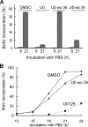
Early G1 phase ERK activity is dispensable for S phase entry. (A) Quiescent MEFs were preincubated with DMSO (vehicle) or 50 μM U0126 (U0) before being reseeded in 100-mm dishes containing coverslips. The cells were stimulated with 10% FBS with BrdU in the continued presence of U0126. U0126 was washed-out (wo) at the indicated times, and the cells were refed DMEM-10% FBS and BrdU without U0126. Coverslips were collected after 9 and 21 h of total incubation. Entry into S phase was measured by incorporation of BrdU using epifluorescence microscopy. Results show the mean ± SD of three experiments. (B) Quiescent MEFs preincubated with DMSO or U0126 as in A, were reseeded onto 35-mm dishes containing coverslips and stimulated with 10% FBS in the presence of BrdU. U0126 was either maintained throughout the mitogenic stimulation (dotted line) or washed-out after 3 h of mitogenic stimulation (dashed line). In the latter case, the cells were refed DMEM-10% FBS and BrdU without U0126. Coverslips were collected at the indicated times, and S phase entry was determined by BrdU incorporation.
Figure 3.
Figure 3.
Enforced expression of cyclin D1 overcomes the need for ERK signaling in G1 phase. MEFs expressing a tetracycline-repressible cyclin D1 were maintained in the presence of 2 μg/ml tetracycline (Tet). Near-confluent cells were serum-starved, preincubated with DMSO or U0126, replated at subconfluence in dishes containing coverslips, and stimulated with 10% FBS, all in the absence or presence of Tet. (A) Collected cells were analyzed by Western blotting with antibodies to pERK, cyclin D1, cyclin A, or cdk4 (loading control). Cyclin D1 overexpression (U0126-tet relative to +tet; 9–15 h) was determined with Image J and normalized to the matched cdk4 loading control. (B) FBS-stimulated cells incubated without Tet were analyzed by Western blotting for the hyperphosphorylation of Rb by gel-shift (the upper and lower arrows, respectively, show hyper- and hypophosphorylated Rb), and the levels of cyclin D1, pERK, and cdk4 (loading control). The samples in A and B were run and analyzed by Western blotting at the same time, but they were divided into different gels as needed to accommodate the number of samples. The small arrowheads indicate where samples were divided into two identical gels. The thick space between the G0 and 6-h samples in A indicates removal of extraneous information. (C) S phase entry was determined from coverslips collected after 21 h of mitogenic stimulation. The results show the mean ± SD of two experiments. (D) Equal amounts of total cell protein from samples collected at G0 or after a 9-h incubation with FBS and DMSO (D) or FBS and 50 μM U0126 (U) were immunoprecipitated (IP) with anti-cdk4 and then blotted with anti-cyclin D1 and anti-cdk4. ns, nonspecific.
Figure 4.
Figure 4.
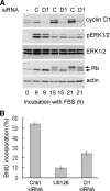
Effect of cyclin D1 siRNA on G1 phase progression. MEFs were either serum-starved and pretreated with 50 μM U0126 or transfected with irrelevant control or cyclin D1 siRNA before serum starvation. The cells were plated at subconfluence in dishes containing coverslips and stimulated with 10% FBS in the presence of BrdU. (A) Western blot of cell lysates treated with control (C) or cyclin D1 (D1) siRNA and probed with antibodies to cyclin D1, pERK1/2, ERK1/2, Rb, and actin (loading control). The upper and lower arrows in the Rb blot indicate hyper- and hypophosphorylated Rb, respectively. (B) BrdU incorporation determined from coverslips collected after 21-h stimulation with 10% FBS.
Figure 5.
Figure 5.
Regulation of p27 at late G1/S phase by ERK. (A) Quiescent MEFs pretreated with DMSO or 50 μM U0126 were plated at subconfluence and stimulated with 10% FBS; DMSO or U0126 remained throughout the FBS incubation period. Cells collected at different times throughout G1 and S phases were analyzed by Western blotting for p27, pERK, and cdk4 (loading control). (B) Serum-starved early passage wild-type or p27187A MEFs pretreated with DMSO or 50 μM U0126 were reseeded at subconfluence and stimulated with 10% FBS for 21 h. Collected cells were analyzed by Western blotting for Skp2, p27, and cdk4 (loading control). (C) MEFs incubated with selected concentrations of U0126 for 12 or 18 h were collected and analyzed by Western blotting for p27, Skp2, Rb, cyclin D1, pERK1/2, and actin (loading control). The upper and lower arrows in the Rb blot show hyper- and hypophosphorylated Rb, respectively. (D) MEFs treated with 10% FBS ± 50 μM U0126 for 21 h were collected and analyzed by QPCR for p27 mRNA, Skp2 mRNA, and 18S rRNA. The figure shows p27 and Skp2 mRNA levels plotted relative to 18S rRNA and normalized to the mRNA levels seen in the FBS-stimulated cells.
Figure 6.
Figure 6.
Regulation of Skp2 and p27 by cyclin D1 RNAi. MEFs transfected with control (C) or cyclin D1 siRNA were serum-starved and then replated at subconfluence with 10% FBS for 20 h. (A) A portion of the collected cells were analyzed by QPCR for Skp2 mRNA and 18S rRNA; the level of Skp2 mRNA plotted relative to 18S rRNA is shown. (B) The remainder of the cells was analyzed by Western blotting for Skp2, cyclin D1, and actin (loading control). (C) Collected cell lysates from an independent experiment were Western blotted for p27, cyclin D1, and actin.
Figure 7.
Figure 7.
Ectopic cyclin D1 rescues Skp2 expression and p27 degradation in MEK/ERK-inhibited cells. (A and B) Serum-starved MEFs infected with 300, 900, or 2700 moi Ad-cyclin D1 (or 2700 moi Ad-LacZ as control; LZ) were pretreated with DMSO or 50 μM U0126, reseeded at subconfluence, and stimulated with 10% FBS for 21 h. Collected cells were analyzed by QPCR for Skp2 mRNA, cyclin E mRNA and 18S rRNA (A), or by Western blotting for Skp2, cyclin D1, and actin (B). The levels of Skp2 and cyclin E mRNAs in A are plotted relative to 18S rRNA and normalized to mRNA levels in the LacZ-infected cells lacking U0126. (C) Serum-starved MEFs infected at 600 moi with Ad-LacZ (LZ) or Ad-cyclin D1 (D1) were serum-starved, pretreated with DMSO or 50 μM U0126, and either immediately collected (0) or replated with 10% FBS for 20 h before collection. The collected cells were analyzed by Western blotting with antibodies to Skp2, p27, cyclin D1, and actin (loading control). Image J was used to quantify the degree of Skp2 expression in Ad-cyclin D1 infected cells treated with U0126, relative to Lac Z–infected cells incubated for 20 h with 10% FBS. The degree of rescue was 40–85%, n = 4.

Similar articles

Cited by

References

    1. Albanese C., Johnson J., Watanabe G., Eklund N., Vu D., Arnold A., Pestell R. G. Transforming p21ras mutants and c-Ets-2 activate the cyclin D1 promoter through distinguishable regions. J. Biol. Chem. 1995;270:23589–23597. - PubMed
    1. Balmanno K., Cook S. J. Sustained MAP kinase activation is required for the expression of cyclin D1, p21Cip1 and a subset of AP-1 proteins in CCL39 cells. Oncogene. 1999;18:3085–3097. - PubMed
    1. Bhatt K. V., Spofford L. S., Aram G., McMullen M., Pumiglia K., Aplin A. E. Adhesion control of cyclin D1 and p27Kip1 levels is deregulated in melanoma cells through BRAF-MEK-ERK signaling. Oncogene. 2005;24:3459–3471. - PubMed
    1. Bohmer R. M., Scharf E., Assoian R. K. Cytoskeletal integrity is required throughout the mitogen stimulation phase of the cell cycle and mediates the anchorage-dependent expression of cyclin D1. Mol. Biol. Cell. 1996;7:101–111. - PMC - PubMed
    1. Bottazzi M. E., Zhu X., Bohmer R. M., Assoian R. K. Regulation of p21(cip1) expression by growth factors and the extracellular matrix reveals a role for transient ERK activity in G1 phase. J. Cell Biol. 1999;146:1255–1264. - PMC - PubMed

Publication types

MeSH terms

LinkOut - more resources