

Dendritic cell expression of the transcription factor T-bet regulates mast cell progenitor homing to mucosal tissue
- PMID: 17296784
- PMCID: PMC2118716
- DOI: 10.1084/jem.20060626
Dendritic cell expression of the transcription factor T-bet regulates mast cell progenitor homing to mucosal tissue
Abstract
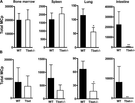
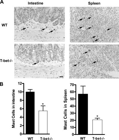
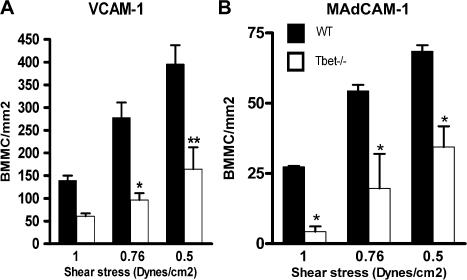
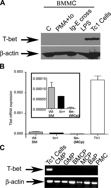
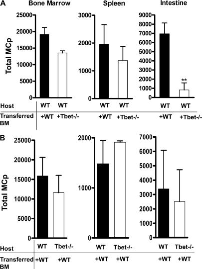
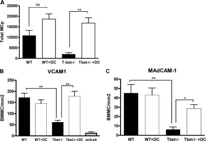
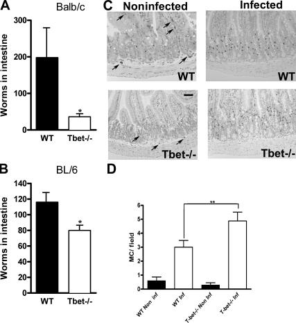
The transcription factor T-bet was identified in CD4(+) T cells, and it controls interferon gamma production and T helper type 1 cell differentiation. T-bet is expressed in certain other leukocytes, and we recently showed (Lord, G.M., R.M. Rao, H. Choe, B.M. Sullivan, A.H. Lichtman, F.W. Luscinskas, and L.H. Glimcher. 2005. Blood. 106:3432-3439) that it regulates T cell trafficking. We examined whether T-bet influences homing of mast cell progenitors (MCp) to peripheral tissues. Surprisingly, we found that MCp homing to the lung or small intestine in T-bet(-/-) mice is reduced. This is reproduced in adhesion studies using bone marrow-derived MCs (BMMCs) from T-bet(-/-) mice, which showed diminished adhesion to mucosal addresin cellular adhesion molecule-1 (MAdCAM-1) and vascular cell adhesion molecule-1 (VCAM-1), endothelial ligands required for MCp intestinal homing. MCp, their precursors, and BMMCs do not express T-bet, suggesting that T-bet plays an indirect role in homing. However, adoptive transfer experiments revealed that T-bet expression by BM cells is required for MCp homing to the intestine. Furthermore, transfer of WT BM-derived dendritic cells (DCs) to T-bet(-/-) mice restores normal MCp intestinal homing in vivo and MCp adhesion to MAdCAM-1 and VCAM-1 in vitro. Nonetheless, T-bet(-/-) mice respond vigorously to intestinal infection with Trichinella spiralis, eliminating a role for T-bet in MC recruitment to sites of infection and their activation and function. Therefore, remarkably, T-bet expression by DCs indirectly controls MCp homing to mucosal tissues.
Figures
Similar articles
-
Integrin alpha4beta7 and its counterreceptor MAdCAM-1 contribute to hematopoietic progenitor recruitment into bone marrow following transplantation.Blood. 2004 Oct 1;104(7):2020-6. doi: 10.1182/blood-2003-12-4157. Epub 2004 May 25. Blood. 2004. PMID: 15161666
-
Intestinal mast cell progenitors require CD49dbeta7 (alpha4beta7 integrin) for tissue-specific homing.J Exp Med. 2001 Nov 5;194(9):1243-52. doi: 10.1084/jem.194.9.1243. J Exp Med. 2001. PMID: 11696590 Free PMC article.
-
Alpha-4 integrins and VCAM-1, but not MAdCAM-1, are essential for recruitment of mast cell progenitors to the inflamed lung.Blood. 2006 Sep 1;108(5):1588-94. doi: 10.1182/blood-2005-12-012781. Epub 2006 May 2. Blood. 2006. PMID: 16670268 Free PMC article.
-
Mast cells: ontogeny, homing, and recruitment of a unique innate effector cell.J Allergy Clin Immunol. 2006 Jun;117(6):1285-91. doi: 10.1016/j.jaci.2006.04.017. J Allergy Clin Immunol. 2006. PMID: 16750988 Review.
-
Mast cell progenitor trafficking and maturation.Adv Exp Med Biol. 2011;716:14-28. doi: 10.1007/978-1-4419-9533-9_2. Adv Exp Med Biol. 2011. PMID: 21713649 Free PMC article. Review.
Cited by
-
T-bet-/- RAG2-/- ulcerative colitis: the role of T-bet as a peacekeeper of host-commensal relationships.Cytokine. 2009 Oct-Nov;48(1-2):144-7. doi: 10.1016/j.cyto.2009.07.007. Epub 2009 Aug 8. Cytokine. 2009. PMID: 19666230 Free PMC article. Review.
-
Innate immune response of alveolar macrophage to house dust mite allergen is mediated through TLR2/-4 co-activation.PLoS One. 2013 Oct 1;8(10):e75983. doi: 10.1371/journal.pone.0075983. eCollection 2013. PLoS One. 2013. PMID: 24098413 Free PMC article.
-
Left Ventricular T-Cell Recruitment Contributes to the Pathogenesis of Heart Failure.Circ Heart Fail. 2015 Jul;8(4):776-87. doi: 10.1161/CIRCHEARTFAILURE.115.002225. Epub 2015 May 28. Circ Heart Fail. 2015. PMID: 26022677 Free PMC article.
-
Isoforms of Vitamin E Differentially Regulate PKC α and Inflammation: A Review.J Clin Cell Immunol. 2013 Mar 14;4(137):1000137. doi: 10.4172/2155-9899.1000137. J Clin Cell Immunol. 2013. PMID: 23977443 Free PMC article.
-
Increased T-bet+ cytotoxic effectors and type I interferon-mediated processes in chronic graft-versus-host disease of the oral mucosa.Blood. 2009 Apr 9;113(15):3620-30. doi: 10.1182/blood-2008-07-168351. Epub 2009 Jan 23. Blood. 2009. PMID: 19168793 Free PMC article.
References
-
- Rodewald, H.R., M. Dessing, A.M. Dvorak, and S.J. Galli. 1996. Identification of a committed precursor for the mast cell lineage. Science. 271:818–822. - PubMed
-
- Williams, C.M., and S.J. Galli. 2000. The diverse potential effector and immunoregulatory roles of mast cells in allergic disease. J. Allergy Clin. Immunol. 105:847–859. - PubMed
-
- Metcalfe, D.D., D. Baram, and Y.A. Mekori. 1997. Mast cells. Physiol. Rev. 77:1033–1079. - PubMed
Publication types
MeSH terms
Substances
Grants and funding
- U19 AI031599/AI/NIAID NIH HHS/United States
- R01 HL053993/HL/NHLBI NIH HHS/United States
- P01 HL036028/HL/NHLBI NIH HHS/United States
- R01 CA112663/CA/NCI NIH HHS/United States
- AI56296/AI/NIAID NIH HHS/United States
- HL56985/HL/NHLBI NIH HHS/United States
- AI 031599/AI/NIAID NIH HHS/United States
- HL 036110/HL/NHLBI NIH HHS/United States
- P50 HL056985/HL/NHLBI NIH HHS/United States
- CA112663/CA/NCI NIH HHS/United States
- P01 AI056296/AI/NIAID NIH HHS/United States
- P01 AI031599/AI/NIAID NIH HHS/United States
- P01 HL036110/HL/NHLBI NIH HHS/United States
- HL36028/HL/NHLBI NIH HHS/United States
- G108/380/MRC_/Medical Research Council/United Kingdom
- HL53993/HL/NHLBI NIH HHS/United States
LinkOut - more resources
Full Text Sources
Other Literature Sources
Medical
Molecular Biology Databases
Research Materials
Miscellaneous
