. 2007 Apr;27(8):3131-42.
doi: 10.1128/MCB.02190-06. Epub 2007 Feb 12.

The human Tim/Tipin complex coordinates an Intra-S checkpoint response to UV that slows replication fork displacement

Affiliations

The human Tim/Tipin complex coordinates an Intra-S checkpoint response to UV that slows replication fork displacement

Keziban Unsal-Kaçmaz et al. Mol Cell Biol. 2007 Apr.

Abstract

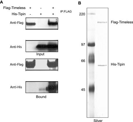
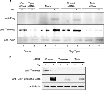
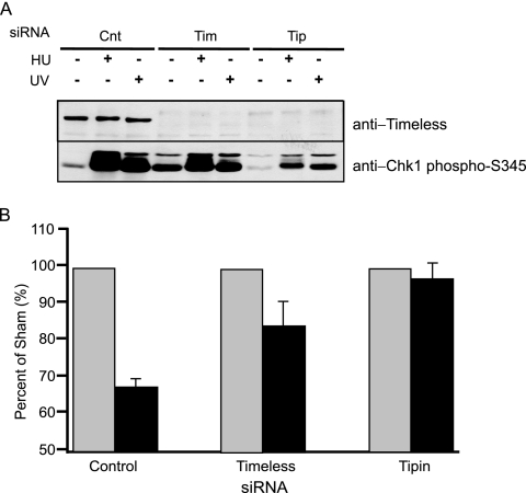
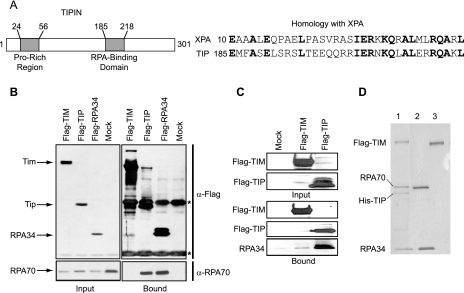
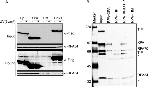
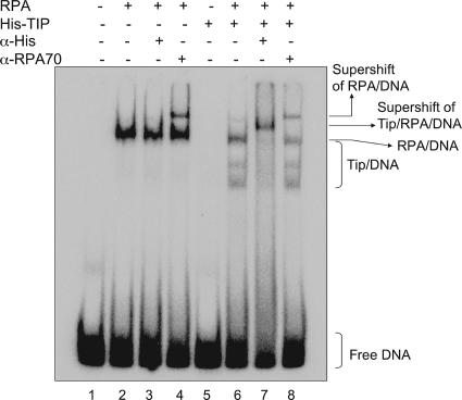
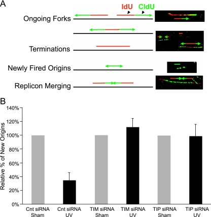
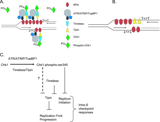
UV-induced DNA damage stalls DNA replication forks and activates the intra-S checkpoint to inhibit replicon initiation. In response to stalled replication forks, ATR phosphorylates and activates the transducer kinase Chk1 through interactions with the mediator proteins TopBP1, Claspin, and Timeless (Tim). Murine Tim recently was shown to form a complex with Tim-interacting protein (Tipin), and a similar complex was shown to exist in human cells. Knockdown of Tipin using small interfering RNA reduced the expression of Tim and reversed the intra-S checkpoint response to UVC. Tipin interacted with replication protein A (RPA) and RPA-coated DNA, and RPA promoted the loading of Tipin onto RPA-free DNA. Immunofluorescence analysis of spread DNA fibers showed that treating HeLa cells with 2.5 J/m(2) UVC not only inhibited the initiation of new replicons but also reduced the rate of chain elongation at active replication forks. The depletion of Tim and Tipin reversed the UV-induced inhibition of replicon initiation but affected the rate of DNA synthesis at replication forks in different ways. In undamaged cells depleted of Tim, the apparent rate of replication fork progression was 52% of the control. In contrast, Tipin depletion had little or no effect on fork progression in unirradiated cells but significantly attenuated the UV-induced inhibition of DNA chain elongation. Together, these findings indicate that the Tim-Tipin complex mediates the UV-induced intra-S checkpoint, Tim is needed to maintain DNA replication fork movement in the absence of damage, Tipin interacts with RPA on DNA and, in UV-damaged cells, Tipin slows DNA chain elongation in active replicons.


Figures

FIG. 1.
FIG. 1.
Tim forms a complex with Tipin. (A) Tim-Tipin interaction in mammalian cells. HEK293T cells were transfected either with Flag-Tim or with His-Tipin or were cotransfected with Flag-Tim and His-Tipin and then immunoprecipitated (IP) with anti-Flag antibodies. Immunoprecipitates were separated by SDS-polyacrylamide gel electrophoresis (PAGE) and then immunoblotted with antibodies to Flag and His as indicated. Input represents 1/30 of the cell lysates used for immunoprecipitation. (B) Purification of Tim-Tipin complex. Insect (Sf21) cells were coinfected with baculoviruses expressing His-Flag-Tim and His-Tipin, and proteins from extracts were first collected with Ni-NTA agarose and eluted with 200 mM imidazole. The eluates were then collected with anti-Flag agarose, and proteins were eluted by Flag peptide. The profile of the purified complex was visualized on a silver-stained SDS-polyacrylamide gel.
FIG. 2.
FIG. 2.
Loss of Tipin downregulates the expression level of Tim and attenuates Chk1 activation. (A) Tet-inducible expression of Flag-Tipin. Proteins were induced in the presence of Tet (1 mM) for 24 h and detected with anti-Flag antibodies (upper panel). HEK293T cells with inducible expression of Flag-Tipin and the control cell line expressing vector only were transfected with scrambled control or Tipin siRNA two times over a 3-day period. Forty-eight hours after the initial transfection, cells were treated with Tet or left untreated. A vector-expressing cell line (Mock) was used as a negative control for inducible protein expression and positive control for the siRNA treatment, where reduced expression of Tim in Tipin siRNA-treated cells can be seen as a result of successful knockdown of endogenous Tipin (lane 2). Cell lysate proteins (80 μg) were immunoblotted with anti-Tim, anti-Flag, and anti-actin antibodies (lower panel). Cnt, control. (B) Tipin is required for HU-induced Chk1 activation. HeLa cells were transfected with control, Tim, or Tipin siRNA two times over a 3-day period. Seventy-two hours after the initial transfection, cells were treated with 10 mM HU for 1.5 h or left untreated. Cell lysate proteins (150 μg) were immunoblotted with anti-Tim, anti-Chk1-phosphoS345, and antiactin antibodies.
FIG. 3.
FIG. 3.
Knockdown of Tipin attenuates the intra-S checkpoint response to UV. (A) Tipin is required for UV-induced Chk1 activation. HeLa cells were transfected with scrambled control (Cnt), Tim, or Tipin siRNA two times over a 3-day period. Seventy-two hours after the initial transfection, cells were treated with either 10 mM HU for 1.5 h or UV (6 J/m2) for 1 h or were left untreated. Cell lysate proteins (150 μg) were immunoblotted with anti-Tim and anti-Chk1-phosphoS345. (B) HeLa cells that were transfected with scrambled control, Tim, and Tipin siRNA were grown in the presence of [14C]thymidine for 40 h to label DNA uniformly until the second transfection and then grown in nonradioactive medium for an additional 24 h. Cells were exposed to UV (2.5 J/m2) or left untreated, incubated at 37°C for 30 min, and then labeled for 15 min in medium containing [3H]thymidine. Relative DNA synthesis was estimated from the incorporated [3H]thymidine normalized to total DNA by the 14C radioactivity. Data are expressed as percentages of the control samples (no UV irradiation) and plotted as means ± standard deviations (SD; n = 3). 3H/14C ratios in untreated cells were 67 ± 21% and 41 ± 21% (mean ± SD) of the scrambled control cells for Tim and Tipin knockdown cells, respectively.
FIG. 4.
FIG. 4.
Interaction of Tipin with RPA. (A) Schematic representation of human Tipin protein with proline-rich and RPA-binding domains and protein sequence alignment of XPA and Tipin homology regions. (B) Tipin-RPA interaction in mammalian cells. HEK293T cells were transfected with vector (Mock; lane 4) or plasmids expressing Flag-Tim (Flag-TIM; lane1), Flag-Tipin (Flag-TIP; lane 2), and Flag-RPA34 (lane 3). Flag immunoprecipitates (Bound) as well as the whole-cell extract (Input) were separated by SDS-PAGE and blotted with anti-Flag and anti-RPA70 antibodies. Asterisks (*) indicate the heavy and light chains of the anti-Flag antibody. Input represents 1/30 of the cell lysates used for immunoprecipitation. (C) Tipin-RPA interaction in vitro. Insect cells (Sf21) were either uninfected (Mock; lane 1) or infected with viruses expressing Flag-Tim (lane 2) and Flag-Tipin (lane 3). Flag-purified proteins were incubated with 3.5 μg of RPA while still on the beads overnight at 4°C, and then the Flag agarose beads were washed three times. The bound proteins were eluted with Flag peptide and separated by SDS-PAGE and blotted with anti-Flag and anti-RPA34 antibodies. (D) Formation of Tim-Tipin-RPA complexes. Insect cells were either coinfected with viruses expressing Flag-Tim and His-Tipin (lane 1) or infected only with Flag-Tim virus (lane 3). The Tim-Tipin complex was purified by chromatograpy with Ni-NTA-agarose and anti-Flag agarose as described in Materials and Methods. Tim was purified by chromatography with anti-Flag agarose. Proteins bound to beads were incubated with RPA (lanes 1 and 3) on ice for one hour. After extensive washing, the proteins bound to beads were eluted with Flag peptide, and an aliquot of the first elution was analyzed by SDS-PAGE followed by silver staining. The second lane contains 1/15 of the RPA used in the binding assay.
FIG. 5.
FIG. 5.
Tipin and XPA bind to RPA with comparable affinities. (A) UV damage does not stimulate the interactions between either XPA and RPA or Tipin and RPA. HEK293T cells transfected with vector (Cnt), Flag-Tipin (Tip), Flag-XPA (XPA), or Flag-Chk1 (Chk1) were either left untreated or treated with UV and then lysed 1 h later. An equal amount of cell lysate (2 mg) was immunoprecipitated with anti-Flag agarose and separated by SDS-PAGE. Flag immunoprecipitates (Bound) as well as the whole cell extract (Input) were blotted with anti-Flag and anti-RPA34 antibodies. Input represents 1/30 of the whole-cell lysate used for immunoprecipitation. (B) Tipin and XPA bind to RPA in vitro with comparable affinities. RPA protein (10 μg) was either mixed with 2 μg of XPA (RPA+XPA; lane 3), 2 μg of Tipin (RPA+TIP; lane 4), 2 μg of Tipin and 2 μg XPA (RPA+XPA+TIP; lane 5), or 2 μg of Tim (RPA+TIM; lane 6). Immunoprecipitation experiments were carried out with anti-RPA34 antibody. A fraction of bound proteins was separated by SDS-PAGE and visualized by silver staining. Input lane contains 1/10 of the amount of XPA, Tipin, and Tim and 1/40 of that of RPA used in the binding assay (lane 2). Asterisks (*) indicate the heavy and light chains of the anti-RPA34 antibody.
FIG. 6.
FIG. 6.
RPA recruits Tipin to DNA. Terminally labeled 50-bp duplex DNA was incubated with ∼200 ng of RPA, 150 ng of His-Tipin, or both proteins, and the DNA-protein complexes were separated on a 5% polyacrylamide gel. Anti-His and anti-RPA70 antibodies were added to the samples during the reaction where indicated. Lane 1 is DNA only.
FIG. 7.
FIG. 7.
Knockdown of Tim and Tipin reverses the inhibition of replicon initiation in UV-treated HeLa cells. (A) Left, schematic illustration of results expected when a 10-min pulse of asynchronous cells with IdU is followed by a 20-min pulse with CldU and individual replication units are visualized by immunofluorescence detection of the incorporated halogenated nucleotides in DNA spreads. Right, the presence and relative positions of single and dual labeling in continuous replication tracks are used to interpret their representations of various stages of DNA synthesis. (B) Percentage (compared to the sham-treated control [Cnt]) of new origin initiation (green tracks). Tipin (Tip) or Tim protein levels were knocked down by siRNA prior to the exposure of HeLa cells to 2.5 J/m2 of UV (Table 1). The data are the means from three independent experiments (plus one SD). The results showed a significant UV-induced inhibition in green-only tracks in cells transfected with scrambled control siRNA (two-sided P value, <0.0001) and insignificant UV response in cells with knockdown of Tim (two-sided P value, 0.47) or Tipin (two-sided P value, 0.94).
FIG. 8.
FIG. 8.
Theoretical model depicting potential interactions in the intra-S checkpoint signaling pathway. (A) After UV-induced DNA damage, the Tim-Tipin complex brings Chk1 to sites of ssDNA coated with RPA, where the ATR/ATRIP/TopBP1 complex phosphorylates multiple molecules of Chk1 that diffuse away from the stalled fork to transduce signal throughout the nucleus (3). This model differs from that of Gotter et al. (18), as DNA damage did not destabilize the interaction between Tipin and RPA in checkpoint-competent HEK293T cells (Fig. 5A). Thus, with uncoupling of helicase and polymerase activities at sites of stalled replication forks, the increased amount of RPA-coated DNA increases the opportunity for Tim/Tipin/Chk1 complexes to interact with ATR/ATRIP/TopBP1 complexes to phosphorylate and activate Chk1. (B) Tipin molecules may be loaded onto DNA by RPA. When Tim is depleted by siRNA, loading of Tipin at replication forks may inhibit DNA chain elongation. (C) Tipin inhibits DNA chain elongation and replication fork progression, and this activity is inhibited by Tim. Chk1 also preserves replication fork progression (52), and this may be through an effect on Tipin or the Tim/Tipin complex (indicated by “?”).

Similar articles

Cited by

References

    1. Aten, J. A., P. J. Bakker, J. Stap, G. A. Boschman, and C. H. Veenhof. 1992. DNA double labelling with IdUrd and CldUrd for spatial and temporal analysis of cell proliferation and DNA replication. Histochem. J. 24:251-259. - PubMed
    1. Bassett, E., N. M. King, M. F. Bryant, S. Hector, L. Pendyala, S. G. Chaney, and M. Cordeiro-Stone. 2004. The role of DNA polymerase eta in translesion synthesis past platinum-DNA adducts in human fibroblasts. Cancer Res 64:6469-6475. - PubMed
    1. Bekker-Jensen, S., C. Lukas, R. Kitagawa, F. Melander, M. B. Kastan, J. Bartek, and J. Lukas. 2006. Spatial organization of the mammalian genome surveillance machinery in response to DNA strand breaks. J. Cell Biol. 173:195-206. - PMC - PubMed
    1. Byun, T. S., M. Pacek, M. C. Yee, J. C. Walter, and K. A. Cimprich. 2005. Functional uncoupling of MCM helicase and DNA polymerase activities activates the ATR-dependent checkpoint. Genes Dev. 19:1040-1052. - PMC - PubMed
    1. Calzada, A., B. Hodgson, M. Kanemaki, A. Bueno, and K. Labib. 2005. Molecular anatomy and regulation of a stable replisome at a paused eukaryotic DNA replication fork. Genes Dev. 19:1905-1919. - PMC - PubMed

Publication types

MeSH terms

LinkOut - more resources