Comparative Study
. 2007 Feb 21;26(4):1163-75.
doi: 10.1038/sj.emboj.7601568. Epub 2007 Feb 8.

Disease mutations in RUNX1 and RUNX2 create nonfunctional, dominant-negative, or hypomorphic alleles

Affiliations
Comparative Study

Disease mutations in RUNX1 and RUNX2 create nonfunctional, dominant-negative, or hypomorphic alleles

Christina J Matheny et al. EMBO J. .

Abstract

Monoallelic RUNX1 mutations cause familial platelet disorder with predisposition for acute myelogenous leukemia (FPD/AML). Sporadic mono- and biallelic mutations are found at high frequencies in AML M0, in radiation-associated and therapy-related myelodysplastic syndrome and AML, and in isolated cases of AML M2, M5a, M3 relapse, and chronic myelogenous leukemia in blast phase. Mutations in RUNX2 cause the inherited skeletal disorder cleidocranial dysplasia (CCD). Most hematopoietic missense mutations in Runx1 involve DNA-contacting residues in the Runt domain, whereas the majority of CCD mutations in Runx2 are predicted to impair CBFbeta binding or the Runt domain structure. We introduced different classes of missense mutations into Runx1 and characterized their effects on DNA and CBFbeta binding by the Runt domain, and on Runx1 function in vivo. Mutations involving DNA-contacting residues severely inactivate Runx1 function, whereas mutations that affect CBFbeta binding but not DNA binding result in hypomorphic alleles. We conclude that hypomorphic RUNX2 alleles can cause CCD, whereas hematopoietic disease requires more severely inactivating RUNX1 mutations.


Figures
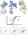
Figure 1
Figure 1
Effects of missense mutations on RD stability. (A) Ribbon diagram of the RD:CBFβ:DNA ternary complex (Bravo et al, 2001; Tahirov et al, 2001) and mutated residues. The RD and CBFβ are shown in gray and blue, respectively, and DNA is purple. Amino acids mutated in Runx2 in CCD (Lee et al, 1997; Quack et al, 1999; Zhou et al, 1999; Yoshida et al, 2002; Zhang et al, 2003) are yellow, whereas green indicates amino acids mutated in Runx1 in FPD/AML, AML M0 subtype, radiation-associated and therapy-related myelodysplastic syndrome and AML, AML M2, M5a, M3 relapse, and chronic myelogenous leukemia in blast phase (Osato et al, 1999; Song et al, 1999; Preudhomme et al, 2000; Buijs et al, 2001; Michaud et al, 2002; Walker et al, 2002; Harada et al, 2003; Roumier et al, 2003; Harada et al, 2004). R139 and R174, which are mutated in both CCD and in hematopoietic diseases, are indicated with yellow and green labels. Shown in gray is T161, an energetic hot spot at the CBFβ interface (Zhang et al, 2003). (B) The fraction of unfolded RD in the presence of increasing concentrations of urea. The boxes indicate proteins that (from right to left) had unaltered, moderately compromised, and more severely compromised stability. (C) 15N-1H-HSQC spectra of WT and mutated RDs. Each panel contains the spectrum of the WT RD (black) overlaid on the RD mutant spectrum (red). The R174Q+WT RD spectra were recorded in the absence (left panel) and presence (middle panel) of DNA. L148F was recorded in the presence of DNA. Arrows on the L148F spectra indicate several examples of peaks specific to the DNA-bound form of the RD that are shifted in the L148F spectrum relative to the WT spectrum, indicative of a conformational change in the DNA-bound L148F protein.
Figure 2
Figure 2
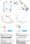
DNA and CBFβ binding by mutant RDs. (A) Schematic diagram of the potential interactions between the RD (gray), CBFβ (blue), and DNA (purple). These interactions can be described by four equilibrium constants: K1, the dissociation constant for the RD-CBFβ heterodimer in the absence of DNA; K2, the dissociation constant for the RD–DNA complex; K3, the dissociation constant for CBFβ binding to the RD–DNA complex; and K4 which describes binding of the RD–CBFβ heterodimer to DNA. (B) FRET assay to determine K1. Cerulean (Cer), an optimized version of the cyan fluorescent protein (Rizzo et al, 2004), was fused to the N-terminus of the RD, and the YFP derivative Venus (Rizzo et al, 2004) was fused to the N-terminus of CBFβ. Cerulean was excited at 433 nm and emission from Cerulean and Venus detected at 474 and 525 nm, respectively. (C) Fluorescence spectra of Cerulean-RD and Venus-CBFβ showing the FRET effect. The black curve is the spectrum of Cerulean-RD+Venus-CBFβ at a concentration of 25 nM (4.5-fold below K1). The red curve is the spectrum of Cerulean-RD+Venus-CBFβ at a concentration of 400 nM (3.6-fold above K1). (D) FRET assay of the WT RD binding to CBFβ. The samples were excited at 433 nm, and the ratio of Venus-CBFβ/Cerulean-RD emission peaks (525/474 nm) was plotted at different protein concentrations to generate a binding curve. (E) Yeast one-hybrid assay for RD binding to three core sites driving lacZ expression. Mutations that increase K2 by ⩾10-fold decrease β-galactosidase activity in a yeast one-hybrid filter assay to undetectable levels (Li et al, 2003). Visible but decreased β-galactosidase activity in the filter assay reflects K2 increases in the 3–10-fold range. (F) Modified yeast one-hybrid assay to measure binding of the RD:CBFβ heterodimer to DNA. Although the Gal4 DNA-binding domain is fused to CBFβ in the modified yeast one-hybrid assay, there are no Gal4-binding sites on the promoter driving lacZ and therefore CBFβ's activity is mediated only through the core sites. CBFβ increases the affinity of the RD for DNA by approximately 10-fold. RD mutants that can bind CBFβ and have K2 increases in the 10–90-fold range and correspondingly no β-galactosidase activity in the one-hybrid assay, yield β-galactosidase signals in the modified one-hybrid assay (Li et al, 2003). On the other hand, RD mutants with K2 values that are >100-fold higher than the WT RD produce very weak or no β-galactosidase activity in the modified yeast one-hybrid assay (Li et al, 2003). (G) EMSA measuring the affinity of the WT RD (top) and the L148F RD (bottom) for DNA (K2). Triangles indicate decreasing concentrations of RDs (WT RD, 2 × 10⁻⁶ to 4 × 10⁻¹⁵ M; L148F, 1 × 10⁻⁵ to 3 × 10⁻¹⁴ M). Arrows indicate lanes in which the RD concentration approximates K2. (H) EMSA measuring the affinity of the WT RD:DNA complex (top) and L148F RD:DNA complex (bottom) for CBFβ (K3). Triangles indicate decreasing concentrations of CBFβ (for the WT RD, 6 × 10⁻⁶–4 × 10⁻¹⁴ M; L148F, 2 × 10⁻⁵–4 × 10⁻¹² M). Arrows indicate lanes in which the CBFβ concentration approximates K3.
Figure 3
Figure 3
Generation and analysis of mutant Runx1 alleles. (A) Targeting vector. Point mutations were engineered into exon 4 of Runx1. A neomycin resistance gene flanked by loxP sites is in intron 4. The location of probes and restriction length fragments from the WT and targeted Runx1 alleles are indicated. B, BamHI; Xb, XbaI; S, SalI; Av, AvrII; Af, AflII; Xh, XhoI. (B) Southern blot screening of L148F/+ ES cell clones with the 3′ probe. (C) Other Runx1 alleles used in these experiments, which include a floxed locus to control for the presence of Neo in intron 4 (Runx1f) (Growney et al, 2005), and an exon 4-deleted allele (Runx1ΔE4) (Wang et al, 1996a). (D) Western blot of nuclear extracts from COS cells transfected with cDNAs encoding mutant Runx1 proteins (left and middle panels), and from thymus extracts prepared from 6-week-old R177X/+ mice and +/+ littermates (right panel). No truncated R177X protein (expected position of the 17.9 kDa protein indicated by arrow) was detected in thymus extracts. (E) R177X/R177X 12.5 d.p.c. fetuses, post-natal day 7 (P7) T161A/T161A mice, and P14 T149A/T149A mice. WT littermates are shown to the right (R177X and T161A) or on top (T149A). Note the pale liver (arrow) and hemorrhages (arrowheads) in the R177X/R177X fetuses, which is characteristic of Runx1 deficiency (Okuda et al, 1996; Wang et al, 1996a). (F) Histologic sections of lung from 2-day-old T161A/T161A and +/+ littermates (× 400). Arrows indicate an airway with infiltration of neutrophils in the T161A/T161A animal and an unaffected airway in the +/+ animal. Bronchopneumonia was diagnosed in three of the four T161A/T161A neonates analyzed. The accumulation of red blood cells in the +/+ animal is an artifact caused by decapitation.
Figure 4
Figure 4
AGM and fetal liver CFU-C assays from 11.5 d.p.c. fetuses. (A) Total number of CFU-Cs (erythroid+granulocyte macrophage+granulocyte erythrocyte monocyte megakaryocyte) per AGM region of Runx1m/m fetuses. Error bars represent 95% confidence intervals. All m/m fetuses were significantly different from +/+ (*). P-values were determined by unpaired two-tailed Student's t-test. n+/+=38; nf/f=9; nR174Q/R174Q=4; nR177X/R177X=3; nL148F/L148F=5; nT161A/T161A=5; nT149A/T149A=10. +/+ values are pooled from all m/+ intercrosses. (B) Total number of fetal liver CFU-Cs (11.5 d.p.c.). All m/m values were significantly different from +/+ (*). n+/+=59; nR174Q/R174Q=3; nR177X/R177X=4; nL148F/L148F=5; nT161A/T161A=5; nT149A/T149A=12. (C) Total number of CFU-Cs per AGM region from Runx1m/+ fetuses. Significant differences from ΔE4/+ (*) are indicated. The difference between R174Q/+ and ΔE4/+ was significant at P=0.06 by unpaired two-tailed Student's t-test, and at P=0.03 by unpaired one-tailed Student's t-test. n+/+=38; nΔE4/+=15; nR174Q/+=14; nR177X/+=10; nL148F/+=8; nT161A/+=10; nT149A/+=39. (D) Total number of CFU-Cs per 11.5 d.p.c. Runx1m/+ fetal liver. Significant differences from ΔE4/+ (*) are indicated (unpaired two-tailed Student's t-test). n+/+=59; nΔE4/+=24; nR174Q/+=13; nR177X/++19; nL148F/+=9; nT161A/+=9; nT149A/+=41.

Similar articles

Cited by

References

    1. Berardi M, Sun C, Zehr M, Abildgarrd F, Peng J, Speck NA, Bushweller JH (1999) The Ig fold of the core binding factor a Runt domain is a member of a family of structurally and functionally related Ig-fold DNA-binding domains. Structure 10: 1247–1256 - PubMed
    1. Bravo J, Li Z, Speck NA, Warren AJ (2001) The leukemia-associated AML1 (Runx1)–CBFβ complex functions as a DNA-induced molecular clamp. Nat Struct Biol 8: 371–378 - PubMed
    1. Buijs A, Poddighe P, van Wijk R, van Solinge W, Borst E, Verdonck L, Hagenbeek A, Perason P, Lokhorst H (2001) A novel CBFA2 single-nucleotide mutation in familial platelet disorder with propensity to develop myeloid malignancies. Blood 98: 2856–2858 - PubMed
    1. Bullock AN, Fersht AR (2001) Rescuing the function of mutant p53. Nat Rev Cancer 1: 68–76 - PubMed
    1. Corazza A, Pettirossi F, Viglino P, Verdone G, Garcia J, Dumy P, Giorgetti S, Mangione P, Raimondi S, Stoppini M, Bellotti V, Esposito G (2004) Properties of some variants of human beta2-microglobulin and amyloidogenesis. J Biol Chem 279: 9176–9189 - PubMed

Publication types

MeSH terms