Comparative Study
. 2007 Jan 31;27(5):981-92.
doi: 10.1523/JNEUROSCI.4810-06.2007.

A Drosophila model of mutant human parkin-induced toxicity demonstrates selective loss of dopaminergic neurons and dependence on cellular dopamine

Affiliations
Comparative Study

A Drosophila model of mutant human parkin-induced toxicity demonstrates selective loss of dopaminergic neurons and dependence on cellular dopamine

Tzu-Kang Sang et al. J Neurosci. .

Abstract

Mutations in human parkin have been identified in familial Parkinson's disease and in some sporadic cases. Here, we report that expression of mutant but not wild-type human parkin in Drosophila causes age-dependent, selective degeneration of dopaminergic (DA) neurons accompanied by a progressive motor impairment. Overexpression or knockdown of the Drosophila vesicular monoamine transporter, which regulates cytosolic DA homeostasis, partially rescues or exacerbates, respectively, the degenerative phenotypes caused by mutant human parkin. These results support a model in which the vulnerability of DA neurons to parkin-induced neurotoxicity results from the interaction of mutant parkin with cytoplasmic dopamine.


Figures

Figure 1.
Figure 1.
Domain structure of human parkin and Myc-tagged wild-type and FLAG-tagged mutant constructs used to generate transgenic flies.
Figure 2.
Figure 2.
Mutant parkin produces age-dependent deficits in motor performance. Three independent assays were used to analyze motor behavior. A, Using the ddc-GAL4 driver, parkinQ311X and parkinT240R produce age-dependent impairments in climbing performance compared with control and parkinwt. For each genotype at each time point, at least five cohorts consisting of 16–18 flies each were tested. B, At 5 weeks, the righting reflex test demonstrates postural instability of parkinQ311X and parkinT240R flies compared with parkinwt or control. Twenty flies of each genotype were analyzed. C, Rotarod performance demonstrates normal postural stability for all genotypes at 2 weeks. D, By 4 weeks, postural instability is apparent in parkinQ311X and parkinT240R compared with parkinwt and control flies; the <1 score for the arbitrary index at higher angles indicates flies that fall more frequently. For each genotype at each time point, ≥12 flies were tested. Values shown represent mean ± SEM. **p < 0.01, ***p < 0.001 relative to control ddc [two-way ANOVA with Bonferroni's multiple comparison test (A, C, D) or unpaired t test (B)].
Figure 3.
Figure 3.
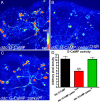
Analysis of activity-dependent G-CaMP signal in living brains at 1 week after eclosion. GFP signal intensity was converted into thermally coded color images. A, In the control ddc::G-CaMP brain, intense GFP signal was detected. B, parkinT240R expressed in ddc::G-CaMP flies resulted in markedly reduced GFP signal. C, Expression with parkinwt resembled the control. D, Analysis of GFP pixel density of at least five brains for each genotypes showed decreased G-CaMP activity in the parkinT240R brain (one-way ANOVA with Bonferroni's multiple comparison test; n ≥ 5; **p < 0.01). The images are represented using a pseudocolor scale as indicated in A, where blue is low intensity and yellow–red is high intensity. Scale bar, 40 μm.
Figure 4.
Figure 4.
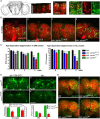
Expression of mutant human parkin in Drosophila DA neurons causes age-dependent neurodegeneration. A, Schematic representation of DA neurons in the central brain of Drosophila. a, The projected confocal image shows anti-TH and phalloidin staining spanning optical sections 40 μm in depth from posterior to anterior. Two major DA clusters, DM (circles) and DL (rectangles), are indicated. b, A projected image shows anti-TH (red) and anti-GFP (green) colocalized in all DA neurons in both DM and DL (enlarged images to the right) clusters. Arrows indicate 5-HT neurons expressing GFP but not TH. B, Reductions in TH immunoreactivity at 5 weeks after eclosion are apparent in ddc::parkinQ311X (b) and ddc::parkinT240R (c) but not ddc control (a) or ddc::parkinwt (d) brains. Circles, DM; rectangles, DL. C, Quantification of DA neurons at 0, 3, and 5 weeks after eclosion in DM and DL clusters. Progressive loss of TH-immunoreactive neurons is induced in both clusters by ddc::parkinQ311X (blue bars) and ddc::parkinT240R (red bars) compared with the ddc control (black bars) and ddc::parkinwt (green bars). Values represent the mean ± SEM; n = 12. *p < 0.05, **p < 0.01, ***p < 0.001 relative to the ddc control. D, At 4 weeks, mCD8-GFP signal (green) is reduced in parkinT240R (b) compared with parkinwt (a) brain, which is similar to changes observed in corresponding images showing reduced TH immunoreactivity. Measurement of GFP pixel density (corresponding to the left y-axis) and GFP cell counts in the DM cluster (corresponding to the right y-axis) from 4-week-old brains of both genotypes showed a significant decrease (unpaired t test; n = 5; *p < 0.05) of GFP signal in parkinT240R brains compared with parkinwt (c). The GFP signal at eclosion was indistinguishable between parkinT240R and parkinwt (data not shown). d, e, Confocal images of ddc::parkinT240R (e) and ddc::parkinwt (d) brains stained with an anti-parkin antibody (green) reveals reduced parkin signals in ddc::parkinT240R compared with ddc::parkinwt brains. f, Analysis of brains at 4 weeks indicates that cell counts stained with anti-parkin for parkinT240R are decreased compared with parkinwt (unpaired t test; n = 6; *p < 0.05 for DM; ***p < 0.001 for DL). Parkin staining at eclosion was indistinguishable between parkinT240R and parkinwt (data not shown). a, b, d, and e are confocal images of adjacent brains imaged simultaneously in the same slide but reoriented for presentation. E, Anti-5-HT staining at 5 weeks after eclosion shows reduced neuritic immunoreactivity in 5-HT neurons expressing mutant parkin (b, c) compared with those expressing wild-type parkin (d) or controls (a). Scale bar, 40 μm.
Figure 5.
Figure 5.
Pan-neuronal expression of parkinT240R induces motor dysfunction and degeneration of DA neurons, whereas photoreceptor neurons are resistant. A, Rotarod assays at 5 weeks demonstrate postural instability of appl:: parkinT240R flies, which fall more frequently at higher angle positions compared with the appl-GAL4 control and appl::parkinwt. appl:: parkinQ311X flies show a modest trend toward postural instability. For each genotype at each time point, >12 flies were scored. The values shown represent mean ± SD. ***p < 0.001 relative to control appl (two-way ANOVA with Bonferroni's multiple-comparison test). Ba–Bd, Confocal images of brains aged 5 weeks stained with anti-TH (green) and phalloidin (red). Circles, DM; rectangles, DL. Reductions in TH immunoreactivity are apparent in the DL cluster in appl::parkinT240R brain (c) compared with appl (a), appl::parkinQ311X (b), and appl::parkinwt (d). Arrowheads indicate reduced TH-immunoreactive processes (c) in brains expressing mutant parkin compared with wild type (d). Be–Bh, No differences in morphology of rhabdomeres within individual ommatidia morphology occur in ddc control (e) or flies expressing parkinQ311X (f), parkinT240R (g), or parkinwt (h). Scale bar, 40 μm.
Figure 6.
Figure 6.
Cholinergic neurons and indirect flight muscle are resistant to mutant parkin. A, Images show signals for TRITC (tetramethylrhodamine isothiocyanate)-phalloidin (red) and GFP (green). a–d, Confocal images of chat::GFP brains coexpressing mutant or wild-type parkin transgenes at 6 weeks. Expression of either parkinQ311X (b) or parkinT240R (c) has no effect on GFP signal that is distinguishable from the expression of parkinwt (d) or control (a). e, f, In contrast to the robust fluorescence of mutant parkin brains (a–d), expression of a toxic polyglutamine construct, Q108 (f), markedly reduces GFP signal in midpupal brains compared with controls (e). (Images show pupal brains for Q108 as expression of this transgene is late pupal lethal.) B, Staining of larval muscle verified expression of human parkin under control of 24B-GAL4. a–c, The driver-alone control (a) shows only background staining with PRK8 monoclonal antibody, whereas a robust signal in larval muscle is observed for both mutant (b) and wild-type human parkin transgenes (c). C, Confocal image of sarcomeres from indirect flight muscle stained with phalloidin. a, b, 24B-GAL4 control (a) and 24B::parkinT240R (b) show normal organization of sarcomeres. Black arrowheads show normal Z-bands. c, The control dpark null mutant shows abnormal deposition of actin-containing debris in indirect fly muscle (white arrows) with irregularly organized sarcomeres. d, 24B-GAL4::parkinwt fails to rescue the muscle phenotype of the dpark null mutant. The white arrow shows abnormal debris. Scale bar, 40 μm.
Figure 7.
Figure 7.
DVMAT modulates mutant parkin-induced degeneration and motor phenotypes. A, Rotarod performance reveals early onset of postural instability in ddc::parkinT240R::DVMATi flies at 2 weeks (gray lines) compared with ddc::parkinT240R (red lines) and ddc::parkinT240R::DVMAT (green lines). At least six flies were scored for each genotype. B, The righting reflex demonstrates earlier onset of postural instability in ddc::parkinT240R::DVMATi flies (gray bars at 1 week) compared with ddc::parkinT240R (red bar at 1 week). At 2 weeks, the progressively impaired righting ability of ddc::parkinT240R (red bar) was ameliorated in ddc::parkinT240R::DVMAT flies (green bar), whereas ddc::parkinT240R::DVMATi flies continued to show a more pronounced motor deficit compared with ddc::parkinT240R (red bar). The values represent the mean ± SEM; n = 20. *p < 0.05, **p < 0.01 relative to control ddc::parkinT240R by one-way ANOVA with Bonferroni's multiple comparison correction. All genotypes at individual stages were compared with ddc::parkinT240R. C, Modulation of DVMAT affects the incomplete pupal lethality phenotype produced using two copies of ddc-GAL4 driver to express mutant parkin. Overexpression of parkinT240R (solid red bar) or parkinQ311X (solid blue bar) causes incomplete pupal lethality, whereas parkinwt (solid green bar) produces an ∼100% eclosion rate. Knockdown of DVMAT significantly worsens the parkinT240R lethality phenotype (red checked bar; *p < 0.05), whereas overexpressing DVMAT significantly suppresses both parkinT240R-induced (red striped bar; **p < 0.01) and parkinQ311X-induced (blue striped bar; *p < 0.05) pupal lethality. Modulation of DVMAT expression has no effect on wild-type parkin (green bars; one-way ANOVA with Bonferroni multiple comparison test; n ≥ 4). D, Confocal images of ddc::parkinT240R (a, b) or ddc::parkinwt (d, e) coexpressed with either DVMAT (a, d) or DVMATi (b, e) brains at 2 weeks. A marked reduction of TH immunoreactivity is observed in ddc::parkinT240R::DVMATi (b) compared with ddc::parkinT240R::DVMAT (a). At this stage, ddc::parkinT240R::DVMAT and ddc::parkinT240R brains are indistinguishable (data not shown). Quantitation of TH-positive neurons shows significant decreases in both DM and DL clusters in mutant parkin brains coexpressing DVMATi (c) (mean ± SEM; unpaired t test; n = 4; *p < 0.05; **p < 0.01). Modulation of DVMAT expression had no evident effect on wild-type parkin brains (df). Confocal images were reoriented from adjacent brains imaged simultaneously for paired comparison (a, b, d, e). E, Confocal images of ddc::parkinT240R (a) coexpressed with DVMAT (b) brains at 4 weeks. Significant increases in TH immunoreactivity are observed in ddc::parkinT240R::DVMAT (b) compared with ddc::parkinT240R (a). Quantitation of TH-positive neurons showed significant increases in both DM and DL clusters in mutant parkin brains coexpressing DVMAT (c) (mean ± SEM; unpaired t test; n = 8; **p < 0.01; ***p < 0.001). Confocal images were reoriented from adjacent brains imaged simultaneously for paired comparison.

Similar articles

Cited by

References

    1. Ardley HC, Scott GB, Rose SA, Tan NG, Markham AF, Robinson PA. Inhibition of proteasomal activity causes inclusion formation in neuronal and non-neuronal cells overexpressing Parkin. Mol Biol Cell. 2003;14:4541–4556. - PMC - PubMed
    1. Auluck PK, Bonini NM. Pharmacological prevention of Parkinson disease in Drosophila . Nat Med. 2002;8:1185–1186. - PubMed
    1. Auluck PK, Chan HY, Trojanowski JQ, Lee VM, Bonini NM. Chaperone suppression of alpha-synuclein toxicity in a Drosophila model for Parkinson's disease. Science. 2002;295:865–868. - PubMed
    1. Benzer S. Behavioral mutants of Drosophila isolated by countercurrent distribution. Proc Natl Acad Sci USA. 1967;58:1112–1119. - PMC - PubMed
    1. Bonifati V, Rizzu P, van Baren MJ, Schaap O, Breedveld GJ, Krieger E, Dekker MC, Squitieri F, Ibanez P, Joosse M, van Dongen JW, Vanacore N, van Swieten JC, Brice A, Meco G, van Duijn CM, Oostra BA, Heutink P. Mutations in the DJ-1 gene associated with autosomal recessive early-onset parkinsonism. Science. 2003;299:256–259. - PubMed

Publication types

MeSH terms

LinkOut - more resources