. 2007 Apr;81(8):3797-806.
doi: 10.1128/JVI.02383-06. Epub 2007 Jan 31.

Molecular bases of viral RNA targeting by viral small interfering RNA-programmed RISC

Affiliations

Molecular bases of viral RNA targeting by viral small interfering RNA-programmed RISC

Vitantonio Pantaleo et al. J Virol. 2007 Apr.

Abstract

RNA silencing is conserved in a broad range of eukaryotes and operates in the development and maintenance of genome integrity in many organisms. Plants have adapted this system for antiviral defense, and plant viruses have in turn developed mechanisms to suppress RNA silencing. RNA silencing-related RNA inactivation is likely based on target RNA cleavage or translational arrest. Although it is widely assumed that virus-induced gene silencing (VIGS) promotes the endonucleolytic cleavage of the viral RNA genome, this popular assumption has never been tested experimentally. Here we analyzed the viral RNA targeting by VIGS in tombusvirus-infected plants, and we show evidence that antiviral response of VIGS is based on viral RNA cleavage by RNA-induced silencing effector complex (RISC) programmed by virus-specific small interfering RNAs (siRNAs). In addition, we found that the RISC-mediated cleavages do not occur randomly on the viral genome. Indeed, sequence analysis of cloned cleavage products identified hot spots for target RNA cleavage, and the regions of specific RISC-mediated cleavages are asymmetrically distributed along the positive- and negative-sense viral RNA strands. In addition, we identified viral siRNAs containing high-molecular-mass protein complexes purified from the recovery leaves of the silencing suppressor mutant virus-infected plants. Strikingly, these large nucleoproteins cofractionated with microRNA-containing complexes, suggesting that these nucleoproteins are silencing related effector complexes.


Figures

FIG. 1.
FIG. 1.
Schematic representation of the cloning strategy used to generate sensor sequences. The 190-bp-long amplified product corresponding to the positions 4446 to 4635 (referred to as the CymRSV genome) was placed downstream of the GFP reporter ORF in both orientations, thus generating GFP-Cym(+) (A) and GFP-Cym(−) (B) sensor sequences. By analogy, the 205-bp-long amplified product corresponding to positions 4006 to 4210 of the PoLV genome was cloned in the same way, thus generating the sensor GFP-PoLV(+) (C). The cloned PoLV and CymRSV fragment showed a level of similarity below 15% (data not shown). Sensor sequences were under the control of the 35S promoter and Nopaline synthase terminator in a pBIN-based binary vector. NCR, noncoding region; NOS, nopalin synthase.
FIG. 2.
FIG. 2.
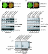
In vivo analysis of GFP-Cym(+) and GFP-PoLV(+) sensor sequences. Spots in half leaves were infiltrated with Agrobacterium containing the indicated sensor sequences and CIRV p19 into noninfected and Cym19stop-recovering plants (A and B, respectively). Leaves were viewed at 3 dai under visible and long-wavelength UV illumination (A and B, upper panels). Time course of protein- and RNA-normalized extracts at 1, 2, and 3 dai subjected to immunoblot analysis or to RNA blot analysis using a radiolabeled GFP sequence probe. Total protein loading control was stained by Ponceau reagent, whereas the 25S RNA was used as a loading control for total RNA (A and B, lower panels). Total RNA extracts at 3 dai were submitted to a low-molecular-mass RNA analysis (C). 32P-labeled RNA probes raised against GFP and CymRSV (positions 4446 to 4635) were used to detect siRNAs deriving from the GFP part and the viral sequences, respectively. Decade markers (Ambion) were used as small RNA molecular markers. *, position of 5′ cleavage product target RNA.
FIG. 3.
FIG. 3.
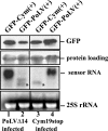
In vivo analysis of the accumulation of GFP-Cym(+) and GFP-PoLV(+) in homologous and heterologous virus-infected N. benthamiana. Total proteins and RNAs were extracted at 3 dai from Cym19stop-recovering (lanes 3 and 4) or PolVΔ14-recovering (lanes 1 and 2) N. benthamiana leaves agro-infiltrated with GFP-Cym(+) and GFP-PoLV(+) sensors. Protein extracts were subjected to immunoblot analysis using GFP-specific antibody, and RNA samples were analyzed by Northern blotting using a radiolabeled GFP sequence probe. The extracted total RNAs are visualized in an ethidium bromide staining gel at the bottom panels; the 25S ribosomal RNAs were used as loading controls. *, position of 5′ cleavage product target RNA.
FIG. 4.
FIG. 4.
In vivo analysis of GFP-Cym(+) and GFP-Cym(−) sensor sequences. Noninfected (A) and Cym19stop-recovering (B) N. benthamiana plants were agro-infiltrated with the indicated sensor constructs, and the GFP, p19 proteins, and sensor mRNAs were analyzed at the indicated time points. Normalized extracts were prepared at 1, 2, and 3 dai and subjected to immunoblot analysis with anti-GFP antibody and anti-CIRV p19 antiserum or subjected to RNA blot analysis using a radiolabeled GFP sequence probe. The extracted total RNAs are visualized in an ethidium bromide staining gel at the bottom panels; the 25S ribosomal RNAs were used as loading controls. *, position of 5′ cleavage product target RNA.
FIG. 5.
FIG. 5.
In vivo analysis of VIGS and miR171-mediated sensor sequences cleavage. Schematic illustration of the GFP-Cym(+) and GFP-Cym(−) deriving from the in vivo 3′-end analysis of the uncleaved and cleaved mRNAs. The GFP ORF is represented by a gray open box, and the noncoding regions are indicated by thick lines (black for viral target sequence and pale gray for nopalin synthase [NOS] terminator-deriving sequence). Arrows indicate the sites of cleavage on the schematic enlargement of viral target sequences (+ and −) in the sensor sequences and in the viral Cym19stop and CymRSV context (gray lines) (A). The position and the frequency of cleavage sites are not shown. (B) RNA gel blot analysis of siRNAs deriving from Cym19stop. Total RNA extracted from Cym19stop-infected plants were separated on 12% acrylamide gels, blotted, and hybridized with specific riboprobes to detect siRNAs deriving from the sensor sequences (referred to as GFP) and from specific viral regions, as indicated in each panel of the picture. (C) An agro-infiltrated N. benthamiana leaf viewed at 3 dai under long-wavelength UV illumination and the corresponding RNA gel blot analysis of RNA extracted from tissues infiltrated with GFP 171.1 and GFP 171.2 constructs are shown. A riboprobe for the GFP ORF was used to detect sensor mRNAs. (D) Southern blot analysis of the PCR products obtained from the 3′-end analysis performed on the sensor mRNAs. Two blots of the same sample were hybridized with a riboprobe raised against the GFP ORF or with 32P-oligonucleotide complementary to the miR171 target sequence (lanes 1 and 2, respectively). (E) Schematic illustration of the GFP 171.1 deriving from the in vivo 3′-end analysis of the uncleaved and the cleaved mRNAs. The GFP ORF is represented by a gray open box, and the noncoding regions are indicated by thick lines (black for miRNA target sequence and pale gray for 35S terminator-deriving sequence). Arrows indicate the sites of cleavage on the miR171 target sequence.
FIG. 6.
FIG. 6.
Cofractionation of viral siRNAs and plant miRNAs containing complexes derived from virus-infected N. benthamiana plants. The protein extract was prepared at 7 dpi from systemic leaves of Cym19stop-infected N. benthamiana plants and size separated by a Superdex-200 gel filtration column. RNA was extracted from each fraction and analyzed on a gel blot. The gel blot was hybridized first with CymRSV probe, and then the membrane was stripped and reprobed with [γ-32P]ATP-labeled oligonucleotide complementary to miR159. The elution positions of protein molecular markers are shown above the panels and are as follows: 669 kDa, thyroglobulin; 443 kDa, ferritin, 150 kDa, aldolase; 66 kDa, bovine serum albumin; 29 kDa, carbonic anhydrase. RNA size markers are shown at the right side.

Similar articles

Cited by

References

    1. Ahlquist, P. 2002. RNA-dependent RNA polymerases, viruses, and RNA silencing. Science 296:1270-1273. - PubMed
    1. Aukerman, M. J., and H. Sakai. 2003. Regulation of flowering time and floral organ identity by a MicroRNA and its APETALA2-like target genes. Plant Cell 15:2730-2741. - PMC - PubMed
    1. Baumberger, N., and D. C. Baulcombe. 2005. Arabidopsis ARGONAUTE1 is an RNA slicer that selectively recruits microRNAs and short interfering RNAs. Proc. Natl. Acad. Sci. USA 102:11928-11933. - PMC - PubMed
    1. Bernstein, E., A. A. Caudy, S. M. Hammond, and G. J. Hannon. 2001. Role for a bidentate ribonuclease in the initiation step of RNA interference. Nature 409:363-366. - PubMed
    1. Bevan, M., W. M. Barnes, and M. D. Chilton. 1983. Structure and transcription of the nopaline synthase gene region of T-DNA. Nucleic Acids Res. 11:369-385. - PMC - PubMed

Publication types

MeSH terms

LinkOut - more resources