

Increase in circulating Foxp3+CD4+CD25(high) regulatory T cells in nasopharyngeal carcinoma patients
- PMID: 17262084
- PMCID: PMC2360054
- DOI: 10.1038/sj.bjc.6603580
Increase in circulating Foxp3+CD4+CD25(high) regulatory T cells in nasopharyngeal carcinoma patients
Abstract
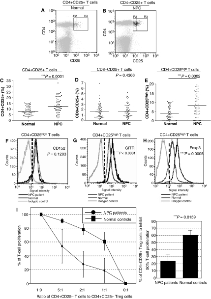
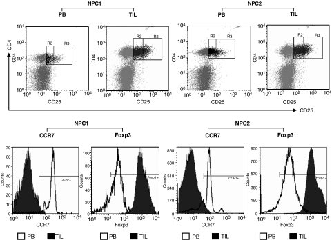
Nasopharyngeal carcinoma (NPC) is an Epstein-Barr virus-associated disease with high prevalence in Southern Chinese. Using multiparametric flow cytometry, we identified significant expansions of circulating naïve and memory CD4+CD25(high) T cells in 56 NPC patients compared with healthy age- and sex-matched controls. These were regulatory T cells (Treg), as they overexpressed Foxp3 and GITR, and demonstrated enhanced suppressive activities against autologous CD4+CD25- T-cell proliferation in functional studies on five patients. Abundant intraepithelial infiltrations of Treg with very high levels of Foxp3 expression and absence of CCR7 expression were also detected in five primary tumours. Our current study is the first to demonstrate an expansion of functional Treg in the circulation of NPC patients and the presence of infiltrating Treg in the tumour microenvironment. As Treg may play an important role in suppressing antitumour immunity, our findings provide critical insights for clinical management of NPC.
Figures
Similar articles
-
Absence of amplification of CD4+CD25(high) regulatory T cells during in vitro expansion of tumor-infiltrating lymphocytes in melanoma patients.Exp Dermatol. 2008 May;17(5):436-45. doi: 10.1111/j.1600-0625.2007.00681.x. Epub 2008 Feb 27. Exp Dermatol. 2008. PMID: 18312383
-
Effect of nasopharyngeal carcinoma-derived exosomes on human regulatory T cells.J Natl Cancer Inst. 2014 Dec 12;107(1):363. doi: 10.1093/jnci/dju363. Print 2015 Jan. J Natl Cancer Inst. 2014. PMID: 25505237
-
CD4(+)CD25(+)CD127(low/-) regulatory T cells express Foxp3 and suppress effector T cell proliferation and contribute to gastric cancers progression.Clin Immunol. 2009 Apr;131(1):109-18. doi: 10.1016/j.clim.2008.11.010. Epub 2009 Jan 18. Clin Immunol. 2009. PMID: 19153062
-
IL-2-independent generation of FOXP3(+)CD4(+)CD8(+)CD25(+) cytotoxic regulatory T cell lines from human umbilical cord blood.Exp Hematol. 2007 Feb;35(2):287-96. doi: 10.1016/j.exphem.2006.10.011. Exp Hematol. 2007. PMID: 17258077
-
CD4(+) CD25(low) GITR(+) cells: a novel human CD4(+) T-cell population with regulatory activity.Eur J Immunol. 2011 Aug;41(8):2269-78. doi: 10.1002/eji.201040943. Epub 2011 Jul 4. Eur J Immunol. 2011. PMID: 21557210
Cited by
-
Lessons from rare tumors: hepatic lymphoepithelioma-like carcinomas.World J Gastroenterol. 2015 Mar 28;21(12):3472-9. doi: 10.3748/wjg.v21.i12.3472. World J Gastroenterol. 2015. PMID: 25834311 Free PMC article. Review.
-
Anti-glucocorticoid-induced Tumor Necrosis Factor-Related Protein (GITR) Therapy Overcomes Radiation-Induced Treg Immunosuppression and Drives Abscopal Effects.Front Immunol. 2018 Sep 20;9:2170. doi: 10.3389/fimmu.2018.02170. eCollection 2018. Front Immunol. 2018. PMID: 30294332 Free PMC article.
-
Lessons learned from SMAD4 loss in squamous cell carcinomas.Mol Carcinog. 2019 Sep;58(9):1648-1655. doi: 10.1002/mc.23049. Epub 2019 May 29. Mol Carcinog. 2019. PMID: 31140647 Free PMC article. Review.
-
Cytolytic T lymphocytes from HLA-B8+ donors frequently recognize the Hodgkin's lymphoma associated latent membrane protein 2 of Epstein Barr virus.Herpesviridae. 2011 Feb 11;2(1):4. doi: 10.1186/2042-4280-2-4. Herpesviridae. 2011. PMID: 21429247 Free PMC article.
-
Immune escape of γ-herpesviruses from adaptive immunity.Rev Med Virol. 2014 Nov;24(6):365-78. doi: 10.1002/rmv.1791. Epub 2014 Apr 15. Rev Med Virol. 2014. PMID: 24733560 Free PMC article. Review.
References
-
- Baecher-Allan C, Viglietta V, Hafler DA (2004) Human CD4+CD25+ regulatory T cells. Semin Immunol 16: 89–98 - PubMed
-
- Baratelli F, Lin Y, Zhu L, Yang SC, Heuze-Vourc'h N, Zeng G, Reckamp K, Dohadwala M, Sharma S, Dubinett SM (2005) Prostaglandin E2 induces FOXP3 gene expression and T regulatory cell function in human CD4+ T cells. J Immunol 175: 1483–1490 - PubMed
-
- Beyer M, Kochanek M, Giese T, Endl E, Weihrauch MR, Knolle PA, Classen S, Schultze JL (2006) In vivo peripheral expansion of naive CD4+CD25high FoxP3+ regulatory T cells in patients with multiple myeloma. Blood 107: 3940–3949 - PubMed
-
- Bromley SK, Thomas SY, Luster AD (2005) Chemokine receptor CCR7 guides T cell exit from peripheral tissues and entry into afferent lymphatics. Nat Immunol 6: 895–901 - PubMed
-
- Chua D, Huang J, Zheng B, Lau SY, Luk W, Kwong DL, Sham JS, Moss D, Yuen KY, Im SW, Ng MH (2001) Adoptive transfer of autologous Epstein–Barr virus-specific cytotoxic T cells for nasopharyngeal carcinoma. Int J Cancer 94: 73–80 - PubMed
MeSH terms
Substances
LinkOut - more resources
Full Text Sources
Other Literature Sources
Research Materials
