

Contributions of pneumolysin, pneumococcal surface protein A (PspA), and PspC to pathogenicity of Streptococcus pneumoniae D39 in a mouse model
- PMID: 17261599
- PMCID: PMC1865719
- DOI: 10.1128/IAI.01384-06
Contributions of pneumolysin, pneumococcal surface protein A (PspA), and PspC to pathogenicity of Streptococcus pneumoniae D39 in a mouse model
Abstract
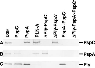
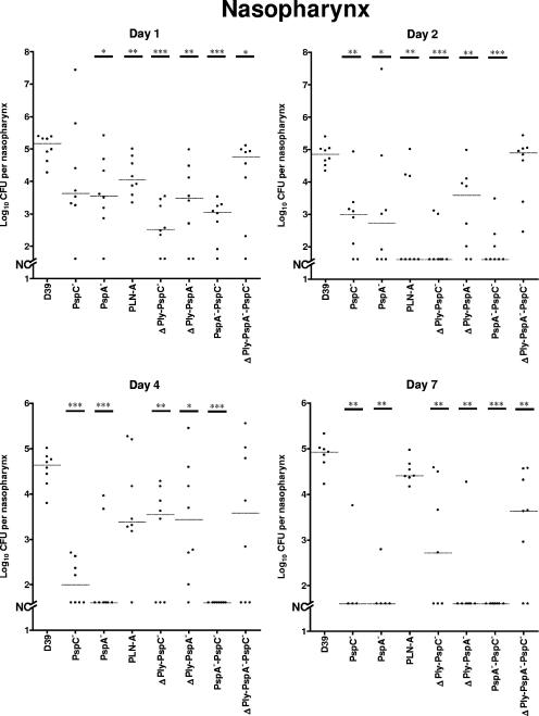
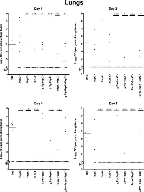
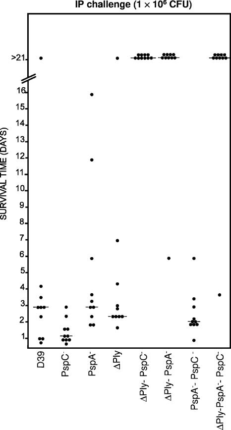
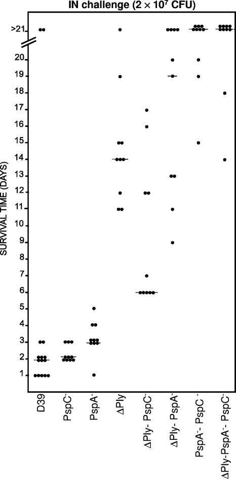
Successful colonization of the upper respiratory tract by Streptococcus pneumoniae is an essential first step in the pathogenesis of pneumococcal disease. However, the bacterial and host factors that provoke the progression from asymptomatic colonization to invasive disease are yet to be fully defined. In this study, we investigated the effects of single and combined mutations in genes encoding pneumolysin (Ply), pneumococcal surface protein A (PspA), and pneumococcal surface protein C (PspC, also known as choline-binding protein A) on the pathogenicity of Streptococcus pneumoniae serotype 2 (D39) in mice. Following intranasal challenge with D39, stable colonization of the nasopharynx was maintained over a 7-day period at a level of approximately 10(5) bacteria per mouse. The abilities of the mutant deficient in PspA to colonize the nasopharynx and to cause lung infection and bacteremia were significantly reduced. Likewise, the PspC mutant and, to a lesser extent, the Ply mutant also had reduced abilities to colonize the nasopharynx. As expected, the double mutants colonized less well than the parent to various degrees and had difficulty translocating to the lungs and blood. A significant additive attenuation was observed for the double and triple mutants in pneumonia and systemic disease models. Surprisingly, the colonization profile of the derivative lacking all three proteins was similar to that of the wild type, indicating virulence gene compensation. These findings further demonstrate that the mechanism of pneumococcal pathogenesis is highly complex and multifactorial but ascribes a role for each of these virulence proteins, alone or in combination, in the process.
Figures
Similar articles
-
Tissue-specific contributions of pneumococcal virulence factors to pathogenesis.J Infect Dis. 2004 Nov 1;190(9):1661-9. doi: 10.1086/424596. Epub 2004 Sep 21. J Infect Dis. 2004. PMID: 15478073
-
Pneumolysin, PspA, and PspC contribute to pneumococcal evasion of early innate immune responses during bacteremia in mice.Infect Immun. 2007 Apr;75(4):2067-70. doi: 10.1128/IAI.01727-06. Epub 2007 Jan 12. Infect Immun. 2007. PMID: 17220305 Free PMC article.
-
Surface Proteins and Pneumolysin of Encapsulated and Nonencapsulated Streptococcus pneumoniae Mediate Virulence in a Chinchilla Model of Otitis Media.Front Cell Infect Microbiol. 2016 May 18;6:55. doi: 10.3389/fcimb.2016.00055. eCollection 2016. Front Cell Infect Microbiol. 2016. PMID: 27242973 Free PMC article.
-
The role of Streptococcus pneumoniae virulence factors in host respiratory colonization and disease.Nat Rev Microbiol. 2008 Apr;6(4):288-301. doi: 10.1038/nrmicro1871. Nat Rev Microbiol. 2008. PMID: 18340341 Review.
-
From nose to lung: the regulation behind Streptococcus pneumoniae virulence factors.Mol Microbiol. 2003 Nov;50(4):1103-10. doi: 10.1046/j.1365-2958.2003.03764.x. Mol Microbiol. 2003. PMID: 14622402 Free PMC article. Review.
Cited by
-
Sucrose metabolism contributes to in vivo fitness of Streptococcus pneumoniae.Mol Microbiol. 2007 Oct;66(1):1-13. doi: 10.1111/j.1365-2958.2007.05878.x. Mol Microbiol. 2007. PMID: 17880421 Free PMC article.
-
Immunization with recombinant Streptococcus pneumoniae PgdA protects mice against lung invasion.Exp Biol Med (Maywood). 2024 Oct 14;249:10119. doi: 10.3389/ebm.2024.10119. eCollection 2024. Exp Biol Med (Maywood). 2024. PMID: 39469203 Free PMC article.
-
Future perspective on host-pathogen interactions during bacterial biofilm formation within the nasopharynx.Future Microbiol. 2012 Feb;7(2):227-39. doi: 10.2217/fmb.11.160. Future Microbiol. 2012. PMID: 22324992 Free PMC article. Review.
-
Pneumococcal Vaccines.Microbiol Spectr. 2019 Nov;7(6):10.1128/microbiolspec.gpp3-0028-2018. doi: 10.1128/microbiolspec.GPP3-0028-2018. Microbiol Spectr. 2019. PMID: 31858954 Free PMC article. Review.
-
Joint sequencing of human and pathogen genomes reveals the genetics of pneumococcal meningitis.Nat Commun. 2019 May 15;10(1):2176. doi: 10.1038/s41467-019-09976-3. Nat Commun. 2019. PMID: 31092817 Free PMC article.
References
-
- Alexander, J. E., R. A. Lock, C. C. A. M. Peeters, J. T. Poolman, P. W. Andrew, T. J. Mitchell, D. Hansman, and J. C. Paton. 1994. Immunization of mice with pneumolysin toxoid confers a significant degree of protection against at least nine serotypes of Streptococcus pneumoniae. Infect. Immun. 62:5683-5688. - PMC - PubMed
-
- Amsbaugh, D. F., C. T. Hansen, B. Prescott, P. W. Stashak, D. R. Barthold, and P. J. Baker. 1972. Genetic control of the antibody response to type III pneumococcal polysaccharide in mice. I. Evidence that an X-linked gene plays a decisive role in determining responsiveness. J. Exp. Med. 136:931-949. - PMC - PubMed
-
- Balachandran, P., A. Brooks-Walter, A. Virolainen-Julkunen, S. K. Hollingshead, and D. E. Briles. 2002. Role of pneumococcal surface protein C in nasopharyngeal carriage and pneumonia and its ability to elicit protection against carriage of Streptococcus pneumoniae. Infect. Immun. 70:2526-2534. - PMC - PubMed
Publication types
MeSH terms
Substances
LinkOut - more resources
Full Text Sources
Other Literature Sources
Medical
Research Materials
