

Increased oxidative stress is associated with balanced increases in hepatocyte apoptosis and proliferation in glycerol-3-phosphate acyltransferase-1 deficient mice
- PMID: 17258706
- PMCID: PMC1865130
- DOI: 10.1016/j.yexmp.2006.12.004
Increased oxidative stress is associated with balanced increases in hepatocyte apoptosis and proliferation in glycerol-3-phosphate acyltransferase-1 deficient mice
Abstract
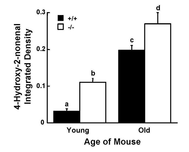
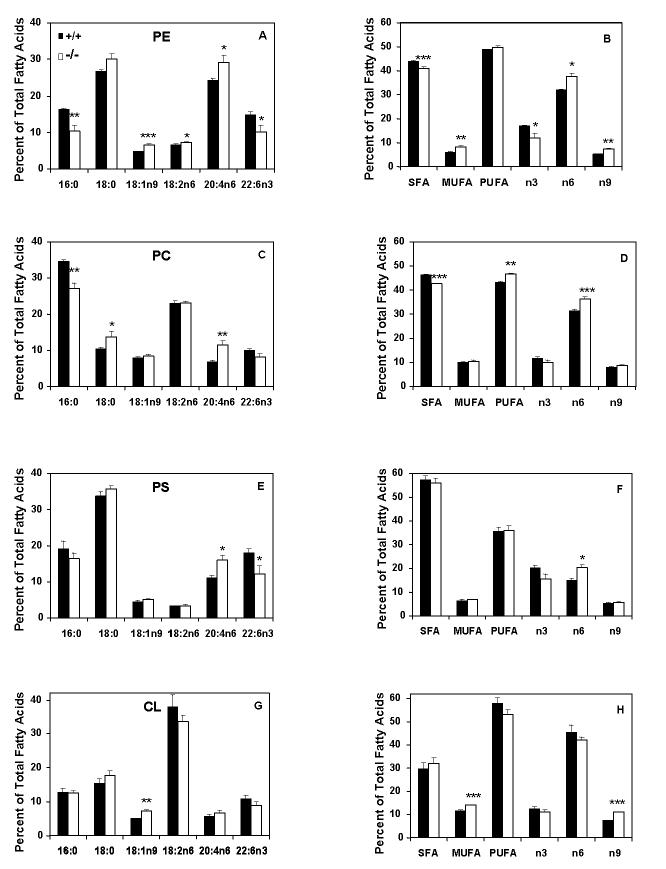
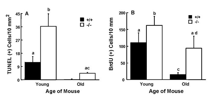
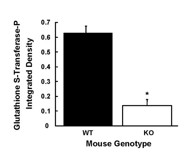
The absence of mouse mitochondrial glycerol-3-phosphate acyltransferase-1 (Gpat1-/-) increases the amount of arachidonate in liver phospholipids and increases beta-hydroxybutyrate and acyl-carnitines, suggesting an elevated rate of liver fatty acid oxidation. We asked whether these alterations might increase reactive oxygen species (ROS), apoptosis, or hepatocyte proliferation. Compared to wildtype controls, liver mitochondria from Gpat1-/- mice showed a 20% increase in the rate of ROS production and a markedly increased sensitivity to the induction of the mitochondrial permeability transition. Mitochondrial phosphatidylethanolamine and phosphatidylcholine from Gpat1-/- liver contained 21% and 67% more arachidonate, respectively, than wildtype controls, and higher amounts of 4-hydroxynonenal, a product of arachidonate peroxidation. Oxidative stress was associated with an increase in apoptosis, and with 3-fold and 15-fold higher TUNEL positive cells in liver from young and old Gpat1-/- mice, respectively, compared to age-matched controls. Compared to controls, bromodeoxyuridine labeling was 50% and 7-fold higher in livers from young and old Gpat1-/- mice, respectively, but fewer glutathione-S-transferase positive cells were present. Thus, Gpat1-/- liver exhibits increased oxidative stress and sensitivity of the mitochondrial permeability transition pore, and a balanced increase in apoptosis and proliferation.
Figures

Similar articles
-
Mice deficient in mitochondrial glycerol-3-phosphate acyltransferase-1 have diminished myocardial triacylglycerol accumulation during lipogenic diet and altered phospholipid fatty acid composition.Biochim Biophys Acta. 2008 Jun-Jul;1781(6-7):352-8. doi: 10.1016/j.bbalip.2008.05.001. Epub 2008 May 15. Biochim Biophys Acta. 2008. PMID: 18522808 Free PMC article.
-
Glycerol-3-phosphate acyltransferase (GPAT)-1, but not GPAT4, incorporates newly synthesized fatty acids into triacylglycerol and diminishes fatty acid oxidation.J Biol Chem. 2013 Sep 20;288(38):27299-27306. doi: 10.1074/jbc.M113.485219. Epub 2013 Aug 1. J Biol Chem. 2013. PMID: 23908354 Free PMC article.
-
Mice deficient in glycerol-3-phosphate acyltransferase-1 have a reduced susceptibility to liver cancer.Toxicol Pathol. 2012 Apr;40(3):513-21. doi: 10.1177/0192623311432298. Epub 2012 Jan 3. Toxicol Pathol. 2012. PMID: 22215515 Free PMC article.
-
Regulation of Triglyceride Metabolism. II. Function of mitochondrial GPAT1 in the regulation of triacylglycerol biosynthesis and insulin action.Am J Physiol Gastrointest Liver Physiol. 2007 May;292(5):G1195-9. doi: 10.1152/ajpgi.00553.2006. Epub 2006 Dec 7. Am J Physiol Gastrointest Liver Physiol. 2007. PMID: 17158253 Free PMC article. Review.
-
Mitochondrial glutathione transferases involving a new function for membrane permeability transition pore regulation.Drug Metab Rev. 2011 May;43(2):292-9. doi: 10.3109/03602532.2011.552913. Epub 2011 Mar 23. Drug Metab Rev. 2011. PMID: 21428695 Review.
Cited by
-
Puerarin ameliorates nonalcoholic fatty liver in rats by regulating hepatic lipid accumulation, oxidative stress, and inflammation.Front Immunol. 2022 Jul 25;13:956688. doi: 10.3389/fimmu.2022.956688. eCollection 2022. Front Immunol. 2022. PMID: 35958617 Free PMC article.
-
Mice deficient in mitochondrial glycerol-3-phosphate acyltransferase-1 have diminished myocardial triacylglycerol accumulation during lipogenic diet and altered phospholipid fatty acid composition.Biochim Biophys Acta. 2008 Jun-Jul;1781(6-7):352-8. doi: 10.1016/j.bbalip.2008.05.001. Epub 2008 May 15. Biochim Biophys Acta. 2008. PMID: 18522808 Free PMC article.
-
Biochemistry, physiology, and genetics of GPAT, AGPAT, and lipin enzymes in triglyceride synthesis.Am J Physiol Endocrinol Metab. 2009 Jun;296(6):E1195-209. doi: 10.1152/ajpendo.90958.2008. Epub 2009 Mar 31. Am J Physiol Endocrinol Metab. 2009. PMID: 19336658 Free PMC article. Review.
-
Glycerol-3-phosphate acyltransferase (GPAT)-1, but not GPAT4, incorporates newly synthesized fatty acids into triacylglycerol and diminishes fatty acid oxidation.J Biol Chem. 2013 Sep 20;288(38):27299-27306. doi: 10.1074/jbc.M113.485219. Epub 2013 Aug 1. J Biol Chem. 2013. PMID: 23908354 Free PMC article.
-
Early hepatic insulin resistance in mice: a metabolomics analysis.Mol Endocrinol. 2010 Mar;24(3):657-66. doi: 10.1210/me.2009-0152. Epub 2010 Feb 11. Mol Endocrinol. 2010. PMID: 20150186 Free PMC article.
References
-
- Albright CD, Friedrich CB, Brown EC, Mar MH, Zeisel SH. Maternal dietary choline availability alters mitosis, apoptosis and the localization of TOAD-64 protein in the developing fetal rat septum. Brain Res. Dev. Brain Res. 1999;115:123–129. - PubMed
-
- Albright CD, Liu R, Bethea TC, Da Costa KA, Salganik RI, Zeisel SH. Choline deficiency induces apoptosis in SV40-immortalized CWSV-1 rat hepatocytes in culture. FASEB J. 1996;10:510–516. - PubMed
-
- Albright CD, Salganik RI, Van Dyke T. Dietary depletion of vitamin E and vitamin A inhibits mammary tumor growth and metastasis in transgenic mice. J. Nutr. 2004;134:1139–1144. - PubMed
-
- Ameen C, Linden D, Larsson BM, Mode A, Holmang A, Oscarsson J. Effects of gender and GH secretory pattern on sterol regulatory element-binding protein-1c and its target genes in rat liver. Am. J. Physiol. Endocrinol. Metab. 2004;287:E1039–1048. - PubMed
Publication types
MeSH terms
Substances
Grants and funding
- ES11660/ES/NIEHS NIH HHS/United States
- P01 DK59340/DK/NIDDK NIH HHS/United States
- K22 ES011660/ES/NIEHS NIH HHS/United States
- P30 DK056350-050001/DK/NIDDK NIH HHS/United States
- DK37034/DK/NIDDK NIH HHS/United States
- P30 CA016086/CA/NCI NIH HHS/United States
- F31 GM20920/GM/NIGMS NIH HHS/United States
- R56 DK037034/DK/NIDDK NIH HHS/United States
- R01 DK056598/DK/NIDDK NIH HHS/United States
- R01 DK56598/DK/NIDDK NIH HHS/United States
- R01 DK037034/DK/NIDDK NIH HHS/United States
- R01 DK073336/DK/NIDDK NIH HHS/United States
- U19 ES011391/ES/NIEHS NIH HHS/United States
- F31 GM020920/GM/NIGMS NIH HHS/United States
- P30 DK056350/DK/NIDDK NIH HHS/United States
- CA16086/CA/NCI NIH HHS/United States
- ES11391/ES/NIEHS NIH HHS/United States
- R01 DK056598-25/DK/NIDDK NIH HHS/United States
- P01 DK059340/DK/NIDDK NIH HHS/United States
- R37 DK037034/DK/NIDDK NIH HHS/United States
LinkOut - more resources
Full Text Sources
