. 2007 Mar 9;354(2):372-8.
doi: 10.1016/j.bbrc.2007.01.044. Epub 2007 Jan 17.

Erythropoietin protects cardiac myocytes against anthracycline-induced apoptosis

Affiliations

Erythropoietin protects cardiac myocytes against anthracycline-induced apoptosis

Ping Fu et al. Biochem Biophys Res Commun. .

Abstract

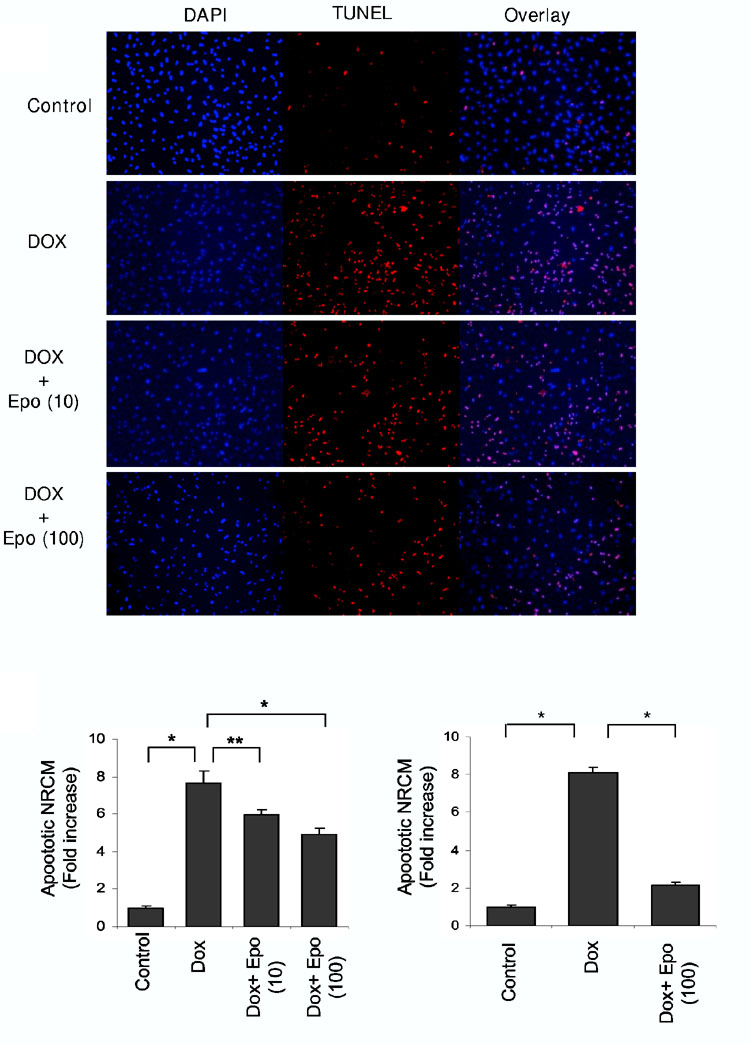
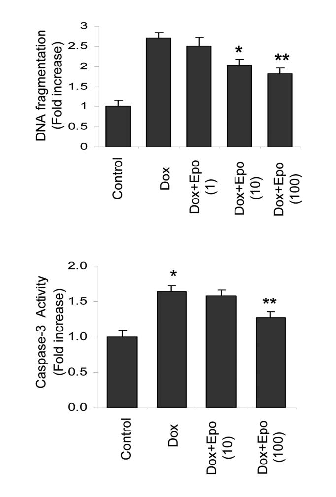
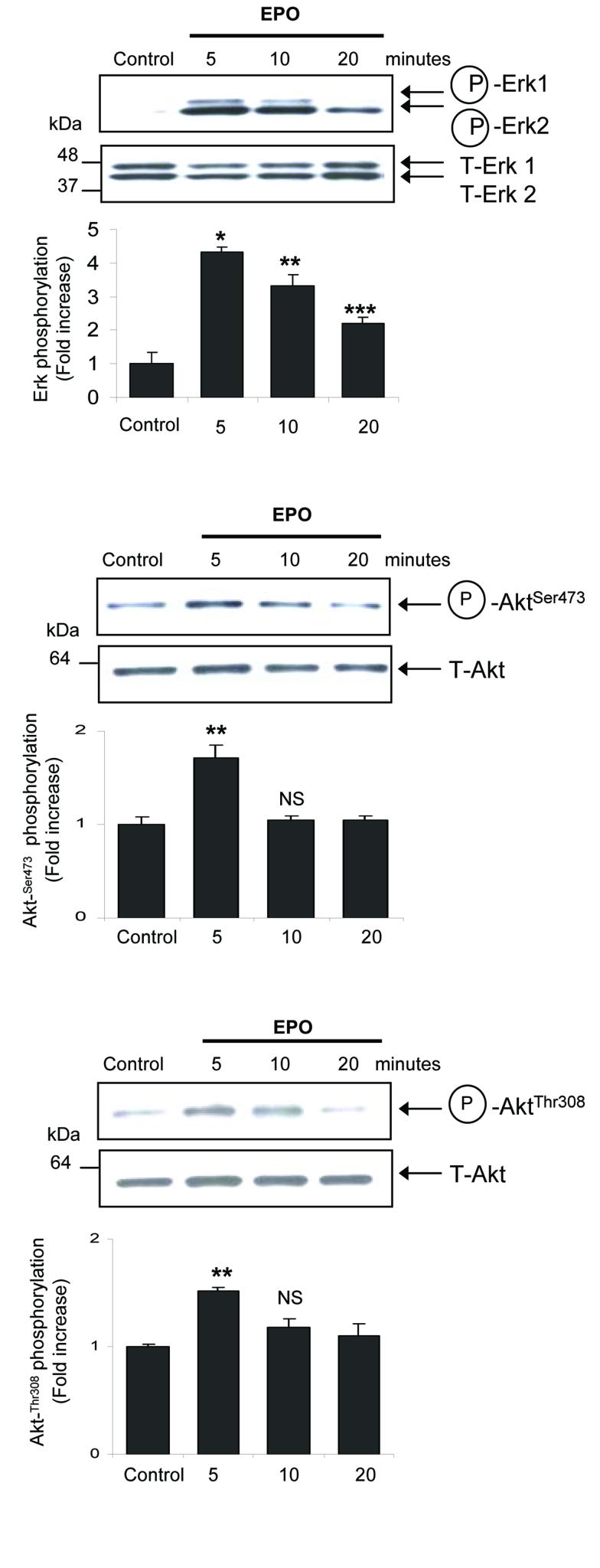
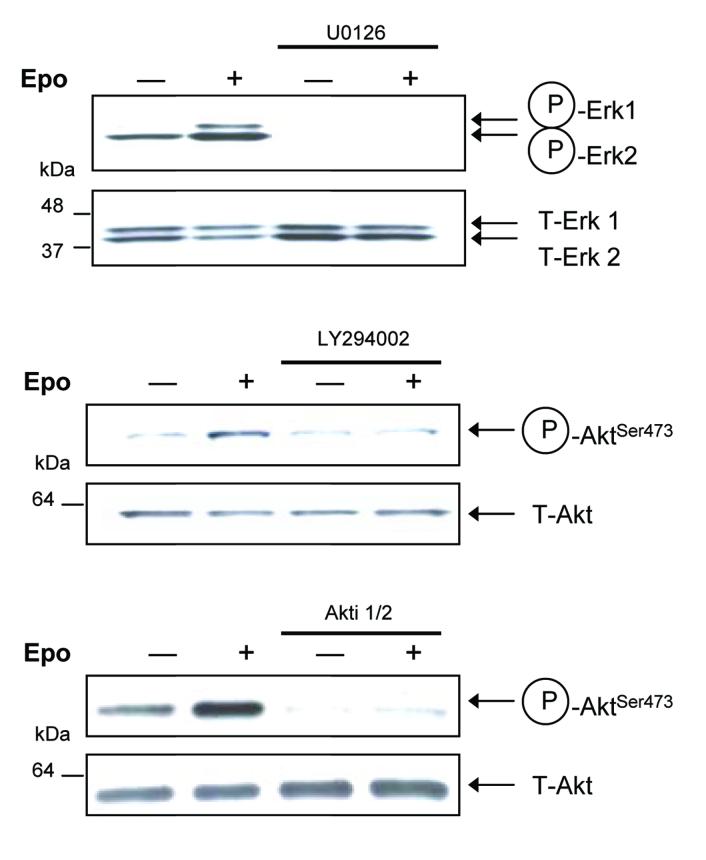
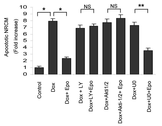
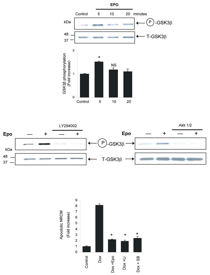
The cardiotoxic adverse effects of anthracycline antibiotics limit their therapeutic utility as essential components of chemotherapy regimens for hematologic and solid malignancies. Here we show that the hematopoietic cytokine erythropoietin attenuates doxorubicin-induced apoptosis of primary neonatal rat ventricular cardiomyocytes in a dose-dependent manner. Erythropoietin treatment induced rapid, time-dependent phosphorylation of MAP kinases (MAPK) Erk1/2 and the phosphatidylinositol 3-kinase substrate Akt. Treatment of cardiomyocytes with inhibitors of phosphatidylinositol 3-kinase (LY294002) or Akt (Akti-1/2) abolished the protective effect of erythropoietin, whereas treatment with MAPK kinase (MEK1) inhibitor U0126 did not. Erythropoietin also induced the phosphorylation of GSK-3beta, a downstream target of PI3K-Akt. Because phosphorylation is known to inactivate GSK-3beta, we investigated whether GSK-3beta inhibition is cardioprotective. We found that GSK-3beta inhibitors SB216763 or lithium chloride blocked doxorubicin-induced cardiomyocyte apoptosis in a manner similar to erythropoietin, suggesting that GSK-3beta inhibition is involved in erythropoietin-mediated cardioprotection. Erythropoietin may serve as a novel cardioprotective agent against anthracycline-induced cardiotoxicity.


Figures

Fig. 1
Fig. 1
EPO inhibits DOX-induced apoptosis of primary cardiomyocytes in a dose-dependent manner. A.-Apoptotic cells were detected by TUNEL assays for labeling apoptotic nuclei (red), and DAPI counter-staining for all nuclei (blue). NRCMs were incubated in serum-free-medium and then treated with DOX in the presence or absence of Epo (10 or 100 units/ml). Control cells cultured in serum-free medium were left untreated. Photomicrographs of representative images are illustrated. B.-Quantification of apoptotic NRCMs following DOX and Epo treatments. NRCMs were incubated in serum-free-medium for either 18 hours (left panel) or 10 hours (right panel) prior to DOX treatment in the presence of indicated concentration of Epo. The fraction of apoptotic cells was determined in 6 or 7 random microscopic fields totaling at least 1,500 cells/group and expressed as fold increase over control. Results are representative of four independent experiments. (*p<0.001,**p<0.05). C.-Histone-associated DNA fragmentation generated in apoptotic NRCMs was quantified by ELISA assays. NRCMs were either left untreated (control) or treated with DOX in the absence or presence of increasing concentrations of Epo as indicated (1, 10 or 100 units/ml). The optical density (405nm) of the untreated control samples was arbitrarily designated 1. (n=11 replicates in each group). *p<0.05,**p<0.01 compared to DOX group. Data is representative of six independent experiments. D.-Caspase-3 activity was determined in NRCMs following DOX treatment in the presence or absence of Epo (10 or 100 units/ml) using ELISA assays. The optical density of the untreated control samples was arbitrarily designated 1. *p<0.01 compared to control and **p<0.05 compared to DOX group (n=6 in each group).
Fig. 1
Fig. 1
EPO inhibits DOX-induced apoptosis of primary cardiomyocytes in a dose-dependent manner. A.-Apoptotic cells were detected by TUNEL assays for labeling apoptotic nuclei (red), and DAPI counter-staining for all nuclei (blue). NRCMs were incubated in serum-free-medium and then treated with DOX in the presence or absence of Epo (10 or 100 units/ml). Control cells cultured in serum-free medium were left untreated. Photomicrographs of representative images are illustrated. B.-Quantification of apoptotic NRCMs following DOX and Epo treatments. NRCMs were incubated in serum-free-medium for either 18 hours (left panel) or 10 hours (right panel) prior to DOX treatment in the presence of indicated concentration of Epo. The fraction of apoptotic cells was determined in 6 or 7 random microscopic fields totaling at least 1,500 cells/group and expressed as fold increase over control. Results are representative of four independent experiments. (*p<0.001,**p<0.05). C.-Histone-associated DNA fragmentation generated in apoptotic NRCMs was quantified by ELISA assays. NRCMs were either left untreated (control) or treated with DOX in the absence or presence of increasing concentrations of Epo as indicated (1, 10 or 100 units/ml). The optical density (405nm) of the untreated control samples was arbitrarily designated 1. (n=11 replicates in each group). *p<0.05,**p<0.01 compared to DOX group. Data is representative of six independent experiments. D.-Caspase-3 activity was determined in NRCMs following DOX treatment in the presence or absence of Epo (10 or 100 units/ml) using ELISA assays. The optical density of the untreated control samples was arbitrarily designated 1. *p<0.01 compared to control and **p<0.05 compared to DOX group (n=6 in each group).
Fig. 2.
Fig. 2.
Epo-induced phosphorylation of Erk1/2 and Akt in primary cardiomyocytes. Isolated NRCMs were either left untreated (Control) or treated with Epo (10 units/ml) for the indicated durations. Whole cell lysates were analyzed by Western blotting using phospho-specific antibodies for A.-Erk1/2 (Thr202/Tyr204), B.-Akt (Ser473), C.-Akt (Thr308). The phosphorylated proteins are indicated by the arrows (upper panels). Comparable loading in each lane and protein integrity were confirmed by stripping the blots and hybridizing to respective antibodies to detect total Erk1/2 or Akt protein (lower panels). The relative positions of the molecular weight markers are indicated in each blot. Quantitative representation of phosphorylated proteins using densitometry are shown (mean±sem values from 3 independent experiments, *p<0.001,**p<0.01,***p<0.05, NS: not significant compared to control group). D-F.-Isolated NRCMs were either left untreated (−) or treated ( + ) with Epo (10 units/ml) for 5 minutes in the presence or absence of D.-MEK1 inhibitor U0126, E.-PI3K inhibitor LY294002, or F.-Akt inhibitor Akti-1/2. Western blotting was performed using phospho-specific antibodies as indicated (upper panels). The blots were stripped and hybridized to antibodies against total Erk1/2 or Akt (lower panels).
Fig. 2.
Fig. 2.
Epo-induced phosphorylation of Erk1/2 and Akt in primary cardiomyocytes. Isolated NRCMs were either left untreated (Control) or treated with Epo (10 units/ml) for the indicated durations. Whole cell lysates were analyzed by Western blotting using phospho-specific antibodies for A.-Erk1/2 (Thr202/Tyr204), B.-Akt (Ser473), C.-Akt (Thr308). The phosphorylated proteins are indicated by the arrows (upper panels). Comparable loading in each lane and protein integrity were confirmed by stripping the blots and hybridizing to respective antibodies to detect total Erk1/2 or Akt protein (lower panels). The relative positions of the molecular weight markers are indicated in each blot. Quantitative representation of phosphorylated proteins using densitometry are shown (mean±sem values from 3 independent experiments, *p<0.001,**p<0.01,***p<0.05, NS: not significant compared to control group). D-F.-Isolated NRCMs were either left untreated (−) or treated ( + ) with Epo (10 units/ml) for 5 minutes in the presence or absence of D.-MEK1 inhibitor U0126, E.-PI3K inhibitor LY294002, or F.-Akt inhibitor Akti-1/2. Western blotting was performed using phospho-specific antibodies as indicated (upper panels). The blots were stripped and hybridized to antibodies against total Erk1/2 or Akt (lower panels).
Fig. 3
Fig. 3
Protective effect of Epo on DOX-induced apoptosis is blocked by PI3K and Akt inhibitors but not MEK1 inhibitor. NRCMs were incubated in serum-free medium for 10 hours followed by DOX treatment (0.5μM) for 14 hours with or without kinase inhibitors LY294002, Akti-1/2, or U0126, and in the presence or absence of Epo (100 units/ml). Quantitative analysis of apoptotic cardiomyocytes was expressed as fold increase of TUNEL-positive apoptotic NRCMs compared to untreated control. *p<0.001, **p<0.01, NS: not significant.
Fig. 4
Fig. 4
Epo stimulates the phosphorylation of GSK-3β and treatment with GSK-3β inhibitors lithium chloride and SB-21673 are protective. A.-NRCMs were either left untreated (Control) or treated with Epo (10 units/ml). Whole cell lysates were analyzed by Western blotting using phospho-specific (Ser9) antibody for GSK-3β (upper panel) followed by stripping and incubation with total GSK-3β antibody (lower panel). Quantification of phosphorylated GSK-3β is shown (mean±sem values from 3 independent experiments, *p<0.05, NS: not significant compared to control). B.-PI3K and Akt inhibitors block Epo-induced stimulation of GSK-3β phosphorylation. Cells were either left untreated (−) or treated ( + ) with Epo in the presence or absence of LY294002 (left panel) or Akti-1/2 (right panel). Western blots for phosphorylated and total GSK-3β protein were performed. C.-Isolated NRCMs were incubated with Epo, or GSK-3β inhibitors lithium chloride (Li) or SB216763 (SB) during DOX treatment. Quantitative analysis of apoptotic cardiomyocytes was expressed as fold increase of TUNEL-positive apoptotic NRCMs compared to untreated controls.*p<0.001 compared to DOX-treated group.

Similar articles

Cited by

References

    1. Dorr RT. Cytoprotective agents for anthracyclines. Semin Oncol. 1996;23:23–34. - PubMed
    1. Arola OJ, Saraste A, Pulkki K, Kallajoki M, Parvinen M, Voipio-Pulkki LM. Acute doxorubicin cardiotoxicity involves cardiomyocyte apoptosis. Cancer Res. 2000;60:1789–1792. - PubMed
    1. Yamaoka M, Yamaguchi S, Suzuki T, Okuyama M, Nitobe J, Nakamura N, Mitsui Y, Tomoike H. Apoptosis in rat cardiac myocytes induced by Fas ligand: priming for Fas-mediated apoptosis with doxorubicin. J Mol Cell Cardiol. 2000;32:881–889. - PubMed
    1. Kotamraju S, Konorev EA, Joseph J, Kalyanaraman B. Doxorubicin-induced apoptosis in endothelial cells and cardiomyocytes is ameliorated by nitrone spin traps and ebselen. Role of reactive oxygen and nitrogen species. J Biol Chem. 2000;275:33585–33592. - PubMed
    1. Wu H, Lee SH, Gao J, Liu X, Iruela-Arispe ML. Inactivation of erythropoietin leads to defects in cardiac morphogenesis. Development. 1999;126:3597–3605. - PubMed

Publication types

LinkOut - more resources