. 2007 Mar;27(6):2384-97.
doi: 10.1128/MCB.02254-06. Epub 2007 Jan 22.

Coupled release of eukaryotic translation initiation factors 5B and 1A from 80S ribosomes following subunit joining

Affiliations

Coupled release of eukaryotic translation initiation factors 5B and 1A from 80S ribosomes following subunit joining

Jeanne M Fringer et al. Mol Cell Biol. 2007 Mar.

Abstract

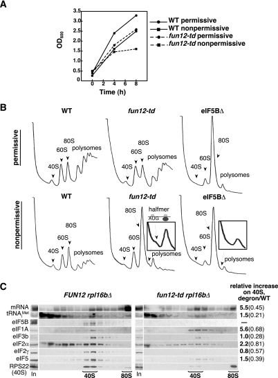
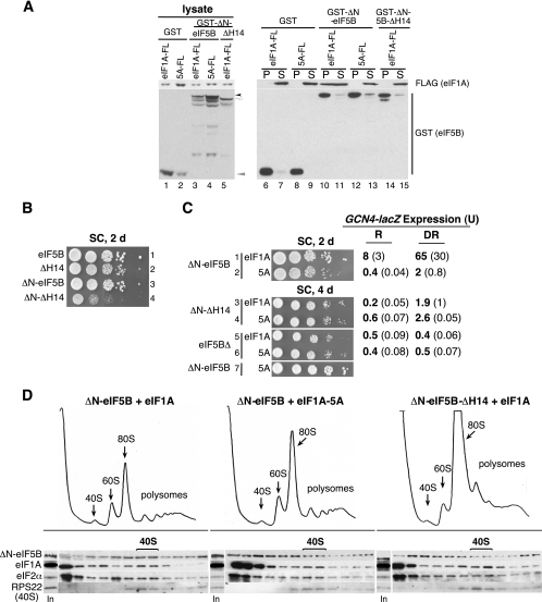
The translation initiation GTPase eukaryotic translation initiation factor 5B (eIF5B) binds to the factor eIF1A and catalyzes ribosomal subunit joining in vitro. We show that rapid depletion of eIF5B in Saccharomyces cerevisiae results in the accumulation of eIF1A and mRNA on 40S subunits in vivo, consistent with a defect in subunit joining. Substituting Ala for the last five residues in eIF1A (eIF1A-5A) impairs eIF5B binding to eIF1A in cell extracts and to 40S complexes in vivo. Consistently, overexpression of eIF5B suppresses the growth and translation initiation defects in yeast expressing eIF1A-5A, indicating that eIF1A helps recruit eIF5B to the 40S subunit prior to subunit joining. The GTPase-deficient eIF5B-T439A mutant accumulated on 80S complexes in vivo and was retained along with eIF1A on 80S complexes formed in vitro. Likewise, eIF5B and eIF1A remained associated with 80S complexes formed in the presence of nonhydrolyzable GDPNP, whereas these factors were released from the 80S complexes in assays containing GTP. We propose that eIF1A facilitates the binding of eIF5B to the 40S subunit to promote subunit joining. Following 80S complex formation, GTP hydrolysis by eIF5B enables the release of both eIF5B and eIF1A, and the ribosome enters the elongation phase of protein synthesis.


Figures

FIG. 1.
FIG. 1.
Depletion of degron-tagged eIF5B leads to polysome runoff and accumulation of halfmer ribosomes with bound mRNA and eIF1A. (A) Cultures of the fun12-td strain J259 and the isogenic WT strain YAJ42 were grown at 25°C under permissive conditions (SC medium containing 2% raffinose and 100 μM CuSO4) to an OD600 of ∼0.2, split in halves, and grown under permissive or nonpermissive conditions (SC medium containing 2% galactose, lacking CuSO4), as indicated. (B) Polysome analysis of WT (YAJ42), fun12-td (J259), and eIF5BΔ (J260) strains. Cultures of cells were grown under permissive conditions as described for panel A to an OD600 of ∼0.2, split in halves, and transferred to either permissive or nonpermissive conditions and grown for 6 h at 25°C. Cells were cross-linked with 1% formaldehyde, and WCEs were separated on 4.5% to 45% sucrose gradients by centrifugation at 39,000 rpm for 2.5 h. Gradients were fractionated while scanning at 254 nm to visualize the indicated ribosomal species. The halfmer shoulder on the 80S peak in the fun12-td strain was magnified for clarity (inset) and is represented in the schematic; the same portion of the 80S peak in the eIF5BΔ strain was also magnified (inset). (C) WCEs prepared from strains J261 (rpl16bΔ) and J262 (rpl16bΔ, fun12-td) were cultured under nonpermissive conditions as described for panel B and then separated on 7 to 30% sucrose gradients by centrifugation at 41,000 rpm for 5 h. One hundred-microliter aliquots (17% of the total aliquot) of the gradient fractions and a portion of the starting WCE (In, input) were subjected to Western analysis using antibodies against the indicated proteins. For Northern analyses, total RNA was isolated from 72% of each fraction and from the WCE and subjected to Northern analysis using probes to detect RPL41A mRNA (mRNA) and Met-formula image, as indicated. The amounts of the various factors and RNAs in the 40S fractions from multiple experiments were quantified, normalized to the amount of RPS22 (40S levels), and then expressed as a ratio of values obtained in the fun12-td versus the WT strain.
FIG. 2.
FIG. 2.
Overexpression of eIF5B suppresses the translation defects associated with the eIF1A-5A mutant. (A) Analysis of cell growth and GCN4 expression. Isogenic WT (J263) and eIF1A-5A mutant (J264) strains expressing eIF5B from single-copy (sc) or high-copy-number (hc) plasmids were grown to saturation, and 4 μl of serial dilutions (at OD600 of 1.0, 0.1, 0.01, 0.001, and 0.0001) were spotted on SC medium or SC medium containing 10 mM 3-AT (SC + 3-AT). Plates were incubated for 2 or 3 days at 30°C, as indicated. The GCN4-lacZ plasmid pC2847 was introduced into the strains, and cells were grown and β-galactosidase activities were determined as described previously (15). R, cells were grown under nonstarvation conditions in SD medium, where GCN4 expression is repressed; DR, cells were grown under starvation conditions (SD + 10 mM 3-AT), where GCN4 expression is derepressed. The β-galactosidase activities represent mean values (with standard errors/deviations in parentheses) of nmol ONPG (o-nitrophenyl-β-d-galactopyranoside) cleaved min−1 mg−1 from three cultures. (B) Polysome analysis. WCEs of the strains described for panel A were separated and analyzed as described for Fig. 1B. The polysome/monosome ratios (P/M) were calculated by measuring the area in the combined polysome fractions and the 80S peak.
FIG. 3.
FIG. 3.
eIF1A-5A and eIF5B-ΔH14 mutations block the eIF1A-eIF5B interaction and impair binding of eIF5B to the 40S subunit in vivo. (A) GST pulldown assay. Yeast strain derivatives of H2971 expressing Flag-tagged eIF1A (eIF1A-FL) or eIF1A-5A (5A-FL) were transformed with plasmids designed to express GST, GST-eIF5B397−1002 (GST-eIF5B), or GST-eIF5B397-974 (GST-5B-ΔH14) under the control of the galactose-regulated GAL1 promoter. Transformants were grown in SC plus galactose medium to induce GST or GST-eIF5B expression, and WCEs were incubated with GST-Sepharose beads. After washing the bound proteins, the pellet (P), supernatant fractions (S; 2%), and 2% of the WCE inputs (lysate) were analyzed by immunoblotting using anti-Flag (eIF1A) and anti-GST (eIF5B) antisera, as indicated. The positions of the various GST fusion proteins are indicated by arrowheads: black, GST-ΔN-eIF5B; white, GST-ΔN-eIF5B-ΔH14; gray, GST. (B) Derivatives of yeast strain H2971 expressing full-length eIF5B1-1002 (row 1), eIF5B1-794 (eIF5B-ΔH14, row 2), eIF5B397-1002 (ΔN-eIF5B, row 3) or eIF5B397-974 (ΔN-eIF5B-ΔH14, row 4), as indicated, were grown to saturation, and 4 μl of serial dilutions (at OD600 of 1.0, 0.1, 0.01, 0.001, and 0.0001) were spotted on SC medium and incubated at 30°C for 2 days. (C) Derivatives of yeast strain H2971 expressing eIF1A or the eIF1A-5A mutant (5A) and either eIF5B397-1002 (ΔN-eIF5B), no eIF5B (eIF5BΔ), or eIF5B397-974 (ΔH14), as indicated, were grown to saturation, spotted on SC medium as described for panel B, and incubated at 30°C for 2 or 4 days. The GCN4-lacZ plasmid pC2847 was introduced into the strains, and cells were grown and β-galactosidase activities were determined as described in the legend to Fig. 2A. (D) Polysome and Western blot analysis. Yeast strains J274 (ΔN-eIF5B + eIF1A), J276 (ΔN-eIF5B + eIF1A-5A), and J275 (ΔN-eIF5B-ΔH14 + eIF1A) were grown in SC medium to an OD600 of ∼1.0 and then fixed with 1% formaldehyde. WCEs were prepared and analyzed for polysome content (upper panels) and factor binding to the 40S subunit (lower panels) as described in the legend to Fig. 1B and C.
FIG. 4.
FIG. 4.
The eIF1A-5A mutation blocks the growth defect and 80S accumulation of the eIF5B-T439A GTPase-defective mutant. (A) eIF5B-T439A accumulates on 80S ribosomes in vivo. Yeast strains J274 (ΔN-eIF5B) and J280 (ΔN-eIF5-T439A) were grown in SC medium to an OD600 of ∼1.0 and fixed with 1% formaldehyde, and then WCEs were separated on 4.5% to 45% sucrose gradients by centrifugation at 39,000 rpm for 2.5 h. Gradients were fractionated while being scanned at 254 nm to visualize the indicated ribosomal species (upper panels). One hundred-microliter aliquots (17% of total) of the gradient fractions and a portion of the starting WCE (In, input) were subjected to Western analysis using antibodies against the indicated proteins. (B) Serial dilutions of derivatives of yeast strain H2971 expressing eIF1A (rows 1 and 3) or the eIF1A-5A mutant (rows 2 and 4) and either no eIF5B (eIF5BΔ, rows 1 and 2) or eIF5B397-1002-T439A (ΔN-eIF5B-T439A, rows 3 and 4) were spotted on SC medium and grown at 30°C for 4 days. (C) WCEs from formaldehyde-fixed yeast strains J280 (ΔN-eIF5B-T439A + eIF1A) and J281 (ΔN-eIF5B-T439A + eIF1A-5A) were prepared, and initiation factor binding to ribosomal species was determined by sucrose gradient and Western analysis, as described for panel A.
FIG. 5.
FIG. 5.
Release of eIF5B and eIF1A following 80S complex formation in vitro is dependent on GTP hydrolysis by eIF5B. (A) Native polyacrylamide gel monitoring incorporation of eIF1A and eIF5B into 43S-mRNA (48S) and 80S complexes. 48S complexes were assembled using purified 40S subunits, Met-formula image, eIF1, eIF1A, eIF2, eIF5, and mRNA in the presence of GTP and the presence (lane 5) or absence (lane 4) of ΔN-eIF5B (see Materials and Methods). 80S complexes were formed from 48S complexes by adding 60S subunits and either WT ΔN-eIF5B (lane 6) or ΔN-eIF5B-T439A (lane 7) in the presence of GDPNP. Purified 40S, 60S, and 80S ribosomes were loaded in lanes 1, 2, and 3, respectively. The native gel was transferred to a nitrocellulose membrane, and Western analysis was carried out using antibodies recognizing yeast eIF1A (upper panel) or eIF5B (middle panel). The positions of 80S and 48S (43S-mRNA) complexes are indicated. (Lower panel) To better resolve the amount of eIF5B on the 80S complexes, 10-fold less of the reaction mixtures from a duplicate experiment was separated on a native gel and subjected to Western analysis to detect eIF5B. (B) SDS-PAGE analysis of 80S and 48S complexes isolated from a native gel. 80S complexes were formed and analyzed as described for panel A, using ΔN-eIF5B or ΔN-eIF5B-T439A in the presence of GTP or GDPNP as indicated. The gel was stained with Coomassie blue to reveal the locations of the 48S and 80S complexes. The complexes were excised, denatured in loading buffer containing SDS, and subjected to SDS-PAGE and immunoblot analysis using the antibodies that recognize eIF5B, eIF1A, 40S subunit protein RPS22, and 60S subunit protein PUB2, as indicated. Lanes 1 to 4 contain the 80S complexes; lanes 5 to 8 contain the 48S complexes. The lower RPS22 panel is a longer exposure of the blot in the upper panel. (Lower panel) The relative amounts of eIF5B and eIF1A in 80S fractions (lanes 1 and 2) of three experiments were quantified and normalized to the amounts obtained in the presence of GDPNP (lane 1).

Similar articles

Cited by

References

    1. Acker, M. G., B. S. Shin, T. E. Dever, and J. R. Lorsch. 2006. Interaction between eukaryotic initiation factors 1A and 5B is required for efficient ribosomal subunit joining. J. Biol. Chem. 281:8469-8475. - PubMed
    1. Algire, M. A., D. Maag, and J. R. Lorsch. 2005. Pi release from eIF2, not GTP hydrolysis, is the step controlled by start-site selection during eukaryotic translation initiation. Mol. Cell 20:251-262. - PubMed
    1. Algire, M. A., D. Maag, P. Savio, M. G. Acker, S. Z. Tarun, Jr., A. B. Sachs, K. Asano, K. H. Nielsen, D. S. Olsen, L. Phan, A. G. Hinnebusch, and J. R. Lorsch. 2002. Development and characterization of a reconstituted yeast translation initiation system. RNA 8:382-397. - PMC - PubMed
    1. Allen, G. S., A. Zavialov, R. Gursky, M. Ehrenberg, and J. Frank. 2005. The cryo-EM structure of a translation initiation complex from Escherichia coli. Cell 121:703-712. - PubMed
    1. Battiste, J. B., T. V. Pestova, C. U. T. Hellen, and G. Wagner. 2000. The eIF1A solution structure reveals a large RNA-binding surface important for scanning function. Mol. Cell 5:109-119. - PubMed

Publication types

MeSH terms