. 2007 Apr;18(4):1220-32.
doi: 10.1091/mbc.e06-10-0960. Epub 2007 Jan 17.

NHERF links the N-cadherin/catenin complex to the platelet-derived growth factor receptor to modulate the actin cytoskeleton and regulate cell motility

Affiliations

NHERF links the N-cadherin/catenin complex to the platelet-derived growth factor receptor to modulate the actin cytoskeleton and regulate cell motility

Christopher S Theisen et al. Mol Biol Cell. 2007 Apr.

Abstract

Using phage display, we identified Na+/H+ exchanger regulatory factor (NHERF)-2 as a novel binding partner for the cadherin-associated protein, beta-catenin. We showed that the second of two PSD-95/Dlg/ZO-1 (PDZ) domains of NHERF interacts with a PDZ-binding motif at the very carboxy terminus of beta-catenin. N-cadherin expression has been shown to induce motility in a number of cell types. The first PDZ domain of NHERF is known to bind platelet-derived growth factor-receptor beta (PDGF-Rbeta), and the interaction of PDGF-Rbeta with NHERF leads to enhanced cell spreading and motility. Here we show that beta-catenin and N-cadherin are in a complex with NHERF and PDGF-Rbeta at membrane ruffles in the highly invasive fibrosarcoma cell line HT1080. Using a stable short hairpin RNA system, we showed that HT1080 cells knocked down for either N-cadherin or NHERF had impaired ability to migrate into the wounded area in a scratch assay, similar to cells treated with a PDGF-R kinase inhibitor. Cells expressing a mutant NHERF that is unable to associate with beta-catenin had increased stress fibers, reduced lamellipodia, and impaired cell migration. Using HeLa cells, which express little to no PDGF-R, we introduced PDGF-Rbeta and showed that it coimmunoprecipitates with N-cadherin and that PDGF-dependent cell migration was reduced in these cells when we knocked-down expression of N-cadherin or NHERF. These studies implicate N-cadherin and beta-catenin in cell migration via PDGF-R-mediated signaling through the scaffolding molecule NHERF.


Figures

Figure 1.
Figure 1.
NHERF-2 is a potential binding partner for β-catenin. Schematic diagrams of β-catenin (A) and NHERF-2 (B). Highlighted are the portion of β-catenin used as bait for phage display and the portion of NHERF-2 that bound to β-catenin in phage display as well as the potential PDZ-binding motif (WFDTDL) in last six AA of β-catenin and the ERM-binding site at the C terminus of NHERF-2. (C) Purified GST fusion proteins including various NHERF-2 domains (or NHERF-1 as a negative control) were resolved by SDS-PAGE and immunoblotted with anti-GST antibodies, mAb 32B6, and mAb 33E2. Highlighted in B are the regions of NHERF-2 recognized by the two mAbs.
Figure 2.
Figure 2.
NHERF-1 and NHERF-2 share sequence homology, and both associate with β-catenin. (A) Schematic diagram of NHERF-1 and NHERF-2. Highlighted are the conserved domains and sequence identities. (B) NHERF coimmunoprecipitates with β-catenin. NHERF-1 and NHERF-2 were immunoprecipitated from total HMEC-1 cell lysate. The immunoprecipitates were resolved by SDS-PAGE and immunoblotted for β-catenin. Lane 1 shows the amount of β-catenin in total cell lysate.
Figure 3.
Figure 3.
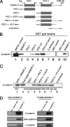
NHERF-2 interacts with β-catenin in vitro and in vivo. (A) Schematic of tagged NHERF-2 proteins, indicating the tag, the two PDZ domains, and the ERM-binding domain (ERM BD). (B) An equal amount of each GST-tagged NHERF protein was immobilized on glutathione resin and incubated with SW707 cell lysate. The bound proteins were resolved by SDS-PAGE and immunoblotted for β-catenin (lanes 2–10). Lane 1 shows β-catenin in the SW707 extract. (C) A431 cells were transfected with 2× myc-tagged versions of the constructs shown in A. Cells were extracted, and lysate was normalized for myc-tagged protein expression, immunoprecipitated with anti-myc, resolved by SDS-PAGE, and immunoblotted for β-catenin. (D) Membrane fractions prepared from A431 cells or HT1080 cells expressing full-length myc-tagged NHERF-2 were extracted with NP-40, immunoprecipitated with anti-NHERF-1 (32B6) or control antibody, resolved by SDS-PAGE, and immunoblotted for β-catenin (15B8), E-cadherin (4A2), N-cadherin (13A9), and α-catenin (1G5).
Figure 4.
Figure 4.
Colocalization of NHERF-2 and β-catenin. A431 cells expressing NHERF-2 and the constructs shown in Figure 2A were fixed on coverslips and stained for β-catenin (red), using rabbit anti-β-catenin and NHERF-2 (green) using 32B6 (against PDZ II) in b, f, h, j, and l, 9E10 in d and n, and rabbit anti-myc in p. A431 cells expressing the C terminus of NHERF-2 were stained for ezrin (red) in o.
Figure 5.
Figure 5.
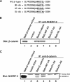
β-catenin/NHERF-2 interaction requires the PDZ-binding motif of β-catenin. (A) Batt cells were transfected with wild-type β-catenin or β-catenin with the C-terminal mutations shown. (B) Extracts of transfected cells were immunoprecipitated with anti-NHERF-2 (32B6), resolved by SDS-PAGE, and immunoblotted with anti-β-catenin (15B8). (C) Transfected Batt cell extracts were immunoprecipitated with anti-β-catenin (15B8)-conjugated Sepharose and resolved by SDS-PAGE. 15B8-conjugated beads alone were loaded in lane 2 to indicate where 15B8 IgG heavy chain migrated in the gel. An immunoblot with anti-NHERF-2 (32B6) showed that endogenous NHERF-2 coimmunoprecipitated with transfected wild-type β-catenin (lane 3) and with β-catenin containing a WF-AA mutation (lane 4) but not the other two mutant forms of β-catenin (lanes 5 and 6).
Figure 6.
Figure 6.
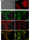
Characterization of membrane ruffles in HT1080 cells. (A) HT-1080 cells were grown on glass coverslips, processed for immunofluorescence, and stained for β-catenin. Phase (a) and fluorescence (b) micrographs are shown. (B) HT-1080 cells expressing NHERF-2 were processed for immunofluorescence and stained for β-catenin (rabbit β-catenin) and NHERF-2 (32B6). Images were deconvolved as described in Materials and Methods. β-Catenin (a) and NHERF-2 (b) were colocalized in junctions (arrowheads) and in membrane ruffles (arrows) in HT1080. A merged micrograph is shown in c. HT1080 cells expressing PDGF-Rβ were processed for immunofluorescence and costained for N-cadherin (e) and PDGF-Rβ (d), which colocalized in membrane ruffles (arrows). N-cadherin also localized to sites of cell–cell contact where PDGF-Rβ did not (arrowhead). A merged micrograph is shown in f.
Figure 7.
Figure 7.
β-Catenin and N-cadherin form a complex with PDGF-Rβ and NHERF-2 in HT1080 cells. (A) HT1080 cells expressing NHERF-2 (a and b) or the first PDZ domain of NHERF-2 (c and d) were fixed on coverslips and costained for β-catenin (rabbit) and NHERF-2 using 33E2 against PDZ I. Deconvolved images are shown. (B) The C terminus of PDGF-Rβ fused to MBP or MBP alone were immobilized on amylose and incubated with SW707 extract in the presence of GST-wild type NHERF-2 (GST-N2) or GST-PDZ I domain of NHERF-2 (PDZ I). The reactions were resolved by SDS-PAGE and immunoblotted for β-catenin (top) or GST (bottom). (C) Extracts of HT1080 cells expressing PDGF-Rβ and full-length NHERF-2 (lanes 1, 2, and 4) or the first PDZ domain of NHERF-2 (lane 3) were immunoprecipitated with control antibody (lane 2) or anti-PDGF receptor (lanes 3 and 4), resolved by SDS-PAGE and immunoblotted for β-catenin. (D) Extracts of HT1080 cells expressing PDGF-Rβ and full-length NHERF-2 (lanes 1, 2, 5, and 6) or the first PDZ domain of NHERF-2 (lanes 3 and 4) were immunoprecipitated with control antibody (lane 2), antibodies against N-cadherin (lanes 4 and 6), or antibodies against β-catenin (lanes 3 and 5) and immunoblotted for PDGF-Rβ.
Figure 8.
Figure 8.
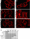
Disruption of the scaffolding activity of NHERF induces formation of actin stress fibers. Parental HT1080 cells (A), HT1080 cells expressing exogenous NHERF-2 (B), HT1080 cells expressing shRNA against N-cadherin (HT + N-cadherin KD; C), HT1080 cells expressing the PDZ I domain of NHERF-2 that cannot bind to β-catenin (D), HT1080 cells expressing shRNA against endogenous NHERF1 (HT + NHERF-1 KD; E), and HT1080 cells expressing the β-catenin mutant unable to bind to NHERF (β-cat DT-AA; F) were fixed on coverslips and stained with rhodamine-phalloidin to visualize filamentous actin. (G) Three hundred cells were evaluated, and the percentage of cells with stress fibers was graphed.
Figure 9.
Figure 9.
The N-cadherin/β-catenin/NHERF/PDGF-R complex contributes to HT1080 cell migration. (A) Confluent monolayers of parental HT1080 cells, HT1080 cells expressing exogenous NHERF-2, (HT + NHERF-2), HT1080 cells expressing the PDZ I domain of NHERF-2 that cannot bind to β-catenin (HT + PDZ I), HT1080 cells expressing shRNA against N-cadherin (HT + N-cad KD), HT1080 cells expressing shRNA against endogenous NHERF1 (HT + NHERF-1 KD), HT1080 cells expressing the β-catenin mutant unable to bind to NHERF (HT + β-cat DT-AA) or parental HT1080 cells treated with PDGF-R inhibitor (HT + PDGF-R inhibitor) were scratched with a sterile pipette tip and allowed to migrate into the wound for 10 h. (B) The above-mentioned assays were quantified by measuring the amount of denuded dish remaining at the specified times. Three independent assays were done and the SD calculated for the final time point (10 h).
Figure 10.
Figure 10.
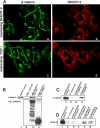
The N-cadherin/β-catenin/NHERF/PDGF-R complex contributes to HeLa cell migration. (A) HeLa cells expressing PDGF-Rβ, HeLa cells expressing PDGF-Rβ together with shRNA against N-cadherin (N-cadherin KD), or HeLa cells expressing PDGF-Rβ together with shRNA against NHERF-1 (NHERF-1 KD) were serum starved for 24 h, scratched with a sterile pipette tip, and treated with (second 3 panels) or without (first 3 panels) PDGF-BB ligand. The right two panels show HeLa cells expressing PDGF-Rβ together with shRNA against NHERF-1 and full-length NHERF-2, or PDGF-Rβ together with shRNA against NHERF-1 and the NHERF-2 PDZ I domain alone. Cells were allowed to migrate into the denuded area, and phase micrographs taken at 0, 18, and 36 h. (B) HeLa cells expressing PDGF-Rβ were extracted, immunoprecipitated with antibodies against N-cadherin, and immunoblotted for PDGF-Rβ (left) or immunoprecipitated with antibodies against PDGF-Rβ and immunoblotted for N-cadherin and β-catenin (right). On both gels, lane 1 is cell extract and lanes 2 and 3 are the immunoprecipitation reactions.

Similar articles

Cited by

References

    1. Ades E. W., Candal F. J., Swerlick R. A., George V. G., Summers S., Bosse D. C., Lawley T. J. HMEC-1, establishment of an immortalized human microvascular endothelial cell line. J. Invest. Dermatol. 1992;99:683–690. - PubMed
    1. Bienz M. beta-Catenin: a pivot between cell adhesion and Wnt signalling. Curr. Biol. 2005;15:R64–R67. - PubMed
    1. Brembeck F. H., Rosario M., Birchmeier W. Balancing cell adhesion and Wnt signaling, the key role of beta-catenin. Curr. Opin. Genet. Dev. 2006;16:51–59. - PubMed
    1. Demoulin J. B., Seo J. K., Ekman S., Grapengiesser E., Hellman U., Ronnstrand L., Heldin C. H. Ligand-induced recruitment of Na+/H+-exchanger regulatory factor to the PDGF (platelet-derived growth factor) receptor regulates actin cytoskeleton reorganization by PDGF. Biochem. J. 2003;376:505–510. - PMC - PubMed
    1. Dobrosotskaya I. Y., James G. L. MAGI-1 interacts with beta-catenin and is associated with cell-cell adhesion structures. Biochem. Biophys. Res. Commun. 2000;270:903–909. - PubMed

Publication types

MeSH terms