Helper T cell IL-2 production is limited by negative feedback and STAT-dependent cytokine signals
- PMID: 17227909
- PMCID: PMC2118423
- DOI: 10.1084/jem.20061198
Helper T cell IL-2 production is limited by negative feedback and STAT-dependent cytokine signals
Abstract
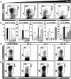
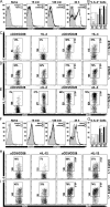
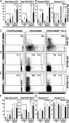
Although required for many fundamental immune processes, ranging from self-tolerance to pathogen immunity, interleukin (IL)-2 production is transient, and the mechanisms underlying this brevity remain unclear. These studies reveal that helper T cell IL-2 production is limited by a classic negative feedback loop that functions autonomously or in collaboration with other common gamma chain (IL-4 and IL-7) and IL-6/IL-12 family cytokines (IL-12 and IL-27). Consistent with this model for cytokine-dependent regulation, they also demonstrate that the inhibitory effect can be mediated by several signal transducer and activator of transcription (STAT) family transcription factors, namely STAT5, STAT4, and STAT6. Collectively, these findings establish that IL-2 production is limited by a network of autocrine and paracrine signals that are readily available during acute inflammatory responses and, thus, provide a cellular and molecular basis for its transient pattern of expression.
Figures


Similar articles
-
STAT4 and STAT6, their role in cellular and humoral immunity and in diverse human diseases.Int Rev Immunol. 2024;43(6):394-418. doi: 10.1080/08830185.2024.2395274. Epub 2024 Aug 26. Int Rev Immunol. 2024. PMID: 39188021 Review.
-
Deregulation in STAT signaling is important for cutaneous T-cell lymphoma (CTCL) pathogenesis and cancer progression.Cell Cycle. 2014;13(21):3331-5. doi: 10.4161/15384101.2014.965061. Cell Cycle. 2014. PMID: 25485578 Free PMC article.
-
STAT4/6-dependent differential regulation of chemokine receptors.Clin Immunol. 2006 Feb-Mar;118(2-3):250-7. doi: 10.1016/j.clim.2003.10.002. Epub 2006 Jan 18. Clin Immunol. 2006. PMID: 16413227
-
Transforming growth factor beta is dispensable for the molecular orchestration of Th17 cell differentiation.J Exp Med. 2009 Oct 26;206(11):2407-16. doi: 10.1084/jem.20082286. Epub 2009 Sep 28. J Exp Med. 2009. PMID: 19808254 Free PMC article.
-
The biology of Stat4 and Stat6.Oncogene. 2000 May 15;19(21):2577-84. doi: 10.1038/sj.onc.1203485. Oncogene. 2000. PMID: 10851056 Review.
Cited by
-
A novel function of IL-2: chemokine/chemoattractant/retention receptor genes induction in Th subsets for skin and lung inflammation.J Autoimmun. 2012 Jun;38(4):322-31. doi: 10.1016/j.jaut.2012.02.001. Epub 2012 Mar 29. J Autoimmun. 2012. PMID: 22464450 Free PMC article.
-
Modulation of the tumor micro-environment by CD8+ T cell-derived cytokines.Curr Opin Immunol. 2021 Apr;69:65-71. doi: 10.1016/j.coi.2021.03.016. Epub 2021 Apr 13. Curr Opin Immunol. 2021. PMID: 33862306 Free PMC article. Review.
-
Aiolos promotes TH17 differentiation by directly silencing Il2 expression.Nat Immunol. 2012 Jul 1;13(8):770-7. doi: 10.1038/ni.2363. Nat Immunol. 2012. PMID: 22751139 Free PMC article.
-
The IL-2/CD25 pathway determines susceptibility to T1D in humans and NOD mice.J Clin Immunol. 2008 Nov;28(6):685-96. doi: 10.1007/s10875-008-9237-9. Epub 2008 Sep 9. J Clin Immunol. 2008. PMID: 18780166
-
In vivo suppression of naive CD4 T cell responses by IL-2- and antigen-stimulated T lymphocytes in the absence of APC competition.J Immunol. 2008 Sep 1;181(5):3323-35. doi: 10.4049/jimmunol.181.5.3323. J Immunol. 2008. PMID: 18714004 Free PMC article.
References
-
- Smith, K.A. 1988. Interleukin-2: inception, impact, and implications. Science. 240:1169–1176. - PubMed
-
- Waldmann, T.A., S. Dubois, and Y. Tagaya. 2001. Contrasting roles of IL-2 and IL-15 in the life and death of lymphocytes: implications for immunotherapy. Immunity. 14:105–110. - PubMed
-
- Malek, T.R., and A.L. Bayer. 2004. Tolerance, not immunity, crucially depends on IL-2. Nat. Rev. Immunol. 4:665–674. - PubMed
-
- Lenardo, M.J. 1991. Interleukin-2 programs mouse alpha beta T lymphocytes for apoptosis. Nature. 353:858–861. - PubMed
Publication types
MeSH terms
Substances
Grants and funding
LinkOut - more resources
Full Text Sources
Other Literature Sources
Molecular Biology Databases
Research Materials
Miscellaneous

