Targeting TACE-dependent EGFR ligand shedding in breast cancer
- PMID: 17218988
- PMCID: PMC1764856
- DOI: 10.1172/JCI29518
Targeting TACE-dependent EGFR ligand shedding in breast cancer
Abstract
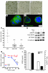
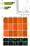
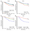
The ability to proliferate independently of signals from other cell types is a fundamental characteristic of tumor cells. Using a 3D culture model of human breast cancer progression, we have delineated a protease-dependent autocrine loop that provides an oncogenic stimulus in the absence of proto-oncogene mutation. Targeting this protease, TNF-alpha-converting enzyme (TACE; also referred to as a disintegrin and metalloproteinase 17 [ADAM17]), with small molecular inhibitors or siRNAs reverted the malignant phenotype in a breast cancer cell line by preventing mobilization of 2 crucial growth factors, TGF-alpha and amphiregulin. We show that TACE-dependent ligand shedding was prevalent in a series of additional breast cancer cell lines and, in all cases examined, was amenable to inhibition. Using existing patient outcome data, we demonstrated a strong correlation between TACE and TGFA expression in human breast cancers that was predictive of poor prognosis. Tumors resulting from inappropriate activation of the EGFR were common in multiple tissues and were, for the most part, refractory to current targeted therapies. The data presented here delineate the molecular mechanism by which constitutive EGFR activity may be achieved in tumor progression without mutation of the EGFR itself or downstream pathway components and suggest that this important oncogenic pathway might usefully be targeted upstream of the receptor.
Figures


Similar articles
-
Hormone-induced expression of tumor necrosis factor alpha-converting enzyme/A disintegrin and metalloprotease-17 impacts porcine cumulus cell oocyte complex expansion and meiotic maturation via ligand activation of the epidermal growth factor receptor.Endocrinology. 2007 Dec;148(12):6164-75. doi: 10.1210/en.2007-0195. Epub 2007 Sep 27. Endocrinology. 2007. PMID: 17901238
-
Clinical significance of heparin-binding epidermal growth factor-like growth factor and a disintegrin and metalloprotease 17 expression in human ovarian cancer.Clin Cancer Res. 2005 Jul 1;11(13):4783-92. doi: 10.1158/1078-0432.CCR-04-1426. Clin Cancer Res. 2005. PMID: 16000575
-
TACE/ADAM-17: a component of the epidermal growth factor receptor axis and a promising therapeutic target in colorectal cancer.Clin Cancer Res. 2008 Feb 15;14(4):1182-91. doi: 10.1158/1078-0432.CCR-07-1216. Clin Cancer Res. 2008. PMID: 18281553 Free PMC article.
-
TACE/ADAM17 processing of EGFR ligands indicates a role as a physiological convertase.Ann N Y Acad Sci. 2003 May;995:22-38. doi: 10.1111/j.1749-6632.2003.tb03207.x. Ann N Y Acad Sci. 2003. PMID: 12814936 Review.
-
The ADAM17-amphiregulin-EGFR axis in mammary development and cancer.J Mammary Gland Biol Neoplasia. 2008 Jun;13(2):181-94. doi: 10.1007/s10911-008-9084-6. Epub 2008 May 10. J Mammary Gland Biol Neoplasia. 2008. PMID: 18470483 Free PMC article. Review.
Cited by
-
Cellular sheddases are induced by Merkel cell polyomavirus small tumour antigen to mediate cell dissociation and invasiveness.PLoS Pathog. 2018 Sep 6;14(9):e1007276. doi: 10.1371/journal.ppat.1007276. eCollection 2018 Sep. PLoS Pathog. 2018. PMID: 30188954 Free PMC article.
-
Architecture Is the Message: The role of extracellular matrix and 3-D structure in tissue-specific gene expression and breast cancer.Pezcoller Found J. 2007 Oct;16(29):2-17. Pezcoller Found J. 2007. PMID: 21132084 Free PMC article.
-
Increased sugar uptake promotes oncogenesis via EPAC/RAP1 and O-GlcNAc pathways.J Clin Invest. 2014 Jan;124(1):367-84. doi: 10.1172/JCI63146. Epub 2013 Dec 9. J Clin Invest. 2014. PMID: 24316969 Free PMC article.
-
Tetraspanins Function as Regulators of Cellular Signaling.Front Cell Dev Biol. 2017 Apr 6;5:34. doi: 10.3389/fcell.2017.00034. eCollection 2017. Front Cell Dev Biol. 2017. PMID: 28428953 Free PMC article. Review.
-
Contribution of ADAM17 and related ADAMs in cardiovascular diseases.Cell Mol Life Sci. 2021 May;78(9):4161-4187. doi: 10.1007/s00018-021-03779-w. Epub 2021 Feb 11. Cell Mol Life Sci. 2021. PMID: 33575814 Free PMC article. Review.
References
-
- Bissell M.J., et al. Tissue structure, nuclear organization, and gene expression in normal and malignant breast. Cancer Res. 1999;59(7 Suppl.):1757s–1763s; discussion 1763s–1764s. - PubMed
-
- Hanahan D., Weinberg R.A. The hallmarks of cancer. Cell. 2000;100:57–70. - PubMed
-
- Wiesen J.F., Young P., Werb Z., Cunha G.R. Signaling through the stromal epidermal growth factor receptor is necessary for mammary ductal development. Development. 1999;126:335–344. - PubMed
-
- Luetteke N.C., et al. Targeted inactivation of the EGF and amphiregulin genes reveals distinct roles for EGF receptor ligands in mouse mammary gland development. Development. 1999;126:2739–2750. - PubMed
-
- Downward J. Targeting RAS signalling pathways in cancer therapy. Nat. Rev. Cancer. 2003;3:11–22. - PubMed
Publication types
MeSH terms
Substances
Grants and funding
LinkOut - more resources
Full Text Sources
Other Literature Sources
Medical
Research Materials
Miscellaneous

