. 2007 Jan 12;25(1):113-26.
doi: 10.1016/j.molcel.2006.11.028.

L13a blocks 48S assembly: role of a general initiation factor in mRNA-specific translational control

Affiliations

L13a blocks 48S assembly: role of a general initiation factor in mRNA-specific translational control

Purvi Kapasi et al. Mol Cell. .

Abstract

Transcript-specific translational control restricts macrophage inflammatory gene expression. The proinflammatory cytokine interferon-gamma induces phosphorylation of ribosomal protein L13a and translocation from the 60S ribosomal subunit to the interferon-gamma-activated inhibitor of translation (GAIT) complex. This complex binds the 3'UTR of ceruloplasmin mRNA and blocks its translation. Here, we elucidate the molecular mechanism underlying repression by L13a. Translation of the GAIT element-containing reporter mRNA is sensitive to L13a-mediated silencing when driven by internal ribosome entry sites (IRESs) that require initiation factor eIF4G, but is resistant to silencing when driven by eIF4F-independent IRESs, demonstrating a critical role for eIF4G. Interaction of L13a with eIF4G blocks 43S recruitment without suppressing eIF4F complex formation. eIF4G attack, e.g., by virus, stress, or caspases, is a well-known mechanism of global inhibition of protein synthesis. However, our studies reveal a unique mechanism in which targeting of eIF4G by mRNA-bound L13a elicits transcript-specific translational repression.


Figures
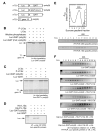
Figure 1
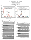
Figure 1. Translational Silencing by Phospho-L13a Blocks 80S Recruitment to Reporter mRNA Containing a GAIT Element
(A) Schematic of Luc reporter RNAs used in translation initiation reactions. m7G-capped, chimeric reporter Luc RNAs with wild-type and mutant (mut.) GAIT element, and with and without a poly(A) tail. T7 gene 10 RNA was used as a specificity control. (B) Translational silencing of GAIT element-containing reporter RNA by phospho-L13a. Luc RNAs (200 ng) were subjected to in vitro translation using [35S]Met and a RRL. T7 gene 10-poly(A) RNA (100 ng) was co-translated in the same reaction as control. Recombinant phospho-L13a (5 μg) made by baculovirus-infected insect cells was treated with shrimp alkaline phosphatase (3 U) for 90 min. Newly translated, [35S]Met-labeled proteins were resolved by 7% SDS-PAGE and detected by fluorography. (C) Poly(A) tail is required for translational silencing by phospho-L13a. Luc RNA with and T7 gene 10 RNAs were subjected to in vitro translation by a RRL. Partially-purified recombinant phosphorylated or unmodified L13a (5 μg) was added to the in vitro translation reaction. [35S]Met-labeled proteins were resolved by SDS-PAGE and detected by fluorography. (D) Poly(A) tail is not required for L13a recruitment to the GAIT element. Translation initiation reactions were reconstituted in RRL, using Luc reporters (500 ng) with or without a poly(A) tail, in the presence of His-tagged, phosphorylated or unmodified L13a (10 μg). L13 was immunoprecipitated (IP) with monoclonal anti-His antibody, and bound RNA detected by RT-PCR with the Luc-specific primers. (E) RNA detection in sucrose density gradient fractions. Initiation reaction containing Luc RNAs (500 ng) and partially purified, His-tagged phosphorylated or unmodified L13a (10 μg) was reconstituted in RRL in the presence of 0.5 mM cycloheximide. The reaction was subjected to sucrose density gradient centrifugation using 10 to 25% gradient and A254 was monitored (top panel). The 80S peak is marked by a horizontal bar. The authenticity of the 80S fractions was confirmed by RT-PCR amplification using 28S- (middle panel) and 18S- (bottom panel) specific rRNA primers. (F) Assembly of 80S ribosome complex. Initiation reactions of a Luc reporter RNA carrying a wild-type or mutant GAIT element (500 ng), and phosphorylated or unmodified L13a (5 μg), were reconstituted in a RRL and subjected to sucrose density gradient centrifugation. Fractions (1 ml) were collected from the top. Total RNA was isolated and subjected to RT-PCR using Luc-specific primers (top 6 panels). The 80S peak is marked by a horizontal bar. Migration of phospho-L13a in the gradient was determined by precipitation of the fractions with trichloroacetic acid (TCA) followed by immunoblot (IB) analysis with anti-His antibody (bottom 2 panels).
Figure 2
Figure 2. Phospho-L13a Blocks Assembly of the 48S Complex
(A) Schematic of α-globin RNA constructs used in translation initiation reactions. Shown are α-globin RNA reporters carrying the wild-type (top) or mutant (middle) GAIT element with a poly(A) tail, and α-globin RNA carrying the wild-type GAIT element without a poly(A) tail (bottom). (B) Ribosomal complex formation on GAIT element-containing RNA. Translation initiation reactions containing 32P-labeled, α-globin-GAIT-poly(A) reporters (300,000 cpm) were reconstituted in RRL in presence of GMP-PNP (black ○, 2 mM) or cycloheximide (red □, CHX, 0.5 mM) and resolved by sucrose density gradient centrifugation. Radioactivity in fractions was determined and peak positions of 48S and 80S complexes are indicated. (C) Phospho-L13a blocks 48S complex formation on a GAIT element-containing RNA in a poly(A)-dependent way. Translation initiation reactions were reconstituted in a RRL with GMP-PNP (2 mM) as in Fig. 2B, but in the presence of His-tagged, phospho-L13a (5 μg). Conditions tested were: α-globin-GAIT-poly(A) (red ▲), α-globin-GAIT-poly(A) + L13a (blue ○), α-globin-GAIT-poly(A) + phospho-L13a (black ●), α-globin-GAIT (mut.)-poly(A) + phospho-L13a (green ×), α-globin-GAIT (no poly(A) tail) + phospho-L13a (orange □). The reaction was subjected to sucrose density gradient centrifugation, fractions (0.75 ml) were collected, and radioactivity determined. (D) Phospho-L13a prevents formation of eIF3-containing 48S complex. Gradient fractions from experiments with α-globin-GAIT-poly(A) reporter and buffer control (top), phospho-L13a (middle), and unmodified L13a (bottom) were subjected to TCA precipitation followed by IB analysis with anti-eIF3 antibody. Two eIF3 subunits (44 and 110 kDa) are indicated. (E) Migration of eIF4G and phospho-L13a in the sucrose gradient. Gradient fractions from Fig. 2C containing α-globin-GAIT-poly(A) reporter and buffer control (top panel) or phospho-L13a (2nd panel) were subjected to TCA-precipitation followed by IB analysis with anti-eIF4G antibody. Migration of phospho-L13a in the presence of α-globin-GAIT-poly(A) (3rd panel), α-globin-GAIT (mut.)-poly(A) (4th panel), or α-globin-GAIT (bottom panel) was determined by IB analysis with anti-His antibody.
Figure 3
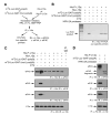
Figure 3. Formation of Cap-binding Complex, eIF4F, is not Disrupted by Phospho-L13a
(A) Schematic of initiation reactions reconstituted on GAIT element-bearing RNA with His-tagged, phospho-L13a, and immunoprecipitation and detection procedures. (B) Binding of phospho-L13a to GAIT element-bearing RNA in RRL. Translation initiation reactions were reconstituted in RRL using Luc reporters (500 ng) in presence of His-tagged, phosphorylated or unmodified L13a (10 μg). L13a was immunopre-cipitated (IP) with anti-His antibody, followed by RT-PCR with Luc-specific primers. (C) Analysis of eIF4F components interacting with RNA-bound phospho-L13a. Translation initiation reaction was reconstituted in RRL. L13a was immunoprecipitated (IP) with anti-His antibody and precipitates subjected to immunoblot analysis with anti-eIF4G (upper panel), anti-eIF4E (2nd panel), anti-eIF4A (3rd panel) and anti-L13a (bottom panel) antibodies. A control lane shows immunoprecipitation with IgG. (D) HRV-2A protease-sensitivity of eIF4F complex assembly. Translation initiation reaction was reconstituted in RRL with or without HRV-2A protease (2 μg). Immunoprecipitation (IP) and immunoblot (IB) procedures were as in Fig. 3C.
Figure 4
Figure 4. Virus IRES-driven RNAs show an eIF4F Component is Required for Translational Silencing by Phospho-L13a
(A) Schematic of ApppG-capped, Luc-GAIT element-poly(A) RNAs with CrP, HCV, or EMCV IRES sequences in the 5′ UTR (top), m7G-Luc-GAIT element-poly(A) (middle), and m7G-T7 gene 10-poly(A) (bottom). (B) In vitro translation of IRES-driven reporters. IRES-driven and m7G-capped reporter RNAs (200 ng) were subjected to in vitro translation in RRL in presence or absence of exogenous cap analog, m7G (200 μM). Newly translated, [35S]Met-labeled Luc and T7 gene 10 were resolved by SDS-PAGE (7%) and detected by fluorography. (C) Sensitivity of IRES-driven reporters to repression by phospho-L13a. ApppG-IRES-Luc-GAIT-poly(A) RNAs (200 ng) were subjected to in vitro translation in RRL with capped, T7 gene 10-poly(A) RNA (100 ng) in presence of recombinant phosphorylated or unmodified L13a (5 μg). Newly synthesized [35S]Met-labeled proteins were resolved by SDS-PAGE and detected by fluorography. (D) Ribosomal RNA detection in sucrose density gradient fractions. Initiation reaction of IRES-driven Luc RNA reporters (500 ng) were reconstituted in RRL in the presence of cycloheximide (0.5 mM). The reaction was subjected to sucrose density gradient centrifugation and A254 was monitored. The 80S peak is indicated (bar). A representative 80S peak obtained from the initiation reaction of EMCV IRES containing RNA in presence of phospho-L13a is shown. (E) Effect of phospho-L13a on 80S assembly on IRES-driven RNAs. Initiation reactions of Luc RNA-GAIT element reporters (500 ng) driven by an m7G cap (top panel), CrPV IRES (2nd panel), HCV IRES (3rd panel), or EMCV IRES (bottom panel) were reconstituted in RRL in the presence of phospho-L13a (10 μg) and cycloheximide (0.5 mM), and resolved by sucrose density gradient centrifugation. Total RNA isolated from 1 ml fractions was subjected to RT-PCR analysis using Luc-specific primers.
Figure 5
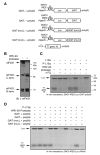
Figure 5. eIF4G C-terminal Domain is Necessary and Sufficient for Translational Silencing by Phospho-L13a
(A) Schematic of ApppG-capped, Luc-GAIT element RNAs with upstream EMCV IRES sequences containing wild-type or mutant GAIT element with and without poly(A) tail (top 4 panels). m7G-T7 gene 10-poly(A) (bottom panel). (B) Cleavage of RRL eIF4G by HRV-2A protease. Recombinant HRV-2A protease (2 μg) was incubated with RRL (100 μg) for 30 min at 37 °C, and subjected to SDS-PAGE and immunoblot analysis with anti-eIF4G antibody. The antibody is a 1:1 mixture of antisera raised against peptide sequences from the N- (amino acids 175-200) and C-(amino acids 1179-1206) termini of eIF4G1. (C) Inhibition of EMCV IRES-driven translation by phospho-L13a in the presence of HRV-2A protease. ApppG-capped Luc RNA (200 ng) containing GAIT element with EMCV IRES and m7G capped T7 gene 10 RNA (above) were translated in RRL with phosphorylated or unmodified L13a, and in presence or absence of exogenous m7G cap and HRV-2A protease. Newly translated, [35S]Met-labeled proteins were resolved by SDS-PAGE and detected by fluorography. (D) Inhibition of EMCV IRES-driven translation by phospho-L13a is poly(A)-independent when eIF4G is cleaved. ApppG-capped Luc RNA (200 ng) with EMCV IRES containing GAIT element with or without the poly(A) tail, and m7G capped T7 gene 10 RNA, were translated in RRL in presence or absence of phosphorylated L13a, and HRV-2A protease. Newly translated, [35S]Met-labeled proteins were resolved by SDS-PAGE and detected by fluorography.
Figure 6
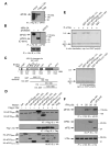
Figure 6. L13a and eIF3 Share a Common eIF4G Binding Site
(A) L13a binds eIF4G in 293 T cells. 293T cells were transfected with pcDNA3-Flag-L13a or pcDNA3-Flag constructs. After 20 h, cell extracts were prepared and subjected to immunoprecipitation (IP) with anti-Flag antibody. The immunoprecipitates were resolved by SDS-PAGE followed by immunoblot (IB) with anti-eIF4G antibody. (B) L13a binds the C-terminal fragment of eIF4G. eIF4G in RRL was cleaved by HRV-2A protease. His-tagged phospho-L13a was incubated with RRL in presence or absence of HRV-2A protease followed by immunoprecipitation with anti-His antibody and immunoblot with anti-eIF4G antibody, or SP2/O as control antibody (upper panel). Immunoprecipitation efficiency was shown by re-probing the blot with anti-L13a antibody (lower panel). (C) Domain organization of eIF4G. Domains of eIF4G responsible for binding of eIF4E, eIF4A, eIF3, PABP, MnK1, and EMCV IRES, and the cleavage site of HRV-2A protease are shown. Schematic showing eIF4G deletion fragments made as GST-fusion proteins or HA-tagged protein is indicated below. (D) Mapping the interacting domain of eIF4G with L13a. 293T cells were co-transfected with Flag-L13a and HA-tagged eIF4G(688–1560), eIF4G(970–1560), or eIF4G(1075–1560). Cell lysates were immunoprecipitated with anti-Flag antibody. Immunoprecipitates were resolved by SDS-PAGE and subjected to immunoblot analysis with anti-HA (upper panel) or with anti-Flag (middle panel) antibodies. Aliquots of lysates without immunoprecipitation were probed by immunoblot with anti-HA antibody (lower panel). (E) Exogenous eIF4G C-terminal fragment overcomes repression by phospho-L13a. Phospho-L13a (5 μg) was pre-incubated with partially-purified GST fusion proteins (50 μg) eIF4G(688–1131), eIF4G(688–1560), and eIF4G(1075–1560), and GST control protein. Pre-incubated solutions were added to RRL and cap-Luc-GAIT element-poly(A) (200 ng) and T7 gene 10-poly(A) were subjected to in vitro translation (upper panel). As a control, partially-purified GST fusion proteins containing eIF4G domains were added to the in vitro translation reaction in the absence of phospho-L13a (lower panel). Newly synthesized [35S]Met-labeled proteins were resolved by SDS-PAGE and detected by fluorography. (F) Overexpression of eIF3e blocks binding of L13a to eIF4G. U937 cells were transfected with HA-tagged eIF3e or eIF3j by electroporation using Amaxa method. 5 x 10⁶ U937 cells were treated with IFN-γ for 4 or 20 h. Cell lysates were subjected to immunoprecipitation (IP) with anti-L13a antibody and immunoblot (IB) with anti-eIF4G (top panel), anti-HA (middle panel), or anti-L13a (bottom panel) antibodies. IP with anti-L13a antibody was done using Sieze-X immunoprecipitation kit (Pierce) to avoid contamination from the antibody light and heavy chains.
Figure 7
Figure 7. Global and Transcript-specific Translational Silencing by Targeting eIF4G
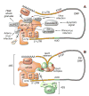
(A) Global inhibition of translation by targeting eIF4G. Virus infection, apoptosis, and cell stress target eIF4G by different mechanisms. (B) L13a targets eIF4G for transcript-specific inhibition of translation. L13a in the 3′ UTR-bound GAIT complex targets eIF4G without cleavage of eIF4G or disruption of cap-binding eIF4F complex. Binding of phospho-L13a to eIF3 binding site of eIF4G blocks interaction of 43S ribosomal complex and represses translation initiation.

Similar articles

Cited by

References

    1. Anthony DD, Merrick WC. Analysis of 40 S and 80 S complexes with mRNA as measured by sucrose density gradients and primer extension inhibition. J Biol Chem. 1992;267:1554–1562. - PubMed
    1. Aragon T, de la Luna S, Novoa I, Carrasco L, Ortin J, Nieto A. Eukaryotic translation initiation factor 4GI is a cellular target for NS1 protein, a translational activator of influenza virus. Mol Cell Biol. 2000;20:6259–6268. - PMC - PubMed
    1. Beckmann K, Grskovic M, Gebauer F, Hentze MW. A dual inhibitory mechanism restricts msl-2 mRNA translation for dosage compensation in Drosophila. Cell. 2005;122:529–540. - PubMed
    1. Bergamini G, Preiss T, Hentze MW. Picornavirus IRESes and the poly(A) tail jointly promote cap-independent translation in a mammalian cell-free system. Rna. 2000;6:1781–1790. - PMC - PubMed
    1. Cho PF, Poulin F, Cho-Park YA, Cho-Park IB, Chicoine JD, Lasko P, Sonenberg N. A new paradigm for translational control: inhibition via 5′-3′ mRNA tethering by Bicoid and the eIF4E cognate 4EHP. Cell. 2005;121:411–423. - PubMed

Publication types

MeSH terms