Comparative Study
. 2007 Jan 24;26(2):600-12.
doi: 10.1038/sj.emboj.7601501. Epub 2007 Jan 11.

Structural insight into the ESCRT-I/-II link and its role in MVB trafficking

Affiliations
Comparative Study

Structural insight into the ESCRT-I/-II link and its role in MVB trafficking

David J Gill et al. EMBO J. .

Abstract

ESCRT (endosomal sorting complex required for transport) complexes orchestrate efficient sorting of ubiquitinated transmembrane receptors to lysosomes via multivesicular bodies (MVBs). Yeast ESCRT-I and ESCRT-II interact directly in vitro; however, this association is not detected in yeast cytosol. To gain understanding of the molecular mechanisms of this link, we have characterised the ESCRT-I/-II supercomplex and determined the crystal structure of its interface. The link is formed by the vacuolar protein sorting (Vps)28 C-terminus (ESCRT-I) binding with nanomolar affinity to the Vps36-NZF-N zinc-finger domain (ESCRT-II). A hydrophobic patch on the Vps28-CT four-helix bundle contacts the hydrophobic knuckles of Vps36-NZF-N. Mutation of the ESCRT-I/-II link results in a cargo-sorting defect in yeast. Interestingly, the two Vps36 NZF domains, NZF-N and NZF-C, despite having the same core fold, use distinct surfaces to bind ESCRT-I or ubiquitinated cargo. We also show that a new component of ESCRT-I, Mvb12 (YGR206W), engages ESCRT-I directly with nanomolar affinity to form a 1:1:1:1 heterotetramer. Mvb12 does not affect the affinity of ESCRT-I for ESCRT-II in vitro. Our data suggest a complex regulatory mechanism for the ESCRT-I/-II link in yeast.


Figures
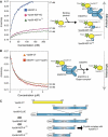
Figure 1
Figure 1
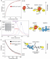
Yeast ESCRT-I binds to ESCRT-II with nanomolar affinity. (A) The fluorescence anisotropy of the isolated recombinant FlAsH-Vps28-CT increases in accordance with the size of the titrated recombinant ESCRT-II binding partner: 9.5 kDa extended Vps36-NZF-N (residues 110–176), 12.5 kDa Vps36-NZF-NC (residues 110–205) and 140 kDa full-length ESCRT-II. Direct fitting of the titration curves shows similar Kd values of Vps36-NZF-N (61 nM), Vps36-NZF-NC (57 nM) and full-length ESCRT-II (27 nM) for FlAsH-Vps28-CT. (B) In a fluorescence anisotropy competition assay, various ESCRT-I proteins are titrated into a solution containing a preformed complex of the FlAsH-Vps28-CT/full-length ESCRT-II. Titration of either ESCRT-I(Δ21-Vps37) or non-labelled Vps28-CT results in a displacement of fluorescent FlAsH-Vps28-CT from ESCRT-II and consequently, in a decrease of anisotropy. Fitting of the titration curves allows determination of Kd values for Vps28-CT (53 nM) and ESCRT-I(Δ21-Vps37) (44 nM) for ESCRT-II. (C) Constructs used to generate ESCRT-I/-II subcomplexes used in this study.
Figure 2
Figure 2
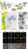
The structure of the Vps28 C-terminus (ESCRT-I) bound to the Vps36-NZF-N domain (ESCRT-II). (A) Ribbon diagrams of Vps28-CT/extended Vps36-NZF-N complex (generated by PyMOL). Two orthogonal views are shown illustrating the ‘fist'-shaped NZF-N contacting the splayed α2/α3 helices on the ‘punchbag'-shaped Vps28-CT four-helix bundle. (B) A schematic of the Vps28-CT/Vps36-NZF-N binding interface highlighting residues important for this interaction. (C) Two close-up representations of the Vps28-CT/Vps36-NZF-N binding interface. Important residues on the Vps28-CT binding interface are coloured green (hydrophobic), blue (Arg190 and Arg193) and orange (Asn210). Upper panel, critical residues in Vps36-NZF-N comprising the Vps28-CT binding motif (Ile122Vps36/Val148Vps36/Leu154Vps36) are shown as pink sticks. Lower panel, flanking polar interactions are shown. Ordered waters at the interface are shown as yellow spheres. (D) The full-length Vps36 with an I122D/V148D double mutation or an I122D/V148D/D151R/L154R quadruple mutation in the NZF-N domain exhibits a strong defect in GFP-CPS cargo sorting in the context of vps36Δ strain. The GFP-CPS cargo is mis-sorted to the limiting membrane of prevacuolar endosomes (class E compartment, arrowheads) and the limiting membrane of the vacuole (both labelled by FM4-64) upon shift to 37°C in the double mutant or at 30°C in the vps36Δ and quadruple mutant.
Figure 3
Figure 3
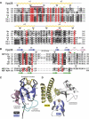
The two Vps36 NZF domains (NZF-N and NZF-C) have the same core fold with distinct binding sites for either ESCRT-I (in NZF-N) or Ub (in NZF-C). (A) Structure-based sequence alignment of the Vps28 C-terminus in ESCRT-I. Species used in this alignment are fungi S. cerevisiae (Sc), Yarrowia lipolytica (Yl) and Schizosaccharomyces pombe (Sp) and metazoa Caenorhabditis elegans (Ce), Xenopus laevis (Xl) and Homo sapiens isoforms 1 (Hs-1) and 2 (Hs-2). Invariant (red), conserved (black), similar (grey) and non-conserved residues (white) are coloured with BOXSHADE using a sequence identity cutoff of 50%. Secondary structure is shown schematically above the Sc sequence. Important functional residues in the Vps28-CT/Vps36-NZF-N binding interface are labelled with asterisks (hydrophobic green, positively charged as blue and polar as orange). The conserved surface patch on Vps28 is marked with a maroon bar. Residues implicated in binding to ScVps20 (Pineda-Molina et al, 2006) are labelled with red asterisks. Polar residues in X. laevis Vps28-CT spatially equivalent to the ScVps28-CT residues that are involved in the Vps36-NZF-N binding site are shown as red circles. (B) Structure-based sequence alignment of the Vps28-CT-binding and Ub-binding NZF domains: Vps36-NZF-N domains of S. cerevisiae (Sc), Candida glabrata (Cg), Aspergillus nidulans (An), Y. lipolytica (Yl), S. pombe (Sp) and Ustilago maydis (Um) against the Ub-binding Sc Vps36-NZF-C domain (NZF-C Sc) and the rat Npl4-NZF domain (NZF Npl4). Important hydrophobic residues in the Vps36-NZF-N and Npl4-NZF domains for binding Vps28-CT and Ub are marked with green and brown bars, respectively. Asp151 in Vps36-NZF-N important for binding Vps28-CT is marked with a red bar. Secondary structure elements of Vps36-NZF-N and rat Npl4-NZF domains are shown above and below the sequence alignment. (C) Structural alignment of the Vps36-NZF-N (coloured ribbon diagram) and Npl4-NZF (black worm) (1Q5W) domains reveal a core common fold with additional features in Vps36-NZF-N (C-terminal extension in pink and extended β2/β3 loop in cyan). Cysteine residues binding to the Zn2+ ion in the Vps36-NZF-N (yellow sticks) and Npl4-NZF domains (black sticks) are shown. (D) Vps36-NZF-N and Vps36-NZF-C domains use distinct binding sites on the rubredoxin knuckles for binding to ESCRT-I and Ub. The NZF domains from the crystal structures of Vps36-NZF-N bound to ESCRT-I and Npl4-NZF bound to Ub (1Q5W) were superimposed, and the Vps28-CT (yellow) and Ub (silver) ligands are shown on the Vps36-NZF-N domain (blue).
Figure 4
Figure 4
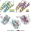
The structural comparison of the isolated S. cerevisiae and X. laevis ESCRT-I Vps28 C-terminus reveals that the four-helix bundle fold is rigid and highly conserved throughout evolution. (A) Alignment of the isolated (cyan) and Vps36-NZF-N-bound (yellow) S. cerevisiae Vps28-CT domains (left panel) and the S. cerevisiae (yellow) and X. laevis (purple) Vps28-CT domains (right panel). (B) Three orthogonal views of conserved surface residues calculated using CONSURF (Landau et al, 2005) displayed on the semi-transparent surface of the isolated S. cerevisiae Vps28-CT structure. Sequence conservation is scored by colour (from most conserved (maroon) to most variable (green)) and displayed by PyMOL. The 238-FDxE-241 sequence motif is shown as black sticks.
Figure 5
Figure 5
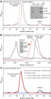
A new ESCRT-I component, Mvb12, forms a stable heterotetrameric ESCRT-I complex in vitro. (A) Gel filtration of recombinant Mvb12 (20 μM), ESCRT-I(Δ21-Vps37) (20 μM) or their mixture (20 μM each) on a Superdex 200 10/300 column. SDS analysis of input ESCRT-I(Δ21-Vps37) and Mvb12 proteins (each diluted to 0.4 mg/ml) is shown alongside a peak fraction from the ESCRT-I(Δ21-Vps37)/Mvb12 complex. (B) Elution profiles of recombinant aggregated heterotrimeric ESCRT-I (≫670 kDa), heterotetrameric ESCRT-I/Mvb12 (313 kDa) and heterotrimeric ESCRT-I(Δ21-Vps37) (233 kDa) (all 6 μM) on a Superdex 200 10/300 column. SDS analysis of a peak fraction from the heterotetrameric ESCRT-I/Mvb12 complex is shown as an inset. (C) Gel filtration of pre-mixtures of ESCRT-I(Δ21-Vps37) (20 μM) with Mvb12 (20 μM: red solid line; 30 μM: grey dashed line; 40 μM: black dashed line) and Mvb12 alone (20 μM: blue solid line) on a Superdex 200 10/300 column.
Figure 6
Figure 6
Mvb12 binds to ESCRT-I with nanomolar affinity and does not affect the affinity of ESCRT-I for recombinant ESCRT-II in vitro. (A) The fluorescence anisotropy of the isolated recombinant FlAsH-Mvb12 increases when titrated with recombinant ESCRT-I complexes. Direct fitting of the titration curves shows that ESCRT-I(Δ21-Vps37) binds to FlAsH-Mvb12 with a Kd of approximately 28 nM. (B) Elution profiles of ESCRT-I/Mvb12 alone (6 μM, peak 1), ESCRT-II alone (6 μM, peak 2) and their mixture (6 μM each, peak 3) on a Superdex 200 10/300 column showing formation of an ESCRT-I/Mvb12/ESCRT-II supercomplex. SDS–PAGE of samples from each gel filtration is shown as an inset. (C) In a fluorescence anisotropy competition assay, both heterotrimeric ESCRT-I(Δ21-Vps37) and heterotetrameric ESCRT-I(Δ21-Vps37)/Mvb12 are equally efficient competitors of an FlAsH-Vps28-CT/ESCRT-II complex (Kd 44 and 30 nM, respectively).

Similar articles

Cited by

References

    1. Adams SR, Campbell RE, Gross LA, Martin BR, Walkup GK, Yao Y, Llopis J, Tsien RY (2002) New biarsenical ligands and tetracysteine motifs for protein labeling in vitro and in vivo: synthesis and biological applications. J Am Chem Soc 124: 6063–6076 - PubMed
    1. Alam SL, Sun J, Payne M, Welch BD, Blake BK, Davis DR, Meyer HH, Emr SD, Sundquist WI (2004) Ubiquitin interactions of NZF zinc fingers. EMBO J 23: 1411–1421 - PMC - PubMed
    1. Babst M, Katzmann DJ, Estepa-Sabal EJ, Meerloo T, Emr SD (2002a) ESCRT-III: an endosome-associated heterooligomeric protein complex required for MVB sorting. Dev Cell 3: 271–282 - PubMed
    1. Babst M, Katzmann DJ, Snyder WB, Wendland B, Emr SD (2002b) Endosome-associated complex, ESCRT-II, recruits transport machinery for protein sorting at the multivesicular body. Dev Cell 3: 283–289 - PubMed
    1. Bache KG, Stuffers S, Malerod L, Slagsvold T, Raiborg C, Lechardeur D, Walchli S, Lukacs GL, Brech A, Stenmark H (2006) The ESCRT-III subunit hVps24 is required for degradation but not silencing of the epidermal growth factor receptor. Mol Biol Cell 17: 2513–2523 - PMC - PubMed

Publication types

MeSH terms

Substances